Key Points
iMC expand independent of the type of antitumor response and are not immunosuppressive in a cell-autonomous fashion
iMC are licensed to become MDSC in vivo in the presence of GM-CSF
Abstract
Tumors frequently induce immature myeloid cells (iMC), which suppress specific and unrelated cytotoxic T lymphocyte (CTL) responses and are termed myeloid-derived suppressor cells (MDSC). Mainly analyzed by in vitro assays in tumor transplantation models, little is known about their function in autochthonous tumor models in vivo. We analyzed iMC in 3 SV40 large T (Tag)-driven conditional autochthonous cancer models with different immune status: (1) Early Tag-specific CTL competence and rare stochastic Tag activation leading to sporadic cancer, which induces an aberrant immune response and CTL tolerance; (2) Cre/LoxP recombinase-mediated hepatocellular carcinoma (HCC) development in neonatal Tag-tolerant mice; and (3) Tag-activation through Cre recombinase-encoding viruses in the liver and HCC development with systemic anti-Tag CTL immunity. In the first but not two latter models, tumors induced CTL hyporesponsiveness to tumor-unrelated antigens. Regardless of the model, tumors produced interleukin-6 and vascular endothelial growth factor but not granulocyte macrophage–colony-stimulating factor (GM-CSF) and induced iMC (CD11b+Gr-1int) that suppressed CTL responses in vitro. None of the iMC from the different tumor models suppressed CTL responses in adoptive cell transfer experiments unless GM-CSF was provided in vivo. Together, iMC expand independent of the type of antitumor response and are not immunosuppressive in a cell-autonomous fashion.
Introduction
Immature myeloid cells (iMC) represent a heterogeneous population of innate immune cells at early stages of differentiation and were first characterized by the expression of the markers CD11b (Mac-1) and Gr-1 (Ly-6C/G). In healthy mice, small numbers of iMC are detected in blood and secondary lymphoid organs, but iMC frequently expand under various pathologic/inflammatory conditions, including cancer.1 Because of their immunosuppressive function in transplantable tumor models, subsets of iMC have been termed myeloid-derived suppressor cells (MDSC).2 MDSC participate in an immunosuppressive network responsible for diminishing cytotoxic T lymphocyte (CTL) responses.1,3-7
Analysis of spontaneous adaptive immune responses during autochthonous tumor development, which means the cancer evolves in the original host and is not induced by cancer cell inoculation, requires the knowledge of a tumor antigen to which the host can efficiently respond before tumor onset. We have established a mouse model of SV40 T antigen (Tag)-driven cancer (LoxP-Tag), in which the Tag gene is separated from an ubiquitously active promotor by a stop cassette.8 CTLs against the dominant Tag epitope peptide IV (pIV)9 are retained and confer life-long protection if induced by vaccination before a tumor develops.8 Without vaccination, sporadic tumors develop as a result of genetic or epigenetic events in individual cells, which induce CTL tolerance and expansion of anergic Tag-specific CTLs. Importantly, the sporadic cancer had retained its high immunogenicity because it was rejected in immunocompetent mice upon transplantation (“regressor” phenotype). Probably because of chronic exposure to Tag and aberrant immune reaction (eg, anti-Tag IgG production), the mice develop a general CTL hyporesponsiveness toward tumor-unrelated antigens during the long period between premalignancy and tumor progression. This latency is associated with the expansion of iMC.10 Because these autochthonous tumors elicit a strong but noneffective immune reaction in the primary host, we define the sporadic cancer here as “pseudo-immunogenic.” Tumors in LoxP-Tag mice can also be induced by Cre recombinase-mediated stop cassette deletion, either genetically (eg, by crossing to Alb-Cre transgenic mice) or through virus infection (eg, with Cre recombinase-encoding adenoviruses, Ad.Cre). Because Tag is the dominant transplantation rejection antigen in this model, and LoxP-Tag × Alb-Cre mice are completely tolerant for Tag because of neonatal Tag expression in the liver and thus do not induce a measurable Tag-specific immune response, tumors in these mice are considered “nonimmunogenic.” The nonimmunogenic tumors expanded iMC but did not inhibit CTL responses to tumor-unrelated antigens.10 Cre/LoxP-mediated Tag activation during viral infection using an adenovirus encoding the Cre recombinase induced pIV-specific CTLs, which initially inhibited tumor development and then remained systemically functional despite subsequent progression of Tag+ tumors.11 Thus, these tumors were defined as “immunogenic” and, as shown here, also induced iMC but no impairment of tumor unrelated CTL responses. Contrasting existing data from tumor transplantation models, we show here that tumor-induced iMC suppressed CTLs in vitro but not in vivo in a cell-autonomous fashion, regardless of the model. Nonetheless, iMC became competent MDSC in vivo following adoptive transfer when exposed to granulocyte macrophage–colony-stimulating factor (GM-CSF).
Materials and methods
Animals
Generation and characterization of LoxP-Tag, LoxP-Tag × Alb-Cre, Rag1−/− × P14, and CD45.1 mice have previously been described.8,10,12-14 Virus-induced hepatocellular carcinoma (HCC) developed after IV injection of 2-month-old LoxP-Tag mice with 1 × 10⁹ plaque-forming units Ad.Cre. Mice considered as positive for autochthonous tumors (advanced stage) had at least a twofold increase in weight of the affected organ. C57BL/6 (B6) mice were obtained from Charles River Laboratories (Wilmington, MA). Animals were housed in our animal facilities and all animal experiments were performed according to governmental guidelines (Landesamt für Gesundheit und Soziales Berlin).
Cell lines and tumor transplantation
In vivo cytotoxicity assays
Mice were immunized by intraperitoneal injection of either 1 × 10⁷ Tag+ 16.113 cells, 5 × 10⁶ male spleen cells, or 1 × 10⁷ MC57-gp33-Hi cells. In vivo cytotoxicity analyses were previously described.10 Briefly, modified Tag pIV (VVYDFLKL)-loaded splenocytes or gp33 (KAVYNFATM)-loaded splenocytes (each 2 µg/mL) were combined with nonloaded splenocytes. For antigen HY–specific assays, male splenocytes were combined with female splenocytes. Each cell population (1 × 10⁷) was labeled with a different amount of carboxyfluorescein succinimidyl ester (CFSE) (0.75 µM, CFSEhigh for peptide-loaded cells; 0.075 µM, CFSElow for non–peptide-loaded cells) and transferred together IV into the recipients. Eighteen hours later, the CFSE-labeled populations were analyzed by flow cytometry. In some cases, two epitopes were analyzed at the same time. In these experiments, spleen cells from CD45.1 mice were labeled with CFSE at final concentrations of 0.75 µM (CFSEhigh), 0.075 µM (CFSEint), and 0.0075 µM (CFSElow), respectively. Eighteen hours after adoptive transfer, target cells were stained with anti-CD45.1 antibodies and were analyzed by flow cytometry.
Flow cytometric analysis and cell sorting
For flow cytometry, single-cell suspensions of spleen cells were stained with monoclonal antibodies specific for CD11b (M1/70), Gr-1 (RB6-8C5), CD8α (53-6.7), and CD45.1 (A20) (BD Biosciences, Heidelberg, Germany).
For in vitro and in vivo analysis, iMC were separated using magnetic MicroBeads (Miltenyi Biotec, Bergisch Gladbach, Germany). iMC of 50% purity (tumor-free mice) or 60% to 93% purity (tumor-bearing mice) were isolated from spleens of tumor-free or tumor-bearing mice using the Myeloid-Derived Suppressor Cell Isolation Kit (Miltenyi Biotec) according to the manufacturer’s instructions. In brief, single-cell preparations were first pretreated with Fc receptor block. Then, CD11b+Gr-1high iMC were separated with an anti-Ly-6G antibody. The CD11b+Gr-1int iMC fraction was then separated from the Ly-6G-negative fraction with an anti-Gr-1 antibody. The samples were separated on LS MACS Columns (Miltenyi Biotec).
In vitro suppression assays
Splenocytes were isolated from B6 mice for polyclonal activation or from Rag1−/−/P14 T-cell receptor (TCR)-transgenic mice for peptide-specific activation and labeled with CFSE (5 µM) for 15 minutes at 37°C in phosphate-buffered saline supplemented with 3% fetal calf serum. Six × 105 CFSE-labeled splenocytes were then activated in vitro with anti-CD3ε (145-2C11; 3 µg/mL, plate-bound) and anti-CD28 (37.51; 2 µg/mL) antibodies, or gp33 peptide (2 µg/mL) in the presence of iMC. After 72 hours, CFSE dilution was analyzed by flow cytometry.
Adoptive transfer of iMC
In vivo function of iMC was analyzed in B6 mice. iMC were separated with the Myeloid-Derived Suppressor Cell Isolation Kit (Miltenyi Biotec) from spleens of tumor-bearing mice as described previously. Five × 106 iMC were injected IV into B6 recipients. Within 1 hour, the mice were immunized subcutaneously with an emulsion containing 100 µg pIV in incomplete Freund’s adjuvant (Sigma, Munich, Germany). Eight to 10 days later, cytotoxicity against pIV was analyzed in vivo as described previously. Control groups (no iMC) for CD11b+Gr-1high and CD11b+Gr-1int iMC transfers were the same.
Cytokine measurements
Cytokine levels were determined in serum samples and cell culture supernatants. Vascular endothelial growth factor (VEGF), GM-CSF and transforming growth factor (TGF)-β1 (MB100) were determined by enzyme-linked immunosorbent assay (R&D Systems Wiesbaden-Nordenstadt, Germany) according to the manufacturer’s instructions. Interleukin (IL)-6 was determined by FlowCytomix multianalyte immunoassays (Bender Medsystems, Vienna, Austria) according to the manufacturer’s instructions.
Generation of iMC in the presence of GM-CSF
For GM-CSF production in vivo, 16.113 tumor cells were retrovirally transduced with pMFG-GM-CSF16 and produced ∼207 ng/mL/106 cells/day mouse GM-CSF without further selection (16.113-GM). One × 107 parental 16.113 tumor cells or 1 × 10⁷ cells of a bulk culture of 16.113-GM cells were injected subcutaneously into 4- to 6-week-old LoxP-Tag × Alb-Cre mice. After 3 weeks, CD11b+Gr-1int iMC were isolated and had a purity of 90% (without GM-CSF; n = 3) and 89% (with GM-CSF; n = 3). At this time point, 16.113 and 16.113-GM tumors had reached a size of 50 to 100 mm3 and primary HCC was detectable microscopically (early HCC). Serum levels of GM-CSF in LoxP-Tag × Alb-Cre mice bearing 16.113-GM tumors were 21+/− 9 pg/mL (n = 10).
Statistic reports
Statistical analyses were performed with help of Andrea Stroux (Department of Biostatistics and Clinical Epidemiology, Charité Campus Benjamin Franklin, Berlin, Germany) using SPSS Software. The overall significance of each graph was calculated with the Kruskal-Wallis test. Comparisons of 2 groups were done by Mann-Whitney U test. P values of less than .05 were regarded as statistically significant.
Results
Pseudo-immunogenic but not nonimmunogenic or immunogenic tumors induce general CTL hyporesponsiveness in vivo
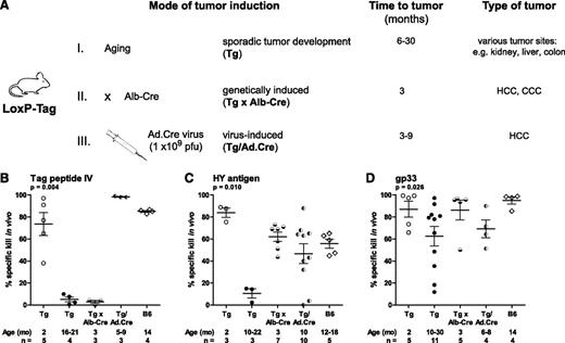
Pseudo-immunogenic sporadic tumors in LoxP-Tag (Tg), nonimmunogenic tumors in LoxP-Tag × Alb-Cre (Tg × Alb-Cre), and immunogenic tumors in Ad.Cre-infected LoxP-Tag mice (Tg/Ad.Cre) mice developed with different kinetics (Figure 1A). Tg × Alb-Cre and, because of viral tropism, Tg/Ad.Cre mice develop HCC; Tg mice usually develop 1 macroscopically visible tumor in various organs including liver; nevertheless, we found that the tumor site did not influence systemic immune responses. We first compared in vivo CTL responses toward the oncogene Tag (pIV) and 2 tumor-unrelated antigens in the 3 tumor models. Although CD8+ T cells exhibited strong responsiveness for pIV upon immunization at young age (2-month-old Tg, 73.6%), no activity was detected in Tg mice with sporadic tumors (16- to 21-month-old Tg). Most likely because of Tag expression early in life, Tg × Alb-Cre mice did not respond to pIV-loaded targets. Notably, Tag-immunized Tg/Ad.Cre mice bearing HCC showed vigorous CTL killing of the pIV-loaded targets (98.3%), comparable to CTL activity in Tag-immunized young Tg controls and 14-month-old C57BL/6 (B6) mice (85.1%; Figure 1B). To investigate tumor-unrelated antigens, CTL reactivity against minor histocompatibility antigens HY and the lymphocytic choriomeningitis virus–derived epitope gp33 was analyzed. Following HY-specific immunization, CTLs from tumor-bearing female Tg mice barely eliminated male splenocytes (10.5%), whereas anti-HY CTL responses in Tg × Alb-Cre (62.1%) and Tg/Ad.Cre (46.7%) mice were comparable to immunized young Tg (84.0%) and 12- to 18-month-old B6 mice (55.8%; Figure 1C). Upon gp33-specific immunization, in accordance with our previous data, some, but not all Tg mice with sporadic tumors showed reduced cytotoxicity toward gp33. Again, Tg/Ad.Cre mice (69.2%) and Tg × Alb-Cre mice (86.3%) as well as young Tg (87.1%) and 14-month-old B6 mice (95.0%) attained gp33-specific killing in vivo (Figure 1D). Together, only pseudo-immunogenic but not nonimmunogenic or immunogenic tumors induce general CTL hyporesponsiveness.
Sporadic, but not virus-induced immunogenic tumors cause general CTL hyporesponsiveness in vivo. (A) Three mouse models of SV40 large T tumor antigen (Tag)-driven cancer development. (i) Sporadic tumor development resulting from genetic or epigenetic events in individual cells. (ii) Tissue-specific expression of Cre recombinase by the albumin promoter induces hepatocellular carcinoma (HCC). (iii) HCC develops after infection with Cre recombinase-expressing adenoviruses (Ad.Cre). (B-D) CTL activities against Tag and 2 tumor-unrelated antigens were analyzed after immunization. Tumor-bearing mice and control mice were immunized with Tag+ 16.113 tumor cells, male splenocytes (female mice only), or MC57-gp33-Hi tumor cells. The percentage of specific killing in vivo is indicated. (B) In vivo cytotoxicity toward Tag peptide IV (pIV) was analyzed in young LoxP-Tag mice (Tg, 2 months), LoxP-Tag mice bearing sporadic tumors (Tg; HCC n = 2, other n = 2), HCC-bearing LoxP-Tag × Alb-Cre mice (Tg × Alb-Cre), Ad.Cre-infected LoxP-Tag mice bearing virus-induced HCC (Tg/Ad.Cre), and old C57BL/6 mice (B6, 14 months). (C) Analysis of CTL activity toward HY antigens in young Tg mice, Tg mice (kidney tumor n = 2, other n = 1), Tg × Alb-Cre mice, Tg/Ad.Cre mice, and old B6 mice. (D) In vivo cytotoxicity toward gp33 was analyzed in young Tg mice, Tg mice (kidney tumor n = 5, HCC n = 4, other n = 2), Tg × Alb-Cre mice, Tg/Ad.Cre mice, and old B6 mice. For each antigen, data were pooled from at least 2 independent experiments. Horizontal bars show means; error bars represent standard error of the mean (SEM). Overall significance for each graph was tested by Kruskal–Wallis 1-way analysis of variance.
Sporadic, but not virus-induced immunogenic tumors cause general CTL hyporesponsiveness in vivo. (A) Three mouse models of SV40 large T tumor antigen (Tag)-driven cancer development. (i) Sporadic tumor development resulting from genetic or epigenetic events in individual cells. (ii) Tissue-specific expression of Cre recombinase by the albumin promoter induces hepatocellular carcinoma (HCC). (iii) HCC develops after infection with Cre recombinase-expressing adenoviruses (Ad.Cre). (B-D) CTL activities against Tag and 2 tumor-unrelated antigens were analyzed after immunization. Tumor-bearing mice and control mice were immunized with Tag+ 16.113 tumor cells, male splenocytes (female mice only), or MC57-gp33-Hi tumor cells. The percentage of specific killing in vivo is indicated. (B) In vivo cytotoxicity toward Tag peptide IV (pIV) was analyzed in young LoxP-Tag mice (Tg, 2 months), LoxP-Tag mice bearing sporadic tumors (Tg; HCC n = 2, other n = 2), HCC-bearing LoxP-Tag × Alb-Cre mice (Tg × Alb-Cre), Ad.Cre-infected LoxP-Tag mice bearing virus-induced HCC (Tg/Ad.Cre), and old C57BL/6 mice (B6, 14 months). (C) Analysis of CTL activity toward HY antigens in young Tg mice, Tg mice (kidney tumor n = 2, other n = 1), Tg × Alb-Cre mice, Tg/Ad.Cre mice, and old B6 mice. (D) In vivo cytotoxicity toward gp33 was analyzed in young Tg mice, Tg mice (kidney tumor n = 5, HCC n = 4, other n = 2), Tg × Alb-Cre mice, Tg/Ad.Cre mice, and old B6 mice. For each antigen, data were pooled from at least 2 independent experiments. Horizontal bars show means; error bars represent standard error of the mean (SEM). Overall significance for each graph was tested by Kruskal–Wallis 1-way analysis of variance.
Tumor-induced expansion of iMC and in vitro CTL suppression by the CD11b+Gr-1int iMC subset does not correlate with CTL suppression in vivo
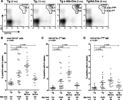
Accepting the different quality of systemic antitumor and tumor-unrelated CTL responses as a reference point in the 3 models and the assumption that T-cell priming occurs in secondary lymphoid organs but not the tumor, we analyzed the role of splenic iMC in CTL suppression. Spleens of young Tg mice comprised 3.7% CD11b+ cells. Tumor-bearing mice of all 3 models showed significantly elevated myeloid populations. Spleens of tumor-bearing Tg mice contained 9.2%, Tg × Alb-Cre mice 13.2% and Tg/Ad.Cre 5.9% CD11b+ cells (Figure 2A-B and supplemental Figure 1). Within the CD11b+ myeloid cells, the CTL suppressive activity of iMC has been attributed to CD11b+Gr-1int but not CD11b+Gr-1high cells.17,18 Therefore, we analyzed iMC in spleens of mice without or with autochthonous tumors according to the brightness in Gr-1 staining and found 0.5% and 1.1% of the respective CD11b+Gr-1high and CD11b+Gr-1int iMC in young Tg mice, 1.4% and 3.9% in Tg mice with sporadic tumors, 1.7% and 7.5% in HCC-bearing Tg × Alb-Cre mice, and 0.9% and 2.2% in Tg/Ad.Cre mice with virus-induced HCC (Figure 2C-D). This indicated that the CD11b+Gr-1int subset of iMC accumulated in mice developing autochthonous tumors, irrespective of the tumor model and the immune status of the mice.
iMC expand upon autochthonous tumor development. Single-cell preparations of spleens were stained with antibodies against CD11b and Gr-1. Spleens were derived from young LoxP-Tag mice (Tg, 2-4 months), LoxP-Tag mice bearing sporadic tumors (Tg; kidney tumor n = 7, HCC n = 5, colon tumor n = 4, others n = 3), HCC-bearing LoxP-Tag × Alb-Cre mice (Tg × Alb-Cre), and Ad.Cre-infected LoxP-Tag mice bearing virus-induced HCC (Tg/Ad.Cre). (A) One representative example per experimental group is shown; numbers indicate the percentage CD11b+Gr-1high and CD11b+Gr-1int iMC of total spleen cells. All data are summarized in (B-D). The percentage of total CD11b+ cells (B), CD11b+Gr-1int iMC (C), and CD11b+Gr-1high iMC (D) in spleens of young Tg mice, tumor-bearing Tg mice, Tg × Alb-Cre mice, and Tg/Ad.Cre mice is indicated. Horizontal bars show means; error bars represent SEM. Overall significance for each graph was tested by Kruskal–Wallis 1-way analysis of variance.
iMC expand upon autochthonous tumor development. Single-cell preparations of spleens were stained with antibodies against CD11b and Gr-1. Spleens were derived from young LoxP-Tag mice (Tg, 2-4 months), LoxP-Tag mice bearing sporadic tumors (Tg; kidney tumor n = 7, HCC n = 5, colon tumor n = 4, others n = 3), HCC-bearing LoxP-Tag × Alb-Cre mice (Tg × Alb-Cre), and Ad.Cre-infected LoxP-Tag mice bearing virus-induced HCC (Tg/Ad.Cre). (A) One representative example per experimental group is shown; numbers indicate the percentage CD11b+Gr-1high and CD11b+Gr-1int iMC of total spleen cells. All data are summarized in (B-D). The percentage of total CD11b+ cells (B), CD11b+Gr-1int iMC (C), and CD11b+Gr-1high iMC (D) in spleens of young Tg mice, tumor-bearing Tg mice, Tg × Alb-Cre mice, and Tg/Ad.Cre mice is indicated. Horizontal bars show means; error bars represent SEM. Overall significance for each graph was tested by Kruskal–Wallis 1-way analysis of variance.
Commonly, in vitro CTL suppression assays are used to analyze suppressive properties of iMC.7,19 To analyze the function of iMC induced by autochthonous tumors in the different types of mice, 2 different coculture experiments were performed. CTL were activated either polyclonally (Figure 3A) or antigen-specifically (gp33) (Figure 3B) in vitro in the presence of titrated amounts of enriched iMC subsets (ranging from 0% to 24%). Splenic CD11b+ cells and CD11b+Gr-1int iMC of tumor-bearing Tg, Tg × Alb-Cre, or Tg/Ad.Cre mice but not of young Tg mice, blocked proliferation of polyclonal (Figure 3C) and gp33-specific (P14) T cells (Figure 3D) in vitro. Suppression could be maintained down to 3% to 6% CD11b+ myeloid cell proportionate and was nitric oxide–dependent (data not shown). For splenic CD11b+ cells, comparable results were obtained when analyzed in a 51Cr release assay (data not shown). Contamination of the enriched CD11b+Gr-1int iMC fractions with either CD11b− cells (4% to 21%), CD11c+ DC (1.5% to 19%), or Gr-1-F4/80+ macrophages (3% to 22%) (data not shown) did not interfere with the CD8+ T-cell suppression. As others have previously shown for the CD11b+Gr-1high iMC subset, this iMC fraction was not suppressive in vitro when obtained from young Tg, tumor-bearing Tg, Tg × Alb-Cre, and Tg/Ad.Cre mice (supplemental Figure 2).
In vitro CTL suppression by the CD11b+Gr-1int iMC subset does not correlate with CTL suppression in vivo. Polyclonally activated B6 T cells or antigen-specifically activated transgenic P14 T cells were cocultured with 24%, 12%, 6%, or 3% CD11b+ cells or CD11b+Gr-1int iMC from young LoxP-Tag mice (Tg [young]), LoxP-Tag mice bearing sporadic tumors (Tg), HCC-bearing LoxP-Tag × Alb-Cre mice (Tg × Alb-Cre), and Ad.Cre-infected LoxP-Tag mice bearing virus-induced HCC (Tg/Ad.Cre). After 72 hours, CTL were stained with anti-mouse CD8α antibodies and CFSE dilution of CD8+ T cells was analyzed by flow cytometry. (A, B) Total CD11b+ cells suppress polyclonally activated and antigen-specifically activated P14 T cells in vitro. One representative example (12% CD11b+ cells) per experimental group is shown for polyclonally activated T cells (A) and suppression at different CD11b+/T cell ratios is shown for antigen-specifically activated P14 T cells (B). (C, D) CTL suppression is mediated by the CD11b+Gr-1int iMC subset that inhibits proliferation of polyclonally activated (C) and antigen-specifically activated P14 T cells (D) in vitro. Overall significance for each graph was tested by Kruskal–Wallis 1-way analysis of variance.
In vitro CTL suppression by the CD11b+Gr-1int iMC subset does not correlate with CTL suppression in vivo. Polyclonally activated B6 T cells or antigen-specifically activated transgenic P14 T cells were cocultured with 24%, 12%, 6%, or 3% CD11b+ cells or CD11b+Gr-1int iMC from young LoxP-Tag mice (Tg [young]), LoxP-Tag mice bearing sporadic tumors (Tg), HCC-bearing LoxP-Tag × Alb-Cre mice (Tg × Alb-Cre), and Ad.Cre-infected LoxP-Tag mice bearing virus-induced HCC (Tg/Ad.Cre). After 72 hours, CTL were stained with anti-mouse CD8α antibodies and CFSE dilution of CD8+ T cells was analyzed by flow cytometry. (A, B) Total CD11b+ cells suppress polyclonally activated and antigen-specifically activated P14 T cells in vitro. One representative example (12% CD11b+ cells) per experimental group is shown for polyclonally activated T cells (A) and suppression at different CD11b+/T cell ratios is shown for antigen-specifically activated P14 T cells (B). (C, D) CTL suppression is mediated by the CD11b+Gr-1int iMC subset that inhibits proliferation of polyclonally activated (C) and antigen-specifically activated P14 T cells (D) in vitro. Overall significance for each graph was tested by Kruskal–Wallis 1-way analysis of variance.
iMC from mice with pseudo-immunogenic, nonimmunogenic, or immunogenic tumors do not transfer CTL suppression in vivo
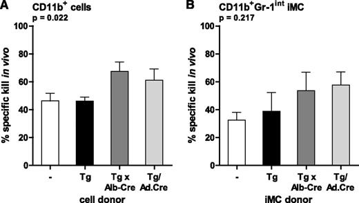
That iMC from mice with immunogenic (Tg/Ad.Cre) and pseudo-immunogenic tumors (Tg) were similarly suppressive in vitro raised the question whether they were also suppressive in vivo, because these mice differed substantially not only in the CTL reactivity against the tumor antigen Tag (pIV) but also tumor-unrelated antigens (HY and gp33). Analysis of adoptively transferred iMC in otherwise healthy B6 mice may serve better to dissect functional differences of iMC from the different tumor models.17,20 Therefore, iMC subsets (CD11b+, CD11b+Gr-1int, and CD11b+Gr-1high cells) were isolated from the different tumor-bearing mice and transferred into B6 mice, which were immunized with pIV peptide within 1 hour. As assessed by in vivo killing assays 8 to 10 days later, CD11b+ cells and CD11b+Gr-1int iMC did not diminish priming of functional endogenous pIV-specific CD8+ T cells, regardless from which mice they were isolated (Figure 4). Similarly, transferred CD11b+Gr-1high iMC did not suppress pIV-specific T cells (supplemental Figure 3). Transferred CD11b+Gr-1int cells also lacked the capacity to interfere with pIV-specific CTL effector function (data not shown), compatible with the persistence of functional pIV-specific CTLs in Tg/Ad.Cre mice (Figure 1B).
iMC from mice bearing Tag-driven autochthonous tumors do not transfer CTL suppression in vivo. (A, B) The suppressive potential of CD11b+ cells (A) and CD11b+Gr-1int iMC (B) from LoxP-Tag mice bearing sporadic tumors (Tg), HCC-bearing LoxP-Tag x Alb-Cre mice (Tg × Alb-Cre), and Ad.Cre-infected LoxP-Tag mice bearing virus-induced HCC (Tg/Ad.Cre) was analyzed upon cell transfer into B6 mice, immunized with Tag pIV in incomplete Freund's adjuvant. In vivo cytotoxicity against Tag peptide IV was analyzed by flow cytometry. The percentage of specific killing in vivo is indicated. Experimental groups were no iMC (ø, n = 7), iMC from Tg mice (n = 3), iMC from Tg × Alb-Cre mice (n = 5), and iMC from Tg/Ad.Cre mice (n = 4). Bars represent means; error bars indicate SEM.
iMC from mice bearing Tag-driven autochthonous tumors do not transfer CTL suppression in vivo. (A, B) The suppressive potential of CD11b+ cells (A) and CD11b+Gr-1int iMC (B) from LoxP-Tag mice bearing sporadic tumors (Tg), HCC-bearing LoxP-Tag x Alb-Cre mice (Tg × Alb-Cre), and Ad.Cre-infected LoxP-Tag mice bearing virus-induced HCC (Tg/Ad.Cre) was analyzed upon cell transfer into B6 mice, immunized with Tag pIV in incomplete Freund's adjuvant. In vivo cytotoxicity against Tag peptide IV was analyzed by flow cytometry. The percentage of specific killing in vivo is indicated. Experimental groups were no iMC (ø, n = 7), iMC from Tg mice (n = 3), iMC from Tg × Alb-Cre mice (n = 5), and iMC from Tg/Ad.Cre mice (n = 4). Bars represent means; error bars indicate SEM.
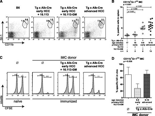
Among other factors, GM-CSF, IL-6, and VEGF are held responsible for inducing and maintaining suppressive iMC.5,17,18,21,22 Primary tumor cell lines derived from the 3 models produced, albeit to a different extent, VEGF and IL-6, which were also detectable in sera of tumor-bearing mice (Figure 5). GM-CSF was not detectable in serum (data not shown) and primary tumor cell lines produced, if at all, only minute amounts of GM-CSF (Figure 5C). To ask whether iMC can transfer CTL suppression in general in our tumor models, we provided GM-CSF exogenously. Therefore, 16.113 cells, a sporadic tumor cell line derived from Tg mice, were engineered to produce ∼207 ng GM-CSF/mL/106 cells per day (16.113-GM; Fig. 5C). 16.113-GM, but not 16.113 tumor cells that both grew subcutaneously in young Tg × Alb-Cre mice bearing early HCC, only detectable after microscopic examination, induced CD11b+Gr-1high and CD11b+Gr-1int iMC subsets, comparable to almost twice as old Tg × Alb-Cre mice bearing advanced macroscopically detectable HCC. These data suggest that CD11b+Gr-1int iMC cells were induced by GM-CSF in this experimental setting. Additionally, 16.113-GM cells also induced CD11b+Gr-1− cells (Figure 6A and supplemental Figure 4). Because increased CD11b+Gr-1int iMC were induced only in the presence of 16.113-GM but not parental 16.113 cells, we subsequently induced CD11b+Gr-1int iMC in Tg × Alb-Cre mice bearing early primary HCC and the transplanted 16.113-GM in comparison with Tg × Alb-Cre mice bearing only advanced primary HCC (but no 16.113 tumor). CD11b+Gr-1int iMC were purified and transferred into B6 mice, which were immunized with pIV peptide on the same day. Eight to 10 days later, in vivo killing assay was performed as before. Only CD11b+Gr-1int iMC from Tg × Alb-Cre mice bearing both early HCC and the transplanted 16.113-GM tumor but not Tg × Alb-Cre mice bearing advanced HCC suppressed the generation of pIV-specific CTL in vivo (Figure 6C-D). The CD11b+Gr-1high iMC subset induced in the presence of GM-CSF was not suppressive (supplemental Figure 4). These data demonstrate that CD11b+Gr-1int iMC in our tumor model can transfer CTL suppression if induced by GM-CSF. However, GM-CSF did not appear to be involved in the induction of iMC by the autochthonous tumors, again irrespective of the immune status of the mice.
Cytokine levels released by cell lines established from autochthonous tumors and in the serum of tumor-bearing mice. (A-C) VEGF and IL-6 are secreted by oncogene-driven autochthonous tumors. VEGF, IL-6, and GM-CSF levels were determined in cell culture supernatants of cell lines established from Tg mice bearing sporadic tumors (9.27, 9.29, 16.30, 16.113), HCC-bearing Tg x Alb-Cre mice (Alb.7, Alb.14, Alb.347), and Tg/Ad.Cre mice bearing virus-induced HCC (Ad.56, Ad.434). Retrovirally transduced 16.113 cells (16.113-GM) were tested for GM-CSF levels only. (D, E) VEGF and IL-6 were determined in sera of young Tg mice (n = 5 and n = 4), Tg mice (n = 8 and n = 38), Tg x Alb-Cre mice (n = 10 and n = 4), and Tg/Ad.Cre mice (n = 4 and n = 13). Note that no GM-CSF could be detected in sera of the mice shown above (data not shown). (F) TGF-β1 levels were determined in sera of young Tg mice (n = 4), Tg mice (n = 7), Tg × Alb-Cre mice (n = 4), and Tg/Ad.Cre mice (n = 4). Bars indicate means; error bars represent SEM; n.d. = not detected.
Cytokine levels released by cell lines established from autochthonous tumors and in the serum of tumor-bearing mice. (A-C) VEGF and IL-6 are secreted by oncogene-driven autochthonous tumors. VEGF, IL-6, and GM-CSF levels were determined in cell culture supernatants of cell lines established from Tg mice bearing sporadic tumors (9.27, 9.29, 16.30, 16.113), HCC-bearing Tg x Alb-Cre mice (Alb.7, Alb.14, Alb.347), and Tg/Ad.Cre mice bearing virus-induced HCC (Ad.56, Ad.434). Retrovirally transduced 16.113 cells (16.113-GM) were tested for GM-CSF levels only. (D, E) VEGF and IL-6 were determined in sera of young Tg mice (n = 5 and n = 4), Tg mice (n = 8 and n = 38), Tg x Alb-Cre mice (n = 10 and n = 4), and Tg/Ad.Cre mice (n = 4 and n = 13). Note that no GM-CSF could be detected in sera of the mice shown above (data not shown). (F) TGF-β1 levels were determined in sera of young Tg mice (n = 4), Tg mice (n = 7), Tg × Alb-Cre mice (n = 4), and Tg/Ad.Cre mice (n = 4). Bars indicate means; error bars represent SEM; n.d. = not detected.
Tumor-induced CD11b+Gr-1int iMC transfer CTL suppression if induced by GM-CSF. (A) Spleen cells of B6 mice, LoxP-Tag × Alb-Cre mice (Tg × Alb-Cre) with early primary HCC and transplanted Tag+ 16.113 tumors, Tg × Alb-Cre mice with early primary HCC and transplanted Tag+ GM-CSF-secreting 16.113 tumors (16.113-GM), and Tg × Alb-Cre mice with advanced primary HCC (but no 16.113 tumor) were double stained with antibodies against CD11b and Gr-1. One representative example per experimental group is shown. Numbers indicate the percentage of CD11b+Gr-1high and CD11b+Gr-1int iMC of total spleen cells. All data for CD11b+Gr-1int iMC are shown in (B). (C) CD11b+Gr-1int iMC from Tg × Alb-Cre mice with early primary HCC and additionally transplanted 16.113-GM tumors and LoxP-Tag × Alb-Cre mice with advanced primary HCC (but no 16.113 tumor) were transferred IV into B6 mice and immunized with pIV subcutaneously on the same day. Naive and immunized B6 mice, which did not receive CD11b+Gr-1int iMC served as control. Eight to 10 days later, in vivo cytotoxicity was analyzed. One representative histogram of each experimental group is shown. All data for transferred CD11b+Gr-1int iMC are shown in (D). The percentage of specific killing in vivo is indicated. Experimental groups were: no iMC (ø, n = 4), iMC from Tg × Alb-Cre with early HCC and 16.113-GM (n = 3), and iMC from Tg × Alb-Cre with advanced HCC (n = 6). Early HCC, microscopically detectable tumors; advanced HCC, macroscopically visible tumors. Bars represent mean, error bars indicate SEM. Overall significance for each graph was tested by Kruskal–Wallis 1-way analysis of variance.
Tumor-induced CD11b+Gr-1int iMC transfer CTL suppression if induced by GM-CSF. (A) Spleen cells of B6 mice, LoxP-Tag × Alb-Cre mice (Tg × Alb-Cre) with early primary HCC and transplanted Tag+ 16.113 tumors, Tg × Alb-Cre mice with early primary HCC and transplanted Tag+ GM-CSF-secreting 16.113 tumors (16.113-GM), and Tg × Alb-Cre mice with advanced primary HCC (but no 16.113 tumor) were double stained with antibodies against CD11b and Gr-1. One representative example per experimental group is shown. Numbers indicate the percentage of CD11b+Gr-1high and CD11b+Gr-1int iMC of total spleen cells. All data for CD11b+Gr-1int iMC are shown in (B). (C) CD11b+Gr-1int iMC from Tg × Alb-Cre mice with early primary HCC and additionally transplanted 16.113-GM tumors and LoxP-Tag × Alb-Cre mice with advanced primary HCC (but no 16.113 tumor) were transferred IV into B6 mice and immunized with pIV subcutaneously on the same day. Naive and immunized B6 mice, which did not receive CD11b+Gr-1int iMC served as control. Eight to 10 days later, in vivo cytotoxicity was analyzed. One representative histogram of each experimental group is shown. All data for transferred CD11b+Gr-1int iMC are shown in (D). The percentage of specific killing in vivo is indicated. Experimental groups were: no iMC (ø, n = 4), iMC from Tg × Alb-Cre with early HCC and 16.113-GM (n = 3), and iMC from Tg × Alb-Cre with advanced HCC (n = 6). Early HCC, microscopically detectable tumors; advanced HCC, macroscopically visible tumors. Bars represent mean, error bars indicate SEM. Overall significance for each graph was tested by Kruskal–Wallis 1-way analysis of variance.
Discussion
iMC are a heterogenous cell population with pleiotropic activities supposed to be involved in tumor progression by immune and nonimmune mechanisms. Mainly investigated in tumor transplantation models,23,24 only a few studies have analyzed the immunosuppressive effect of iMC in autochthonous tumor models.5,22,25,26 We think that the analysis of iMC in primary cancer models better reflects the clinical situation. iMC have not yet been analyzed in mice with autochthonous tumors and differences in tumor-reactive T cells. The 3 related cancer models with the same tumor transplantation rejection antigen but drastically different CTL responses to tumor-related and tumor-unrelated antigens might best uncover phenotypic and functional differences of iMC in vivo. Note that tumor cell lines from all 3 models had a regressor phenotype in transplantation experiments.8,10,11 Surprisingly, the apparently most suppressive iMC population, CD11b+Gr-1int iMC, similarly expanded upon tumor progression and suppressed CTLs in vitro, regardless of whether the mice exhibited tumor-induced tolerance and general CTL hyporesponsiveness (sporadic tumors in LoxP-Tag mice), neonatal tolerance (HCC in LoxP-Tag × Alb-Cre mice), or even systemic tumor immunity (virus-induced HCC in LoxP-Tag mice). These data confirm previous concerns about the relevance of the in vitro assay for the role of iMC in vivo.23 We analyzed spleen iMC because the mice differed in systemic CTL responses; therefore, we cannot exclude functional differences within the tumor microenvironment. Even more surprising, following adoptive transfer the iMC did not suppress CTL priming in vivo in naive B6 mice, again regardless of the immune status of the donor mice. Because iMC were detected for 4 days after transfer, a time when pIV-specific CTL are already detectable, our system might have been able to demonstrate suppressive activity by the iMC (data not shown). In addition, iMC did not inhibit effector function of CTL in vivo because LoxP-Tag mice with virus-induced HCC had persistently functional pIV-specific CTLs despite the increase of iMC. Our data do not dispute that the iMC can be immunosuppressive in vivo, for example in Tg mice with cancer-induced tolerance and general CTL hyporesponsiveness (sporadic cancer model), but apparently iMC do not suppress CTL in a cell-autonomous fashion. Only Tg mice with pseudo-immunogenic sporadic cancer but not Tg × Alb-Cre or Tg/Ad.Cre mice with nonimmunogenic or immunogenic cancer, respectively, have elevated serum TGF-β1 levels and produce anti-Tag IgG antibodies.10 A cell population, termed “barrier” cells and resembling iMC was shown to inhibit third-party CTL responses in a non–cell autonomous fashion by involvement of B cell–derived IgG-TGF-β1 complexes.27,28 These complexes were supposed to be taken up through Fc-receptors by the “barrier” cells, which activated latent TGF-β1 and inhibited CTL responses. Because we detected TGF-β1 producing B cells in Tg mice with pseudo-immunogenic tumors,10 iMC may be involved in general CTL hyporesponsiveness by a similar mechanism in our model. The lack of elevated TGF-β1 in Tg/Ad.Cre mice (Figure 5F) and the lack of both anti-Tag IgG and TGF-β1 in Tg × Alb-Cre mice10 may explain why tumor-unrelated CTL responses were not impaired in these models. However, because of their plasticity (eg, exposure to GM-CSF was sufficient to convert iMC into in vivo suppressive MDSC), the immunoregulatory activity of iMC may vary dependent on the cancer type and/or the factors released within the tumor microenvironment. It also has to be kept in mind that tumor-bearing mice of the different groups differed in age.
One may ask why iMC are so reproducibly induced in cancer-bearing hosts if they do not (in Tg/Ad.Cre) or do not need to suppress tumor-reactive CTLs (in Tg × Alb-Cre mice). Additionally, as noted previously, even in Tg mice with pseudo-immunogenic cancer, the potential contribution of iMC to general CTL hyporesponsiveness appeared to be a symptom, not the cause of cancer, because it occurred subsequent to tumor tolerance, probably resulting from chronic antigen exposure.10 Likely the involvement of iMC in angiogenesis and tumor cell invasion29,30 or fostering a favorable tumor microenvironment by iMC31,32 is of pivotal importance for tumor progression, in particular because iMC also promote tumor growth in the absence of T cells.33,34 IMC have also been shown to accumulate in premalignant skin and spleen without interfering with in vitro polyclonal activation of CD8+ and CD4+ T cells.35 Another question that emerges from this study is how HCC can develop in Tg/Ad.Cre mice with systemic tumor immunity and whether iMC suppress CTLs within the tumor microenvironment. In a parallel study, we found that tumor cells, which were injected into the liver of HCC-bearing Tg/Ad.Cre mice, were selected against the expression of tumor-unrelated neo-antigens, arguing against general CTL suppression by iMC within the tumor. Antigen-specific local tolerance allowing HCC progression in this model was partially mediated by interaction between programmed cell death protein 1 (PD-1) and its ligand PD-L1, because we found that Tag-specific T cells express PD-1 and the virus-induced HCC the respective ligand PD-L1.11 Another conclusion from the current study is that tumor-reactive CTL, functionally activated in the course of viral infection, are not down-regulated by iMC, which expand because of HCC development subsequent to viral Tag activation.
In the Tag-driven tumor models examined in the present study, iMC were licensed to become MDSC in vivo in the presence of GM-CSF. This cytokine was shown to be one of the factors responsible for the preferential expansion of CD11b+Gr-1int/low iMC subsets in vivo17 and to induce the immune-regulatory C/EBPβ transcription factor in vitro.18 The role of GM-CSF in cancer is complicated. For example, opposite effects on immune functions have been observed dependent on the amount of GM-CSF used as vaccine adjuvant.16,19,36 Furthermore, GM-CSF may have different effects at different time points of tumor development.1,35 In conclusion, although iMC are immunosuppressive under various pathological conditions, their role as immune deregulators for cancer progression requires further studies.
The online version of this article contains a data supplement.
The publication costs of this article were defrayed in part by page charge payment. Therefore, and solely to indicate this fact, this article is hereby marked “advertisement” in accordance with 18 USC section 1734.
Acknowledgments
We thank K. Borgwald, S. Horn, K. Retzlaff, and M. Rösch for technical assistance. This work was supported by grants from the Deutsche Forschungsgemeinschaft (Sonderforschungsbereich TR36 and TR54), and the “Alliance” program of the Helmholtz-Gemeinschaft Deutscher Forschungszentren (HA-202).
Authorship
Contribution: K.S. designed and performed experiments, interpreted data, and wrote the manuscript; S.Z. performed experiments; J.C.S. generated and provided GM-CSF producing 16.113; V.B. designed experiments; and T.B. and G.W. designed research, interpreted data, and wrote the manuscript.
Conflict-of-interest disclosure: The authors declare no competing financial interests.
Correspondence: Gerald Willimsky, Institute of Immunology, Charité Campus Buch, Lindenberger Weg 80 13125 Berlin, Germany; e-mail: gerald.willimsky@charite.de


![Figure 3. In vitro CTL suppression by the CD11b+Gr-1int iMC subset does not correlate with CTL suppression in vivo. Polyclonally activated B6 T cells or antigen-specifically activated transgenic P14 T cells were cocultured with 24%, 12%, 6%, or 3% CD11b+ cells or CD11b+Gr-1int iMC from young LoxP-Tag mice (Tg [young]), LoxP-Tag mice bearing sporadic tumors (Tg), HCC-bearing LoxP-Tag × Alb-Cre mice (Tg × Alb-Cre), and Ad.Cre-infected LoxP-Tag mice bearing virus-induced HCC (Tg/Ad.Cre). After 72 hours, CTL were stained with anti-mouse CD8α antibodies and CFSE dilution of CD8+ T cells was analyzed by flow cytometry. (A, B) Total CD11b+ cells suppress polyclonally activated and antigen-specifically activated P14 T cells in vitro. One representative example (12% CD11b+ cells) per experimental group is shown for polyclonally activated T cells (A) and suppression at different CD11b+/T cell ratios is shown for antigen-specifically activated P14 T cells (B). (C, D) CTL suppression is mediated by the CD11b+Gr-1int iMC subset that inhibits proliferation of polyclonally activated (C) and antigen-specifically activated P14 T cells (D) in vitro. Overall significance for each graph was tested by Kruskal–Wallis 1-way analysis of variance.](https://ash.silverchair-cdn.com/ash/content_public/journal/blood/121/10/10.1182_blood-2012-06-436568/4/m_1740f3.jpeg?Expires=1765888070&Signature=Cr~vZekwEsSNy3bEde2WCRkeRL9Z82PpiHNhipakbA7HXkOSZUl1BIk5Jbsamcx3UHiV68gYYmiRHExJpB1PaQMMDqwGb~LxbBr-PI6ztuZPZvLb6cQc5ScJ9NpBPytA8HWWoC4lKFg1Tvg73UQj2GvrPALa7OJUfassXA9G3mH3CoFbtTy46tDMM5m~3-FRI0mLlkxRModfFOuoi-pj0jkDyH4BA0hRYm6zpKPc~eZhZYXwKKGK1LQZFkPTeZBXFCetml-F4-LBFw8Fw3SmXP9UgKUtGZJK67DKe6pMh2SOD1SSAk3JGSgSRcRtFulEKPdDN4YrZD2siWZFevkVlQ__&Key-Pair-Id=APKAIE5G5CRDK6RD3PGA)


