Abstract
The endogenous presentation of the majority of viral epitopes through MHC class I pathway is strictly dependent on the transporter associated with antigen processing (TAP) complex, which transfers the peptide products of proteasomal degradation into the endoplasmic reticulum. A small number of epitopes can be presented through the TAP-independent pathway, the precise mechanism for which remains largely unresolved. Here we show that TAP-independent presentation can be mediated by autophagy and that this process uses the vacuolar pathway and not the conventional secretory pathway. After macroautophagy, the antigen is processed through a proteasome-independent pathway, and the peptide epitopes are loaded within the autophagolysosomal compartment in a process facilitated by the relative acid stability of the peptide-MHC interaction. Despite bypassing much of the conventional MHC class I pathway, the autophagy-mediated pathway generates the same epitope as that generated through the conventional pathway and thus may have a role in circumventing viral immune evasion strategies that primarily target the conventional pathway.
Introduction
In conventional MHC class I antigen presentation, endogenous proteins are degraded by the ubiquitin-proteasome pathway in the cytosol, transported to the endoplasmic reticulum (ER) by the transporter associated with antigen processing (TAP) complex, loaded onto MHC class I molecules, and delivered to the cell surface through the secretory pathway.1 TAP is critical in this process, and cells deficient in TAP have defective peptide loading in the ER and, consequently, low levels of surface MHC class I expression.2 A small number of peptide epitopes, however, can be presented independent of TAP. Approximately half of these are signal peptides that are released directly into the ER during routine protein processing.2-4 Other TAP-independent epitopes that are directly processed within the ER-Golgi compartment include those from the ER resident protein Jaw15 and the HIV-1 envelope protein gp120,6 both processed through as yet undefined proteases; and the hepatitis B secretory core protein, processed by the trans-Golgi network protease furin.7 In addition, a number of hydrophobic peptide epitopes processed conventionally though the cytosolic proteasome pathway can enter the ER and be presented independent of TAP using as yet undefined mechanisms.8 The mechanism of antigen presentation for a significant proportion of TAP-independent peptide epitopes remains unknown.4 Here we describe MHC class I presentation of an endogenous human cytomegalovirus (HCMV) latency-associated protein, pUL138, which can proceed through distinct TAP-dependent and TAP-independent pathways. The TAP-dependent process uses the conventional MHC class I pathway, whereas the TAP-independent process is mediated by macroautophagy and uses the vacuolar pathway. Of note, the 2 distinct pathways generate and present the same epitope.
Autophagy is a process whereby cytoplasmic proteins and organelles can be transported into lysosomes for degradation.9 Endogenous antigens can therefore access the lysosomal compartment through autophagy and be presented on MHC class II through a process similar to the presentation of phagocytosed exogenous antigens.10 More recently, autophagy has been shown to mediate MHC class I presentation of endogenous viral glycoprotein from herpes simplex virus type 1.11 This involved 2 distinct forms of autophagy: a previously described form where intracellular pathogens become surrounded by a double-membrane structure in a process sometimes referred to as “xenophagy,”12 and a form newly described in the report where autophagosomes arose from the nuclear envelope and contained unenveloped viral capsids.11 The autophagy-enhanced MHC class I pathway in this report remained dependent on proteasome and Golgi transport, suggesting that it largely followed the conventional MHC class I pathway.11 Here we demonstrate that there is a separate pathway for autophagy-mediated MHC class I presentation of endogenous antigen. This pathway is proteasome- and TAP-independent, and unlike previously described pathways for endogenous MHC class I presentation, it proceeds through the endocytic rather than the secretory pathway. Despite using minimal components of the conventional MHC class I machinery, this pathway generates the same peptide epitope as that generated from conventional processing. Thus, this autophagy-mediated pathway supplements the conventional pathway, but it may be better positioned to circumvent the multitude of viral immune evasion strategies targeting the MHC class I machinery.
Methods
Cells, viruses, and plasmid
Unless otherwise stated, cells were cultured in RPMI 1640 supplemented with 10% (volume/volume) FCS (JRH Biosciences, Sigma-Aldrich), penicillin (100 units/mL), and streptomycin 100 μg/mL (Invitrogen). The T2.B35 cell line was derived by stable transfection of T2 cells with HLA B*3501.13 Primary fibroblast cell lines were established from human foreskin fibroblasts. Immature monocyte-derived dendritic cells (MoDCs) were generated by plating peripheral blood mononuclear cells in 6-well plates for 90 minutes and treating the adherent monocytes with GM-CSF 1000 IU/mL and IL-4 1000 IU/mL (both from Schering-Plough) for 5 days. CD8+ T cells specific for pUL138 (peptide sequence LPLNVGLPIIGVM; refered to as LPL) were generated from a healthy HLA-B*3501+ HCMV-seropositive person as previously described.14 Bulk EBV LMP1- and LMP2-specific T-cell lines were generated from a healthy HLA-A2+ EBV seropositive person using a recombinant adenovirus, AdE1LMPpoly; and bulk HCMV pp65-specific T cells (HLA-A2/NLV and HLA-B7/RPH) were generated from HCMV-seropositive persons by peptide stimulation as previously described.15 HCMV strain Toledo was propagated in human fetal lung fibroblast cell line, MRC-5 (ATCC). HCMV strain TB40E was a gift from Dr Barry Slobedman (University of Sydney, Sydney, Australia). The recombinant fiber-modified Ad5f35 adenovirus that expresses the UL138 transgene, AdUL138, has been previously described.14 The pUL138-eGFP vector was made by cloning UL138 into the pEGFP-N1 vector (BD Biosciences) and expressed eGFP fused to the carboxyl terminus of pUL138. To generate stable transfectants, T2.B35 cells were electroporated with pUL138-eGFP expression plasmid and GFP-positive cells were selected by repeated rounds of fluorescence-activated cell sorting (FACSAria; BD Biosciences).
Inhibitors and virus infection
Cells were incubated with recombinant adenovirus AdUL138 or HCMV strain Toledo for 90 minutes at the following multiplicity of infection (MOI), unless stated otherwise: AdUL138 and HCMV at MOI of 5 for primary human fibroblasts and AdUL138 at MOI of 100 for T2.B35 cells. MoDCs were infected with HCMV strain TB40E at MOI of 50. The cells were centrifuged for 30 minutes at 800g 32°C to facilitate infection. For treatment with inhibitors, fibroblasts and T2B35 cells were incubated in the following chemical inhibitors starting 1 hour before virus infection: proteasome inhibitors (10μM lactacystin or 1μM epoxomicin; both from Calbiochem), aminopeptidase inhibitor (30μM leucinethiol with 0.5mM dithiothreitol), inhibitors of lysosomal proteases (100μM leupeptin or 80μM chloroquine; both from Sigma-Aldrich), and autophagy inhibitors (10mM 3-methyladenine or 500nM bafilomycin, both from Sigma-Aldrich). In some experiments, additional inhibitors were tested: 10μM pepstatin A, 0.1mM AEBSF (both from Enzo Life Sciences), and 100μM E-64 (Calbiochem). Cells were maintained in inhibitors during and after virus infection until their use in antigen presentation assay 16 hours after infection. For MoDCs, the inhibitors were added 3 hours after infection with HCMV TB40E, and the cells were used in antigen presentation assay at 24 hours after infection. To control for surface MHC class I expression and cell viability, an equal number of chemically treated and untreated cells were pulsed with 0.2 μg/mL of the LPL peptide or other relevant peptide for 1 hour, washed at least 4 times, and used as control stimulators.
Intracellular cytokine secretion assay
Antigen-specific T-cell lines were cocultured with stimulator cells for 4 to 6 hours in the presence of 1 μg/mL brefeldin A (BD Biosciences). The responder-to-simulator ratio, unless otherwise stated, was 4:1 for fibroblasts and 1:1 for T2.B35 cells and MoDCs. Experiments were performed in duplicate or triplicate wells. Cells were stained with anti-CD8 conjugated to PerCP-Cy5.5 (eBioscience) and anti–IFN-γ conjugated to FITC.14 Flow cytometry was performed on FACSCanto II (BD Biosciences) using BD FACSDiva software and results analyzed with FlowJo Version 8.8.3 software (TreeStar). In some studies, cells were costained with allophycocyanin-conjugated multimers: HLA-A*0201/ YLQQNWWTL (EBV LMP1) and HLA-A*0201/CLGGLLTMV (EBV LMP2) pentamers (Proimmune), and HLA-B*3501/LPLNVGLPIIGVM tetramer (National Institutes of Health Tetramer Core Facility).
Transfection by electroporation
T2 or T2.B35 cells (5 × 10⁶) were resuspended in 300 μL of RPMI 1640 90%/FCS 10% and electroporated with 10 μg of expression plasmid in a 0.4-cm cuvette at 260 V 960 μF (Gene Pulser; Bio-Rad).
Silencing of Atg12 with shRNA
Lentivirus-based vector encoding autophagy-related gene 12 (Atg12) short hairpin RNA (shRNA; clone ID NM_004707.2-485s1c1, sequence CCGGTGTTGCAGCTTCCTACTTCAACTCGAGTTGAAGTAGGAAGCTGCAACATTTTT) was obtained from Sigma-Aldrich. Lentivirus was produced in HEK293T cells by cotransfecting the shRNA vector or control vector (pLKO.1-puro) with a packaging vector, pHR8.2ΔR, and an envelope vector, pCMV-VSV-G (vesicular stomatitis virus glycoprotein G). Lentivirus-containing supernatant was collected at 48 and 72 hours after transfection, 0.45 μm filtered, and stored at −80°C. Transduction was performed by resuspending 5 × 10⁵ T2.B35 pUL138-eGFP–transfected cells in 1 mL of lentivirus-containing supernatant and centrifuging for 30 minutes at 800g at 32°C. Transduction of fibroblasts was performed by pretreating adherent fibroblasts for 2 hours in 8 μg/mL polybrene followed by infection in 1 mL lentivirus-containing supernatant supplemented with 8 μg/mL polybrene. Puromycin (1 μg/mL) was added 48 hours after transduction. Cells were used for downstream assays 5 to 7 days after transduction.
Heat shock and cytokine treatment
For heat shock treatment, T2.B35 pUL138-eGFP stable transfectants (5 × 10⁵ cells) were resuspended in 500 μL RPMI 1640 90%/FCS 10% in 10-mL tubes, equilibrated in 6% CO2 for 1 hour, then sealed and cultured for 12 to 14 hours at 37°C for control treatment or 39°C for heat shock treatment. The cells were then rested for 1 hour at 37°C in 6% CO2 and washed before use as antigen-presenting cells in intracellular cytokine secretion assay. For autophagy inhibition with 3-methyladenine (3-MA), cells were pretreated with 10mM 3-MA for 60 hours before heat shock and maintained in 3-MA during heat shock. For cytokine treatment, primary fibroblasts were pretreated for 24 hours with 50 ng/mL TNF-α (R&D Systems), with or without 10mM 3-MA, infected with HCMV strain Toledo for 45 minutes, cultured for a further 14 hours, and then washed and used as antigen-presenting cells in intracellular cytokine secretion assay. TNF-α was maintained during but not after HCMV infection while 3-MA was maintained both during and after HCMV infection. For immunoblotting, MRC-5 primary fibroblast cell line was cultured for 24 hours in the presence of absence of 50 ng/mL TNF-α with or without 40μM chloroquine in the last 12 hours before cell harvest for protein lysate preparation.
Immunoblot
Protein lysates (40 μg) were separated by SDS-PAGE, transferred to nitrocellulose membrane, blocked for 1 hour in Tris-buffered saline/0.1% Tween-20/5% nonfat milk, then incubated overnight at 4°C in 1:500 rabbit anti-Atg12 or rabbit anti-LC3B (both from Cell Signaling Technology) in Tris-buffered saline/0.1% Tween-20/5% BSA. The membrane was washed, probed with polyclonal sheep anti–rabbit conjugated to horseradish peroxidase, and protein bands were detected using Enhanced Chemiluminescence Substrate (Western Lighting Plus ECL; PerkinElmer Life and Analytical Sciences). The membrane was stripped and reprobed with monoclonal mouse anti-GAPDH (clone 6C5; Abcam).
Immunofluorescence microscopy
Cytospins of T2.B35 pUL138-eGFP stable transfectants were air dried, fixed with 4% paraformaldehyde, pH 7.4, for 20 minutes at room temperature, washed twice with PBS, and extracted with 0.5% Triton X-100 for 15 minutes. Slides were blocked for 30 minutes with PBS/5% normal goat serum/5% FCS/3% BSA, then incubated for 1 hour in 1:200 polyclonal rabbit anti–human LC3 (Cell Signaling Technology) and 1:200 monoclonal mouse anti-GFP (ab1218; Abcam), or 1:100 monoclonal mouse anti–human LAMP2 (clone H4B4; Abcam) and 1:300 polyclonal rabbit anti-GFP (ab6556, Abcam) in blocking buffer. Slides were washed 3 times in blocking buffer, incubated with 1:200 goat anti–rabbit or anti–mouse conjugated to AlexaFluor-546 or AlexaFluor-488 (Invitrogen) for 40 minutes, washed 3 times, and mounted with ProLong Gold reagent with 4,6-diamidino-2-phenylindole (Invitrogen). For studies on surface MHC class I internalization, T2.B35 pUL138-eGFP stable transfectants were incubated in 1 μg/mL mouse anti–human MHC class I (clone W6/32) or mouse IgG2aK isotype control (clone eBM2a, eBioscience) in complete medium at 37°C 6% CO2 for 4 hours, washed, then air dried, fixed, extracted, blocked, and stained with goat anti–mouse conjugated to AlexaFluor-546. For colocalization of internalized MHC class I with LC3, nontransfected T2.B35 cells were incubated in the above antibodies with or without 80μM chloroquine for 60 hours. Cells were imaged on an Olympus IX70 microscope using DeltaVision microscopy system, and image deconvolution and colocalization analyses were performed using softWoRx (both from Applied Precision). A minimum of 50 cells were analyzed.
HLA class I stripping
T2.B35 pUL138-eGFP stable transfectants were treated with citrate-phosphate buffer, pH 3.0, for 0, 2, 5, 10, or 30 minutes on ice. The reaction was stopped with 50-fold RPMI 1640/10% FCS, washed twice, fixed in 1% paraformaldehyde for 15 minutes, washed 3 times in PBS, and used as antigen-presenting cells in intracellular cytokine secretion assay. T-cell responses to pUL138 (HLA-B35/LPL), EBV LMP1 (HLA-A2/YLQ), and EBV LMP2 (HLA-A2/CLG) were assessed concurrently by costaining with peptide-MHC multimers.
Results
Presentation of HCMV-encoded pUL138 can occur through 2 distinct pathways
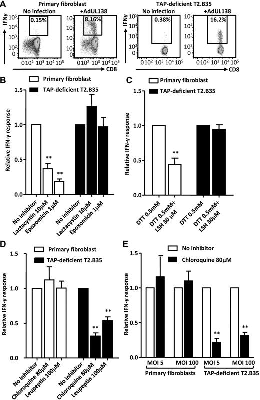
The HCMV latency-associated protein, pUL138, includes a noncanonical 13-amino acid epitope, LPLNVGLPIIGVM(4-16) (referred to as LPL), which is restricted through the HLA-B*3501 allele.14 In characterizing its endogenous presentation, we found that the LPL epitope was not only efficiently processed and presented by TAP-positive primary fibroblasts but also by TAP-deficient T2.B35 cells (Figure 1A), the latter being stable HLA-B*3501 transfectants derived from a TAP1 and TAP2 deficient B xT hybrid cell line known as T2 or 174xCEM.T2.13,16 We next used chemical inhibitors to determine whether antigen processing in the 2 cell types used the same or distinct pathways. The LPL epitope sequence is predicted to be hydrophobic. It has a hydrophobicity score of 5.76, and previous studies have shown that epitopes with hydrophobicity scores greater than 2 to 4 can access the ER through a TAP-independent but proteasome-dependent pathway, the precise mechanism for which remains undefined.8,17 Contrary to these published observations, we found that the presentation of the LPL epitope in TAP-deficient T2.B35 cell line was unaffected by lactacystin and epoxomicin, which are irreversible and selective inhibitors of the proteasome (Figure 1B). Furthermore, treatment with the aminopeptidase inhibitor leucinethiol, with dithiothreitol as a reducing agent, also had no effect (Figure 1C). Hence, the processing and presentation of the LPL epitope can occur through a pathway that is independent of the proteasome, TAP, and ER aminopeptidases, raising the possibility that antigen processing may have occurred in an alternate compartment. Indeed, the presentation of pUL138 by T2.B35 cells was significantly inhibited after treatment with chloroquine (P = .004), which inhibits endosomal acidification, and leupeptin (P = .013), which inhibits cysteine and serine proteases (Figure 1D), thus pointing toward the involvement of an endovacuolar pathway in TAP-independent presentation. In contrast, the endogenous presentation of the LPL epitope in TAP-positive primary fibroblasts was inhibited by lactacystin (P = .014), epoxomicin (P = .002), and leucinethiol (P = .026), and not by chloroquine or leupeptin (Figure 1B-D). Importantly, the presentation of exogenous peptide by surface MHC class I loading was unaffected by chemical inhibitors (data not shown). Because the stress induced by high viral load can affect cellular processes and potentially contribute to the observed differences between cell types, the antigen presentation assays were repeated at high and low MOI. As shown in Figure 1E, chloroquine inhibited the presentation of pUL138 in T2.B35 cells but not fibroblasts, irrespective of MOI. Therefore, in the presence of TAP, the epitope is processed and presented through the conventional MHC class I pathway; and in the absence of TAP, the same epitope can still be generated, although not by proteasomal degradation followed by aminopeptidase trimming in the ER, but in a process that is dependent on lysosomal proteases.
Endogenous presentation of HLA-B*3501–restricted CD8+ T-cell epitope from human cytomegalovirus encoded protein, pUL138, in primary human fibroblasts and TAP-deficient T2.B35 cell line. Cells were infected with recombinant adenovirus encoding the UL138 transgene (AdUL138), and antigen presentation was measured at 16 hours after infection using an intracellular cytokine secretion assay. (A) Flow cytometric dot plots showing IFN-γ secretion by pUL138-specific CD8+ T cells after activation by virus-infected cells. (B-D) T-cell response to virus-infected cells treated with chemical inhibitors of the proteasome (B), aminopeptidases (C), or lysosomal proteases (D). Fibroblasts or T2.B35 cells were treated with individual inhibitors starting at 1 hour before the infection and cultured in the presence of these inhibitors until the time of antigen presentation assay at 16 hours after infection. The T-cell response was normalized to that of untreated controls (B,D) or controls treated with the reducing agent dithiothreitol alone (C). The absolute (mean) percentages of T-cell response to reference cells were 23.3%, 20.7%, and 23.3% for fibroblasts, and 11.0%, 7.5%, and 11.9% for T2.B35 cells in panels B, C, and D, respectively. (E) Fibroblasts and T2.B35 cells were infected by AdUL138 at low and high MOI, with or without treatment with chloroquine. The absolute (mean) percentages of T-cell response to reference cells at MOI 5 and MOI 100 were 9.3% and 15.5% for fibroblasts and 7.4% and 11.0% for T2.B35 cells, respectively. (B-E) Data are from 3 independent experiments, each performed in triplicate samples (mean ± SEM). **P < .05 (2-tailed Student t test).
Endogenous presentation of HLA-B*3501–restricted CD8+ T-cell epitope from human cytomegalovirus encoded protein, pUL138, in primary human fibroblasts and TAP-deficient T2.B35 cell line. Cells were infected with recombinant adenovirus encoding the UL138 transgene (AdUL138), and antigen presentation was measured at 16 hours after infection using an intracellular cytokine secretion assay. (A) Flow cytometric dot plots showing IFN-γ secretion by pUL138-specific CD8+ T cells after activation by virus-infected cells. (B-D) T-cell response to virus-infected cells treated with chemical inhibitors of the proteasome (B), aminopeptidases (C), or lysosomal proteases (D). Fibroblasts or T2.B35 cells were treated with individual inhibitors starting at 1 hour before the infection and cultured in the presence of these inhibitors until the time of antigen presentation assay at 16 hours after infection. The T-cell response was normalized to that of untreated controls (B,D) or controls treated with the reducing agent dithiothreitol alone (C). The absolute (mean) percentages of T-cell response to reference cells were 23.3%, 20.7%, and 23.3% for fibroblasts, and 11.0%, 7.5%, and 11.9% for T2.B35 cells in panels B, C, and D, respectively. (E) Fibroblasts and T2.B35 cells were infected by AdUL138 at low and high MOI, with or without treatment with chloroquine. The absolute (mean) percentages of T-cell response to reference cells at MOI 5 and MOI 100 were 9.3% and 15.5% for fibroblasts and 7.4% and 11.0% for T2.B35 cells, respectively. (B-E) Data are from 3 independent experiments, each performed in triplicate samples (mean ± SEM). **P < .05 (2-tailed Student t test).
TAP-independent presentation of pUL138 requires endogenous antigen synthesis
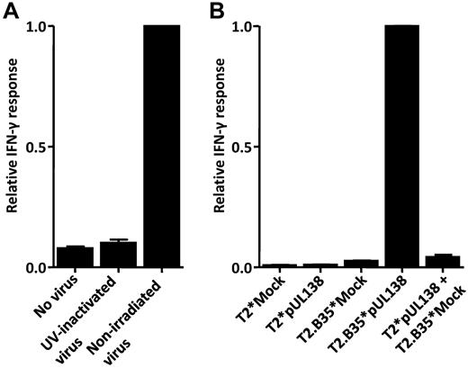
The peptide products of lysosomal degradation are usually presented on MHC class II molecules. An exception to this is in the cross-presentation of exogenous antigens on MHC class I molecules where, in addition to the dominant conventional MHC class I pathway, there exists a minor pathway that is also TAP-independent whereby antigen processing and loading onto MHC class I molecules occur within the endolysosomes.18,19 Endolysosomal processing has not been implicated in MHC class I presentation of endogenous antigens, however; and in describing this new pathway, cross-presentation of exogenous pUL138 contained within the viral cell lysate needed to be ruled out. Data presented in Figure 2A show that inactivation of AdUL138 with ultraviolet radiation abolished T-cell recognition of the LPL epitope, thus demonstrating that active viral infection followed by endogenous antigen synthesis was required for pUL138 presentation by T2.B35 cells. In addition, coculture of T2 cells (HLA-B35-negative) electroporated with pUL138 expression plasmid and mock-electroporated T2.B35 cells did not activate pUL138-specific T cells (Figure 2B). Together, these experiments demonstrated that the LPL epitope presented through the TAP-independent pathway was derived from endogenously synthesized pUL138 and not from cross-presented exogenous antigen.
TAP-independent antigen presentation requires endogenous protein synthesis. (A) T2.B35 cells were infected with AdUL138 with or without UV inactivation and then assessed for endogenous presentation of pUL138 CD8+ T-cell epitope at 16 hours after infection. (B) T2 cells (HLA-B35−) were electroporated with an expression plasmid encoding pUL138 and then cocultured with mock electroporated T2.B35 (HLA-B35+) cells for 18 hours. These cells were then used as antigen-presenting cells in a standard intracellular cytokine secretion assay. The absolute (mean) percentages of T-cell response to reference cells were 20.2% and 35.9% in panels A and B, respectively. Data shown are from 2 independent experiments, each performed in triplicate samples (mean ± SEM).
TAP-independent antigen presentation requires endogenous protein synthesis. (A) T2.B35 cells were infected with AdUL138 with or without UV inactivation and then assessed for endogenous presentation of pUL138 CD8+ T-cell epitope at 16 hours after infection. (B) T2 cells (HLA-B35−) were electroporated with an expression plasmid encoding pUL138 and then cocultured with mock electroporated T2.B35 (HLA-B35+) cells for 18 hours. These cells were then used as antigen-presenting cells in a standard intracellular cytokine secretion assay. The absolute (mean) percentages of T-cell response to reference cells were 20.2% and 35.9% in panels A and B, respectively. Data shown are from 2 independent experiments, each performed in triplicate samples (mean ± SEM).
TAP-independent presentation of pUL138 is mediated by autophagy
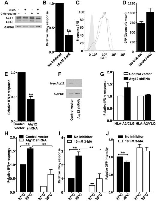
Having established that the TAP-independent presentation of the LPL epitope is dependent on endogenous antigen synthesis and yet occurs through the endovacuolar pathway, we tested the hypothesis that autophagy could be the means by which antigen is transferred into the vacuolar compartment. To investigate this, we generated stable transfectants of TAP-deficient T2.B35 cells made to express a pUL138-eGFP fusion protein. In the first set of experiments, we treated T2.B35 cells expressing pUL138-eGFP protein with 3-MA, which inhibits autophagy through inhibition of class III phosphoinositide 3-kinase. The inhibition of autophagy is demonstrated by a reduction in the absolute amount of LC3-II and a lower LC3-II/LC3-I ratio in 3-MA–treated cells, either in the presence or absence of 80μM chloroquine (Figure 3A). We then assessed the endogenous presentation of the LPL epitope. Data presented in Figure 3B show that treatment with 3-MA significantly reduced the endogenous presentation of the LPL epitope (P = .014) without reducing the amount of pUL138-eGFP protein expression as determined by flow cytometry (Figure 3C-D). Interestingly, there was a trend toward increased pUL138-eGFP intensity after 3-MA treatment (P = .12), potentially reflecting an accumulation of this protein in T2.B35 cells in the absence of autophagy-mediated degradation. To confirm these observations, we specifically inhibited autophagy by silencing autophagy-related gene 12 (Atg12) with RNA interference. The Atg12 system is well conserved in eukaryotic cells, and its covalent conjugation to Atg5 is essential to autophagosome formation and elongation.20,21 Transduction of T2.B35 cells expressing pUL138-eGFP protein with a lentivirus encoding an shRNA targeting Atg12 effectively knocked down Atg12 protein expression and reduced pUL138 presentation (P = .011; Figure 3E-F). To ensure that this inhibitory effect was specific for pUL138 presentation and not the result of global defect in MHC class I presentation, we concurrently assessed the endogenous presentation of MHC class I epitopes from EBV-encoded latent membrane proteins 1 and 2 (LMP1 and LMP2), which have been previously shown to be processed through a proteasome-dependent but TAP-independent pathway. As shown in Figure 3G, the presentation of these antigens remained intact with Atg12 knockdown. Conversely, autophagy up-regulation by 39°C heat shock for 12 to 14 hours significantly enhanced pUL138 presentation by T2.B35 pUL138-eGFP stable transfectants (P = .009 and P = .031 in Figure 3H-I, respectively), and this effect was sensitive to autophagy inhibition by Atg12 knockdown (Figure 3H; P = .016) or 3-MA (Figure 3I; P = .048). The increased antigen presentation after heat shock treatment was also associated with a small but significant reduction (14.9% ± 3.3%; P = .045) in the amount of pUL138-eGFP as determined by flow cytometry, and this reduction was attenuated in the presence of 3-MA (7.5% ± 2.9%; P = .124), consistent with enhanced autophagy-mediated protein degradation (Figure 3J).
Effect of autophagy inhibition and enhancement on pUL138 TAP-independent antigen presentation. (A) LC3 immunoblot on T2.B35 pUL138-eGFP stable transfectants treated with 10mM 3-MA for 14 hours in the presence or absence of 80μM chloroquine. (B-D) T2.B35 pUL138-eGFP stable transfectants were treated with 3-MA for 72 hours and assessed for the presentation of pUL138 CD8+ T-cell epitope by intracellular cytokine secretion assay (B), and the expression of pUL138-eGFP protein by flow cytometry (C-D). (C) Solid line indicates pUL38-eGFP expression in untreated cells; and dotted line, the expression of this protein in 3-MA–treated cells. Gray-shaded area represents nontransfected T2.B35 cells. (E-G) T2.B35 cells stably expressing pUL138-eGFP protein were transduced with recombinant lentivirus encoding Atg12 shRNA or a control vector, rested for 2 days, selected in puromycin for 3 days, and then used as antigen-presenting cells in a standard intracellular cytokine secretion assay. (E) Presentation of pUL138. (F) Immunoblot for Atg12. (G) The cells were concurrently assessed for the presentation of EBV-encoded LMP2 (HLA-A2–restricted CLG epitope) and LMP1 (HLA-A2–restricted YLQ epitope) using CLG- and YLQ-specific T cells in intracellular cytokine secretion assay. (H-I) T2.B35 pUL138-eGFP stable transfectants were treated with heat shock at 39°C for 12 to 14 hours to up-regulate autophagy, and the presentation of pUL138 by heat shock treated cells was compared with control treated cells in the presence or absence of autophagy inhibition with Atg12 knockdown (H) or 3-MA (I). (J) Effect of heat shock treatment on the amount of pUL38-eGFP as measured by flow cytometry. The mean absolute percentages of T-cell response to reference cells were 8.5%, 20.5%, 10.4%, and 11.3% in panels B, E, H, and I, respectively, and 8.9% for HLA-A2/CLG and 90.7% for HLA-A2/YLQ in panel G. Data shown are from 3 independent experiments (B,D,G-J) or 4 independent experiments (E) performed in at least duplicate samples (mean ± SEM). **P < .05 (2-tailed Student t test). (C,F) Representative examples from 3 experiments.
Effect of autophagy inhibition and enhancement on pUL138 TAP-independent antigen presentation. (A) LC3 immunoblot on T2.B35 pUL138-eGFP stable transfectants treated with 10mM 3-MA for 14 hours in the presence or absence of 80μM chloroquine. (B-D) T2.B35 pUL138-eGFP stable transfectants were treated with 3-MA for 72 hours and assessed for the presentation of pUL138 CD8+ T-cell epitope by intracellular cytokine secretion assay (B), and the expression of pUL138-eGFP protein by flow cytometry (C-D). (C) Solid line indicates pUL38-eGFP expression in untreated cells; and dotted line, the expression of this protein in 3-MA–treated cells. Gray-shaded area represents nontransfected T2.B35 cells. (E-G) T2.B35 cells stably expressing pUL138-eGFP protein were transduced with recombinant lentivirus encoding Atg12 shRNA or a control vector, rested for 2 days, selected in puromycin for 3 days, and then used as antigen-presenting cells in a standard intracellular cytokine secretion assay. (E) Presentation of pUL138. (F) Immunoblot for Atg12. (G) The cells were concurrently assessed for the presentation of EBV-encoded LMP2 (HLA-A2–restricted CLG epitope) and LMP1 (HLA-A2–restricted YLQ epitope) using CLG- and YLQ-specific T cells in intracellular cytokine secretion assay. (H-I) T2.B35 pUL138-eGFP stable transfectants were treated with heat shock at 39°C for 12 to 14 hours to up-regulate autophagy, and the presentation of pUL138 by heat shock treated cells was compared with control treated cells in the presence or absence of autophagy inhibition with Atg12 knockdown (H) or 3-MA (I). (J) Effect of heat shock treatment on the amount of pUL38-eGFP as measured by flow cytometry. The mean absolute percentages of T-cell response to reference cells were 8.5%, 20.5%, 10.4%, and 11.3% in panels B, E, H, and I, respectively, and 8.9% for HLA-A2/CLG and 90.7% for HLA-A2/YLQ in panel G. Data shown are from 3 independent experiments (B,D,G-J) or 4 independent experiments (E) performed in at least duplicate samples (mean ± SEM). **P < .05 (2-tailed Student t test). (C,F) Representative examples from 3 experiments.
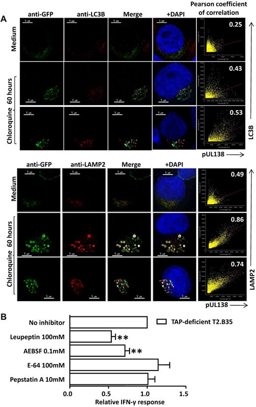
Further confirmation for the role of autophagy was obtained by immunofluorescence microscopy of T2.B35 cells expressing pUL138-eGFP protein. This showed that pUL38-eGFP protein colocalized with LC3, a marker for autophagosomes,22 and lysosome-associated protein 2 (LAMP2), a lysosome marker indicating autophagolysosomes (Figure 4A). Quantification by pixel-by-pixel analysis and Pearson coefficient of correlation are presented in Figure 4A (right panels). Colocalization with LC3, although weaker than that with LAMP2, was readily detectable after chloroquine treatment to inhibit lysosomal acidification, a treatment that also enhanced the degree of colocalization with LAMP2. Collectively, these data demonstrate that TAP-independent presentation is mediated by autophagy through which endogenous pUL138 is transported into autophagolysosomes for degradation by lysosomal proteases.
Subcellular localization of pUL138-eGFP and the effect of protease inhibitors. (A) Subcellular localization of pUL138-eGFP with LC3 and LAMP2 in T2.B35 stable transfectants. T2.B35 cells stably expressing pUL138-eGFP fusion protein were costained with anti-GFP in AlexaFluor-488 and anti-LC3B or anti-LAMP2 in AlexaFluor-546. Cells were either untreated or pretreated with chloroquine (80μM) for 60 hours. Colocalization of pUL138-eGFP (shown in green) and LC3 or LAMP2 (shown in red) is indicated in white. Blue represents nuclei staining with 4,6-diamidino-2-phenylindole (DAPI). Scale bars represent 5 μm. Original magnification ×64 (Olympus IX70 microscope, DeltaVision microscopy system). Image deconvolution and colocalization analysis performed with softWoRx. Right panels: scatter plots and degree of colocalization of pUL138-eGFP with LC3 or LAMP2 by Pearson coefficient of correlation. Results are representative samples from 3 independent experiments. (B) Effect of protease inhibitors on pUL138 TAP-independent antigen presentation. T2.B35 cells were pretreated with protease inhibitors and infected with AdUL138. Data shown are from 3 independent experiments performed in triplicate samples (mean ± SEM). **P < .05 (2-tailed Student t test). The mean absolute percentage of T-cell response to reference cells was 11.3%.
Subcellular localization of pUL138-eGFP and the effect of protease inhibitors. (A) Subcellular localization of pUL138-eGFP with LC3 and LAMP2 in T2.B35 stable transfectants. T2.B35 cells stably expressing pUL138-eGFP fusion protein were costained with anti-GFP in AlexaFluor-488 and anti-LC3B or anti-LAMP2 in AlexaFluor-546. Cells were either untreated or pretreated with chloroquine (80μM) for 60 hours. Colocalization of pUL138-eGFP (shown in green) and LC3 or LAMP2 (shown in red) is indicated in white. Blue represents nuclei staining with 4,6-diamidino-2-phenylindole (DAPI). Scale bars represent 5 μm. Original magnification ×64 (Olympus IX70 microscope, DeltaVision microscopy system). Image deconvolution and colocalization analysis performed with softWoRx. Right panels: scatter plots and degree of colocalization of pUL138-eGFP with LC3 or LAMP2 by Pearson coefficient of correlation. Results are representative samples from 3 independent experiments. (B) Effect of protease inhibitors on pUL138 TAP-independent antigen presentation. T2.B35 cells were pretreated with protease inhibitors and infected with AdUL138. Data shown are from 3 independent experiments performed in triplicate samples (mean ± SEM). **P < .05 (2-tailed Student t test). The mean absolute percentage of T-cell response to reference cells was 11.3%.
Lysosomal proteases mainly consist of cysteine, serine, or aspartate proteases. To characterize the protease involved in the TAP-independent autophagy-dependent pathway, AdUL138 infection of T2.B35 cells was performed with or without treatment with leupeptin (a reversible inhibitor of serine and cysteine proteases), AEBSF (Perfabloc; an irreversible serine protease inhibitor), E64 (an irreversible inhibitor of cysteine proteases), or pepstatin A (a reversible inhibitor of aspartyl proteases). As shown in Figure 4B, leupeptin and AEBSF significantly reduced the presentation of pUL138, whereas E64 and pepstatin A had no significant impact; the presentation of surface-loaded exogenous peptide by control cells was unaffected by chemical inhibitors (data not shown). Hence, pUL138 processing via the vacuolar pathway probably involves lysosomal serine proteases, which include cathepsin A, cathepsin G, and tripeptidyl peptidase I, although involvement of additional proteases is not excluded.
Autophagy-mediated presentation of endogenous pUL138 by HCMV-infected cells
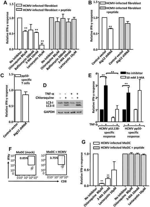
The experiments used to establish the existence of an autophagy-dependent TAP-independent antigen presentation pathway had used a replication-incompetent recombinant adenovirus or expression plasmids rather than natural HCMV infection to minimize the potential for interaction with other cellular events triggered by HCMV. To determine whether autophagy-mediated antigen presentation is operational in naturally infected cells, we investigated the effect of autophagy inhibition on HCMV-infected fibroblasts and MoDCs. We found that, unlike AdUL138-infected fibroblasts, the presentation of pUL138 by HCMV-infected fibroblasts was reduced by inhibitors of the vacuolar pathway and autophagy: chloroquine, bafilomycin, and 3-MA, although the conventional pathway, inhibited by lactacystin, remained dominant (Figure 5A). The presentation of pUL138 by HCMV-infected fibroblasts was also significantly reduced after Atg12 knockdown (P = .004), although there were also small, nonsignificant reductions in the presentation of surface-loaded exogenous LPL peptide and the endogenous presentation of another HCMV-derived epitope (pp50; VTEHDTLLY/HLA-A1; Figure 5B-C). Conversely, the presentation of pUL138 was enhanced when autophagy was up-regulated. Fibroblasts pretreated with 50 ng/mL TNF-α for 24 hours up-regulated autophagy, as demonstrated by an increase in the absolute amount of LC3-II and a higher LC3-II/LC3-I ratio in the presence of lysosomal protease inhibition with chloroquine (Figure 5D). After HCMV infection, TNF-α–pretreated fibroblasts showed increased pUL138 presentation compared with untreated control (Figure 5E). This effect was not entirely autophagy dependent because the presentation of a conventionally processed control antigen, HCMV pp50, was also increased, though to a lesser extent. Importantly, however, the increase in pUL138 presentation was blocked by 3-MA, whereas that of the conventionally processed pp50 was not, indicating that a significant component of the enhanced pUL138 presentation was through autophagy up-regulation. We further assessed the endogenous presentation of pUL138 in human MoDCs. HCMV infection of MoDCs was relatively inefficient compared with fibroblasts, but the presentation of pUL138 could be clearly detected at 24 hours after infection with the clinical HCMV strain TB40E (Figure 5F). The presentation of pUL138 was strongly inhibited by 3-MA and chloroquine, with a trend also for inhibition by lactacystin (Figure 5G), suggesting the coexistence of both the alternative and conventional pathways in naturally infected MoDCs. Together, these observations would support the presence of an autophagy-dependent pathway operating concurrent with the conventional pathway in naturally infected cells.
Effect of autophagy inhibition and enhancement on pUL138 presentation by HCMV-infected cells. (A) T-cell response to HCMV-infected fibroblasts treated with chemical inhibitors. Fibroblasts were pretreated with inhibitors before HCMV infection and cultured in the presence of these inhibitors until the time of antigen presentation assay at 16 hours after infection. (B) T-cell response to HCMV-infected fibroblasts transduced with recombinant lentivirus encoding Atg12 shRNA or a control vector. Antigen presentation assay was performed 14 hours after HCMV infection and 6 to 7 days after lentivirus infection. (A-B) Also shown are T-cell responses to surface-loaded controls. (C) Response of HCMV pp50-specific T cells (VTEHDTLLY/HLA-A1) to HCMV-infected fibroblasts transduced with lentivirus. (D) LC3 immunoblot on primary fibroblast (MRC-5) cell line treated with 50 ng/mL TNF-α for 24 hours in the presence or absence of 40μM chloroquine in the final 12 hours. (E) Effect of autophagy up-regulation on antigen presentation. Fibroblasts were pretreated with 50 ng/mL TNF-α for 24 hours, with or without 3-MA, infected with HCMV, and rested without TNF-α until the time of antigen presentation assay at 14 hours after infection. The presentation of pUL138 was compared with that of a conventionally processed HCMV antigen, pp50. (F) Flow cytometric dot plots showing IFN-γ secretion by pUL138-specific CD8+ T cells in response to mock-infected or HCMV (TB40E strain)–infected MoDCs. (G) T-cell response to HCMV-infected MoDCs treated with chemical inhibitors of antigen-processing pathways from 3 hours to 24 hours after infection. Also shown are T-cell responses to surface-loaded controls. (A-C,E,G) Data are from 3 independent experiments performed in at least duplicate samples (mean ± SEM). **P < .05 (2-tailed Student t test). The mean absolute percentages of T-cell response to reference cells were 10.1%, 11.2%, 21.2%, 2.8%, and 4.2% in panels A, B, C, E, and G, respectively.
Effect of autophagy inhibition and enhancement on pUL138 presentation by HCMV-infected cells. (A) T-cell response to HCMV-infected fibroblasts treated with chemical inhibitors. Fibroblasts were pretreated with inhibitors before HCMV infection and cultured in the presence of these inhibitors until the time of antigen presentation assay at 16 hours after infection. (B) T-cell response to HCMV-infected fibroblasts transduced with recombinant lentivirus encoding Atg12 shRNA or a control vector. Antigen presentation assay was performed 14 hours after HCMV infection and 6 to 7 days after lentivirus infection. (A-B) Also shown are T-cell responses to surface-loaded controls. (C) Response of HCMV pp50-specific T cells (VTEHDTLLY/HLA-A1) to HCMV-infected fibroblasts transduced with lentivirus. (D) LC3 immunoblot on primary fibroblast (MRC-5) cell line treated with 50 ng/mL TNF-α for 24 hours in the presence or absence of 40μM chloroquine in the final 12 hours. (E) Effect of autophagy up-regulation on antigen presentation. Fibroblasts were pretreated with 50 ng/mL TNF-α for 24 hours, with or without 3-MA, infected with HCMV, and rested without TNF-α until the time of antigen presentation assay at 14 hours after infection. The presentation of pUL138 was compared with that of a conventionally processed HCMV antigen, pp50. (F) Flow cytometric dot plots showing IFN-γ secretion by pUL138-specific CD8+ T cells in response to mock-infected or HCMV (TB40E strain)–infected MoDCs. (G) T-cell response to HCMV-infected MoDCs treated with chemical inhibitors of antigen-processing pathways from 3 hours to 24 hours after infection. Also shown are T-cell responses to surface-loaded controls. (A-C,E,G) Data are from 3 independent experiments performed in at least duplicate samples (mean ± SEM). **P < .05 (2-tailed Student t test). The mean absolute percentages of T-cell response to reference cells were 10.1%, 11.2%, 21.2%, 2.8%, and 4.2% in panels A, B, C, E, and G, respectively.
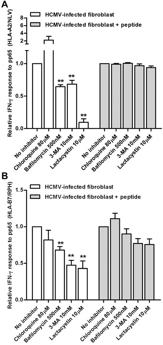
To investigate whether the autophagy-mediated pathway is involved in the presentation of other HCMV antigens, we studied the presentation of the immunodominant pp65 epitopes: HLA-A2 restricted NLV and HLA-B7–restricted RPH. The presentation of both epitopes by HCMV-infected fibroblasts was significantly reduced by chemical inhibitors of autophagy; however, it was not inhibited by chloroquine and therefore probably uses a different pathway from pUL138 (Figure 6A-B).
Effect of autophagy inhibition on the presentation of the pp65 structural antigen by HCMV-infected fibroblasts. Responses of pp65-specific T cells to HCMV-infected fibroblasts treated with chemical inhibitors of the antigen processing pathway. Fibroblasts were treated with inhibitors before and after infection with HCMV strain Toledo. Antigen presentation assays were performed at 16 hours after infection. Shown are the responses to HLA-A2/NLV (A) and HLA-B7/RPH (B) epitopes. Also shown are T-cell responses to surface-loaded controls. Data shown are from 3 independent experiments performed in at least duplicate samples (mean ± SEM). **P < .05 (2-tailed Student t test). The mean absolute percentages of T-cell response to reference cells were 18.1% and 15.2% in panels A and B, respectively.
Effect of autophagy inhibition on the presentation of the pp65 structural antigen by HCMV-infected fibroblasts. Responses of pp65-specific T cells to HCMV-infected fibroblasts treated with chemical inhibitors of the antigen processing pathway. Fibroblasts were treated with inhibitors before and after infection with HCMV strain Toledo. Antigen presentation assays were performed at 16 hours after infection. Shown are the responses to HLA-A2/NLV (A) and HLA-B7/RPH (B) epitopes. Also shown are T-cell responses to surface-loaded controls. Data shown are from 3 independent experiments performed in at least duplicate samples (mean ± SEM). **P < .05 (2-tailed Student t test). The mean absolute percentages of T-cell response to reference cells were 18.1% and 15.2% in panels A and B, respectively.
Endocytic peptide loading and acid stability of peptide-MHC interaction
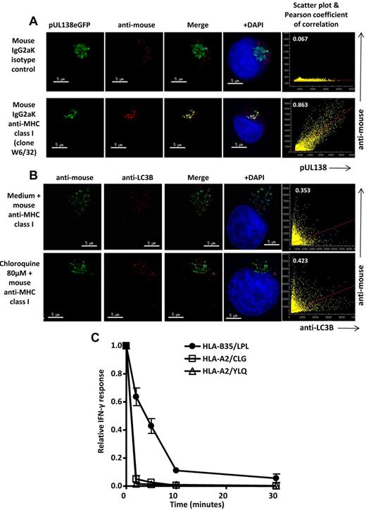
The lack of postlysosomal processing by proteasome and ER-resident aminopeptidases raises the question whether peptide loading and transport to cell surface may have also taken place within the endovacuolar compartment. In cross-presentation of exogenous antigen, the dominant pathway is through retrotranslocation from phagosome to cytosol, which is mediated by ER-resident molecules, such as Sec61, also found on phagosomal membrane,23,24 followed by conventional MHC class I processing.1,25,26 However, an alternative route using the vacuolar pathway is well described.18,19,27,28 Surface MHC class I constitutively undergoes endosomal trafficking,27-29 and exogenous antigens targeted to early endosomes through specific liposomal encapsulation can be cross-presented on MHC class I in a process that is proteasome- and TAP-independent, inhibited by chloroquine, and requires surface MHC class I–stabilizing peptide.18 To investigate whether pUL138 MHC class I loading could have taken place within autophagolysosomes, we studied the colocalization of pUL138 with recirculating MHC class I. T2.B35 cells stably expressing pUL138-eGFP protein were incubated with a purified monoclonal mouse antibody against MHC class I (clone W6/32), or an IgG2aK isotype control, at 37°C 6% CO2 for 4 hours to allow antibody internalization, washed, and then fixed with paraformaldehyde. After staining with a fluorescence-conjugated anti–mouse, colocalization of pUL138-eGFP with internalized MHC class I could be demonstrated (Figure 7A). The internalized MHC class I showed some colocalization with LC3 (Figure 7B). This was less striking than the colocalization of MHC class I with pUL138-eGFP but consistent with the degree of colocalization of LC3 with pUL138-eGFP (Figure 4A).
Colocalization of pUL138-eGFP and LC3 with internalized MHC class I molecules and acid stability of HLA-B35/LPL complexes. (A) T2.B35 pUL138-eGFP stable transfectants were cultured for 4 hours in complete medium containing mouse anti–human MHC class I (clone W6/32) or mouse IgG2aK isotype control, washed, and stained with AlexaFluor-546–conjugated goat anti–mouse IgG (shown in red). Colocalization of pUL138-eGFP with internalized MHC class I is shown in white. (B) Nontransfected T2.B35 cells were cultured in complete medium containing mouse anti–human MHC class I (clone W6/32), in the presence or absence of 80μM chloroquine, for 60 hours, washed, and stained with AlexaFluor-488–conjugated goat anti–mouse IgG (green) and anti-LC3B in AlexaFluor-546 (red). Colocalization of LC3 with internalized MHC class I is shown in white. (A-B) Blue represents nuclei staining with 4,6-diamidino-2-phenylindole (DAPI). Scale bars represent 5 μm. Original magnification ×64 (Olympus IX70 microscope, DeltaVision microscopy system). Image deconvolution and colocalization analysis performed with softWoRx. Right panels: scatter plots and degree of colocalization of anti-MHC class I or isotype control with pUL138 or LC3 by Pearson coefficient of correlation. Results are representative of 3 independent experiments. (C) T2.B35 pUL138-eGFP stable transfectants were treated with citric acid buffer (pH 3.0) for 0 to 30 minutes to dissociate peptide-MHC class I complexes, fixed in paraformaldehyde, and then used as antigen-presenting cells in an intracellular cytokine secretion assay. The acid stability of pUL138 HLA-B35/LPL was assessed in parallel with those of EBV LMP2 HLA-A2–restricted CLG and EBV LMP1 HLA-A2–restricted YLQ epitopes. Data shown were from 3 independent experiments, each performed in duplicate samples (mean ± SEM). The mean absolute percentages of T-cell response at time 0 were 32.0%, 4.5%, and 47.4% for HLA-B35/LPL, HLA-A2/CLG, and HLA-A2/YLQ, respectively.
Colocalization of pUL138-eGFP and LC3 with internalized MHC class I molecules and acid stability of HLA-B35/LPL complexes. (A) T2.B35 pUL138-eGFP stable transfectants were cultured for 4 hours in complete medium containing mouse anti–human MHC class I (clone W6/32) or mouse IgG2aK isotype control, washed, and stained with AlexaFluor-546–conjugated goat anti–mouse IgG (shown in red). Colocalization of pUL138-eGFP with internalized MHC class I is shown in white. (B) Nontransfected T2.B35 cells were cultured in complete medium containing mouse anti–human MHC class I (clone W6/32), in the presence or absence of 80μM chloroquine, for 60 hours, washed, and stained with AlexaFluor-488–conjugated goat anti–mouse IgG (green) and anti-LC3B in AlexaFluor-546 (red). Colocalization of LC3 with internalized MHC class I is shown in white. (A-B) Blue represents nuclei staining with 4,6-diamidino-2-phenylindole (DAPI). Scale bars represent 5 μm. Original magnification ×64 (Olympus IX70 microscope, DeltaVision microscopy system). Image deconvolution and colocalization analysis performed with softWoRx. Right panels: scatter plots and degree of colocalization of anti-MHC class I or isotype control with pUL138 or LC3 by Pearson coefficient of correlation. Results are representative of 3 independent experiments. (C) T2.B35 pUL138-eGFP stable transfectants were treated with citric acid buffer (pH 3.0) for 0 to 30 minutes to dissociate peptide-MHC class I complexes, fixed in paraformaldehyde, and then used as antigen-presenting cells in an intracellular cytokine secretion assay. The acid stability of pUL138 HLA-B35/LPL was assessed in parallel with those of EBV LMP2 HLA-A2–restricted CLG and EBV LMP1 HLA-A2–restricted YLQ epitopes. Data shown were from 3 independent experiments, each performed in duplicate samples (mean ± SEM). The mean absolute percentages of T-cell response at time 0 were 32.0%, 4.5%, and 47.4% for HLA-B35/LPL, HLA-A2/CLG, and HLA-A2/YLQ, respectively.
In cross-presentation, the acidic pH of endosomes and lysosomes is generally considered a barrier to MHC class I peptide loading. However, the stability of peptide-MHC complexes in the acidic environment varies and peptide dissociation and exchange within the acidic endocytic compartment contribute toward an alternate pathway for cross-presentation.18,30,31 To assess the stability of HLA-B35/LPL complexes in the acidic environment, T2.B35 cells expressing pUL138-eGFP protein were treated with citrate phosphate buffer at pH 3, paraformaldehyde fixed, and then used as antigen-presenting cells in an intracellular cytokine secretion assay. We compared the impact of acid treatment on HLA-B35/LPL with that on HLA-A2/YLQ and HLA-A2/CLG, which are TAP-independent epitopes derived from endogenous EBV-encoded LMP1 and LMP2, respectively. Data presented in Figure 7C show that citrate phosphate buffer treatment rapidly reduced the surface presentation of EBV epitopes whereas the presentation of HLA-B35/LPL was significantly more resistant, particularly at early time points (P = .009, .013, .042, and .26 for HLA-B35/LPL vs HLA-A2/YLQ, and P = .014, .019, .015, and .21 for HLA-B35/LPL vs HLA-A2/CLG at 2, 5, 10, and 30 minutes, respectively). T-cell recognition of EBV-encoded epitopes from LMP1(YLQ) and LMP2(CLG) fell by more than 95% after 2 minutes of acid treatment, whereas the response to pUL138 (LPL) was partially preserved, even after 5 to 10 minutes of acid treatment. Collectively, the relative acid stability of HLA-B35/LPL complexes and colocalization of pUL138-eGFP with internalized MHC class I molecules support the hypothesis that peptide loading can take place within the endocytic compartment.
Discussion
HCMV-encoded pUL138 has been shown to be important for the establishment and maintenance of latent infection.32 The protein is not incorporated into virus particles.33 It is a type I integral membrane protein that localizes to the Golgi apparatus of infected cells.33 The majority of the protein resides on the cytoplasmic face, and the LPL epitope resides wholly within the putative transmembrane domain.33 We found that the endogenous processing and presentation of an MHC class I epitope from this protein can occur through both conventional TAP-dependent and nonconventional TAP-independent pathways. Whereas the TAP-dependent pathway involves both proteasomal machinery and ER-resident proteases, the TAP-independent pathway involves neither. Instead, it involves the vacuolar pathway and is mediated by autophagy. Of note, this pathway is distinct from a previously described autophagy-mediated pathway for a structural herpesvirus antigen, which remains dependent on proteasomal processing and Golgi transport, much like the dominant process for cross-presentation of phagocytosed antigens.1,11,26 Conversely, the autophagy-mediated pathway described in this article is similar to the alternate cross-presentation pathway and largely takes place within the endovacuolar compartment. One conceptual difficulty with the alternate vacuolar pathway for cross-presentation is the potential disconnect between peptide epitopes presented during CD8+ T cell cross-priming and those derived from conventionally processed endogenous antigens that effector T cells need to recognize. Hypothetically, autophagy may form a bridge between endogenous and exogenous processing. Of interest, the pUL138 antigen can be also efficiently processed and presented through the conventional MHC class I pathway, and it is noteworthy that both pathways give rise to the same epitope, or possibly slightly different epitopes that are nonetheless recognized by the same CD8+ T cells. This provides an additional means for the recognition of alternatively processed epitopes.
The requirements for alternative processing are as yet unclear. The presentation of the immunodominant pp65 antigen by HCMV-infected cells is also partly autophagy-mediated. However, it is independent of vacuolar processing and therefore more consistent with the pathway described by English et al,11 in which the antigen is processed by the proteasome after autophagy of virion particles or unenveloped viral capsids. It is possible that this proteasome-dependent pathway is critically dependent on the autophagy of assembled virus particles and therefore mainly applies to structural proteins, such as pp65, and not to proteins, such as pUL138, which is encoded by the virus but not incorporated into the virus particles.33 The autophagy-mediated pathway used by pUL138 is operational in HCMV-infected cells; however, but it is not accessible to all antigens, and there are a number of nonstructural HCMV antigens, for example, the immunodominant immediate-early IE1 and IE2 proteins, which remain very poorly presented by cells infected by wild-type HCMV.34-36
Where both the conventional and autophagy-mediated pathways operate in parallel, as in the case of pUL138, the contribution of autophagy is probably relatively small. However, its importance may be heightened in the setting of viral infection where conventional MHC class I pathway is heavily targeted by immune evasion strategies that this and other variant pathways may partly circumvent.37 In this context, a TAP-independent pathway can circumvent HCMV US6, which binds TAP in the ER lumen, and blocks ATP binding, hence depriving TAP of the energy necessary for transporting peptides into the ER.38,39 Autophagy itself can restrict virus replication through direct pathogen degradation. It is induced in virus-infected cells by stress and innate immune sensing pathways, and viruses in turn have evolved an array of inhibitory mechanisms.12 HCMV infection, for example, induces autophagy within 2 hours, in a process that is independent of de novo viral or cellular protein synthesis,40 but the subsequent expression of HCMV viral protein is associated with autophagy inhibition.41 In this respect, it is interesting to note that pUL138 increases TNF-α receptor 1 surface expression.42,43 This may be adaptive for virus spread because TNF-α is known to induce virus reactivation from latency; however, it may also inadvertently enhance TNF-α–induced autophagy with consequences on antigen presentation. Autophagy is therefore a platform for pathogen control and immune activation; it has been shown to contribute to MHC class I presentation through a proteasome-dependent pathway,11 and we now demonstrate that it can also contribute to MHC class I antigen presentation through a TAP- and proteasome-independent pathway.
The publication costs of this article were defrayed in part by page charge payment. Therefore, and solely to indicate this fact, this article is hereby marked “advertisement” in accordance with 18 USC section 1734.
Acknowledgments
This study was supported by the National Health and Medical Research Council Australia. S.-K.T. was supported by a Clinical Research Fellowship from the Leukaemia Foundation of Australia. R.K. was supported by a Senior Principal Research Fellowship from the National Health and Medical Research Council Australia.
Authorship
Contribution: S.-K.T. and R.K. designed the research, analyzed the data, and wrote the manuscript; and S.-K.T. performed the experiments.
Conflict-of-interest disclosure: The authors declare no competing financial interests.
Correspondence: Rajiv Khanna, Tumour Immunology Laboratory, Division of Immunology, Queensland Institute of Medical Research, Bancroft Centre, 300 Herston Road, Brisbane, Australia 4029; e-mail: rajiv.khanna@qimr.edu.au.

This feature is available to Subscribers Only
Sign In or Create an Account Close Modal