On page 6556 in the 15 December 2011 issue, Figure 2 was accidentally presented in an incorrect format, which resulted in difficulty in viewing the data clearly. Furthermore, the labels for genotypes in Figure 2A were blacked out. The errors do not affect any of the results or conclusions of the study. The corrected Figure 2 is shown. The errors have been corrected in the online version, which now differs from the print version.
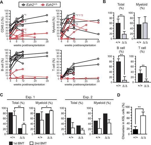
Ezh2-deficient fetal liver cells reconstitute hematopoiesis in BM. (A) Donor chimerism in competitive reconstitution assays using the same number of E12.5 fetal liver test cells and BM competitor cells (ranging from 2 × 10⁵ to 1 × 10⁶ cells, n = 10). The chimerism of CD45.2+ donor-derived cells in PB of each recipient mouse is shown. Lineage contribution of donor cells to myeloid (Gr-1+ and/or Mac-1+), B (B220+), or T (CD4+ and/or CD8+) cells is also shown. Five of 10 recipients infused with Tie2-Cre;Ezh2fl/fl fetal liver cells showed no engraftment of donor cells. The recipients indicated by asterisks were subjected to secondary transplantation at 12 weeks after transplantation. (B) Donor chimerism in PB of recipients with successful engraftment in panel A at 12 weeks after transplantation. Recipient mice with donor cell chimerism > 1.0% for myeloid and for B- and T-lymphoid lineages were considered successfully reconstituted. The data were summarized in bar graphs and presented as the mean ± SD (Tie2-Cre, n = 10; Tie2-Cre;Ezh2fl/fl, n = 5). (C) Donor chimerism in secondary transplantation. A total of 5 × 10⁶ BM cells from the primary recipients indicated by asterisks in panel A were infused into lethally irradiated secondary recipients at 12 weeks after transplantation. Primary recipients *1 and *2 were used for experiments 1 (Exp. 1) and 2 (Exp. 2), respectively. The chimerism in PB is shown. Data are mean ± SD (n = 6). (D) Donor chimerism in BM KSL cells in Exp. 2 in secondary recipients. Data are mean ± SD (n = 6). *P < .05. **P < .005.
Ezh2-deficient fetal liver cells reconstitute hematopoiesis in BM. (A) Donor chimerism in competitive reconstitution assays using the same number of E12.5 fetal liver test cells and BM competitor cells (ranging from 2 × 10⁵ to 1 × 10⁶ cells, n = 10). The chimerism of CD45.2+ donor-derived cells in PB of each recipient mouse is shown. Lineage contribution of donor cells to myeloid (Gr-1+ and/or Mac-1+), B (B220+), or T (CD4+ and/or CD8+) cells is also shown. Five of 10 recipients infused with Tie2-Cre;Ezh2fl/fl fetal liver cells showed no engraftment of donor cells. The recipients indicated by asterisks were subjected to secondary transplantation at 12 weeks after transplantation. (B) Donor chimerism in PB of recipients with successful engraftment in panel A at 12 weeks after transplantation. Recipient mice with donor cell chimerism > 1.0% for myeloid and for B- and T-lymphoid lineages were considered successfully reconstituted. The data were summarized in bar graphs and presented as the mean ± SD (Tie2-Cre, n = 10; Tie2-Cre;Ezh2fl/fl, n = 5). (C) Donor chimerism in secondary transplantation. A total of 5 × 10⁶ BM cells from the primary recipients indicated by asterisks in panel A were infused into lethally irradiated secondary recipients at 12 weeks after transplantation. Primary recipients *1 and *2 were used for experiments 1 (Exp. 1) and 2 (Exp. 2), respectively. The chimerism in PB is shown. Data are mean ± SD (n = 6). (D) Donor chimerism in BM KSL cells in Exp. 2 in secondary recipients. Data are mean ± SD (n = 6). *P < .05. **P < .005.


This feature is available to Subscribers Only
Sign In or Create an Account Close Modal