A 70-year-old woman with renovascular hypertension presented with 8 weeks of worsening chest pain, back and leg pain, dyspnea, and fatigue. She had a faint petechial rash on the abdomen and thighs. Complete blood count showed leukocytosis (26.6 × 10⁹/L), anemia (100 g/L), and thrombocytopenia (114 × 109/L). Manual differential count revealed 5% myeloblasts, 2% myelocytes, 1% metamyelocytes, < 1% eosinophils, and a reticulocyte count of 1.6%. Red cell morphology was unremarkable except for polychromasia.
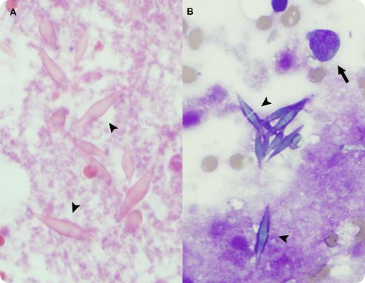
Bone marrow core biopsy revealed numerous bipyramidal Charcot-Leyden crystals (CLCs) (panel A arrowheads) within extensive bone marrow necrosis, precluding definitive diagnosis. Repeat bone marrow aspiration showed CLCs (panel B arrowheads) and 36% myeloblasts (panel B arrow), diagnostic of acute myeloid leukemia. Eosinophils and precursors were not prominent. Despite aggressive management, the patient died of complications before initiation of chemotherapy.
CLCs are composed of a lysophospholipase normally found in eosinophils and may be seen in inflammatory conditions or hematologic malignancies with eosinophilia. CLCs in acute myeloid leukemia are rare and may be accompanied by extensive bone marrow necrosis, as seen in this case. Notably, morphologic evidence of increased eosinophils may or may not be present. The source of lysophospholipase and CLCs in cases lacking eosinophilia is unknown.
For additional images, visit the ASH IMAGE BANK, a reference and teaching tool that is continually updated with new atlas and case study images. For more information visit http://imagebank.hematology.org.


This feature is available to Subscribers Only
Sign In or Create an Account Close Modal