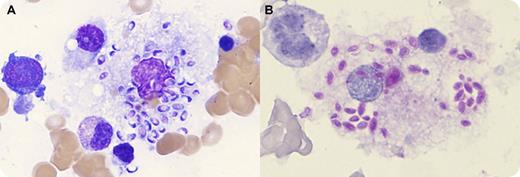
A 44-year-old newly diagnosed HIV-positive man with CD4 count of 47 × 10−6/L (410-1545 × 10−6/L) was admitted to intensive care for management of acute renal failure, profound hypoglycemia, and hemodynamic instability in the setting of pentamidine treatment for Pneumocystis jirovecii pneumonia. A bone marrow biopsy was performed to investigate progressive pancytopenia (hemoglobin 65 g/L, neutrophils 0.41 × 10⁹/L, and platelets 23 × 109/L). Inspection of the bone marrow aspirate revealed innumerable oval/crescent-shaped yeast cells in both intracellular and extracellular distribution, most prominently seen within marrow histiocytes (panel A). Periodic acid-Schiff positively stained the organisms (panel B). Comparison to the published literature and online images yielded a working differential diagnosis of histoplasmosis (Histoplasma capsulatum) or Penicillium marneffei infection. Images of the marrow were referred for expert review and deemed to be most consistent with histoplasmosis. Histoplasma capsulatum was confirmed from fungal culture of the marrow aspirate and PCR for fungal ribosomal RNA. The patient was treated with amphotericin and itraconazole and made a full recovery.
Histoplasma capsulatum and Penicillium marneffei are opportunistic fungi occasionally disseminated in AIDS patients. While an experienced microbiologist may differentiate the 2 infections with microscopy and the time to culture differs, molecular studies are required for definitive diagnosis.
For additional images, visit the ASH IMAGE BANK, a reference and teaching tool that iscontinually updated with new atlas and case study images. For more information visit http://imagebank.hematology.org.


This feature is available to Subscribers Only
Sign In or Create an Account Close Modal