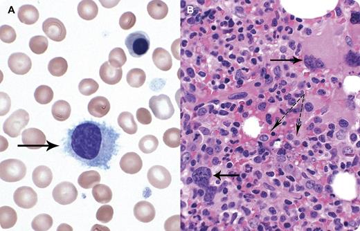
A 59-year-old female was diagnosed 13 years earlier with JAK2 617-positive polycythemia vera (PV) and treated with phlebotomies. Recently she developed progressive splenomegaly and decreased needs for phlebotomy. A diagnosis of post–PV-myelofibrosis was considered, but not confirmed initially by bone marrow (BM) examination. Laboratories tests showed hemoglobin, 92 g/L; platelet count, 119 × 109/L; white blood cell count, 8.6 × 10⁹/L; and lymphocytes, 1.71 × 10⁹/L. Peripheral blood film revealed medium-sized lymphoid cells with indented nucleus, blue cytoplasm, and circumferential projections (panel A solid arrow). BM showed hypercellularity, increased number of abnormally lobated megakaryocytes (panel B solid arrows), and diffuse infiltrates of lymphoid cells (panel B dotted arrows). Flow cytometry showed a λ light chain–restricted clonal B-cell population positive for CD19/20/FMC7/103 and bright CD11c, but negative for CD5/23/10/25/103. Molecular analysis was negative for BRAF V600E mutation. Findings were consistent with hairy cell leukemia variant. A rapid improvement in splenomegaly and cytopenias and disappearance of hairy cells in BM was observed after 1 course of cladribine.
There is increased recognition that patients with myeloproliferative neoplasm (MPN) are at higher risk of lymphoproliferative neoplasms. Physicians should be aware of such association. Even in a patient with typical history suggesting MPN, a peripheral blood smear review should be performed with additional investigations, as needed.
For additional images, visit the ASH IMAGE BANK, a reference and teaching tool that iscontinually updated with new atlas and case study images. For more information visit http://imagebank.hematology.org.


This feature is available to Subscribers Only
Sign In or Create an Account Close Modal