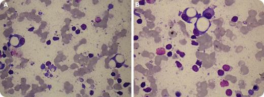
A 70-year-old woman presented with a 4-month history of severe back pain, generalized weakness, and 12-kg weight loss. A spine MRI revealed multiple lytic lesions and collapses of T5 and T7. She had mid-thoracic tenderness and hepatosplenomegaly, but no lymphadenopathy. Hemoglobin was 10.2 g/dL, leukocytes 50.3 × 10⁹/L (88% lymphocytes), and platelets 130 × 109/L. Peripheral smear showed many smudge-cells with an immunophenotype consistent with chronic lymphocytic leukemia (CLL). Serum protein electrophoresis was unremarkable. A bone marrow examination showed marked infiltration with lymphocytes, and increased numbers of plasma cells (18%). Many plasma cells contained 1 to 3 large, clear, PAS-negative cytoplasmic vacuoles (see figure panels). These clinical and morphologic findings raised the suspicion of μ-heavy-chain disease (μ-HCD). Serum immunofixation revealed an M-band typed as IgM without corresponding light chain, thus confirming the diagnosis. A 24-hour urine collection contained 2.84 g of protein consisting of free κ-light chains. The patient received chemotherapy and local radiotherapy with substantial improvement.
μ-HCD is a rare B-cell neoplasm resembling CLL, but differing clinically by lytic bone lesions, hepatosplenomegaly, and the absence of lymphadenopathy. This heavy-chain disorder is characterized by synthesis of monoclonal μ-chain fragments lacking VH region and in part CH1, with an inability to bind light-chain. Associated light-chain proteinuria and skeletal involvement may occur. The absence of M-component on routine electrophoresis may preclude recognition of the disorder. The morphologic clues in this case were the plasma cells with striking vacuoles.
For additional images, visit the ASH IMAGE BANK, a reference and teaching tool that iscontinually updated with new atlas and case study images. For more information visit http://imagebank.hematology.org.


This feature is available to Subscribers Only
Sign In or Create an Account Close Modal