Abstract
Abstract 2827
The myelodysplastic syndromes (MDS) are a group of leukemia characterized by bone marrow failure and increased risk of transformation to acute myelogenous leukemia (AML). Focal adhesion kinase (FAK), a non-receptor protein tyrosine kinase (PTK), plays a key role in the integration of protein-mediated signal transduction. It is a critical mediator of the integrin signaling cascade, which modulates cell proliferation, apoptosis, adhesion, spreading and migration. FAK is upregulated in a wide variety of human cancers. It has been reported that expression of FAK in acute myeloid leukemia (AML) is associated with enhanced blast migration, increased cellularity and poor prognosis. Furthermore, FAK silencing inhibited leukemogenesis in BCR-ABL-transformed hematopoietic cells. FAK has been proposed a potential target in cancer therapy. In this study, we studied the expression patterns of FAK in 98 patients with MDS and performed preclinical studies of FAK inhibition in leukemia cell lines.
CD34+ cells from 98 newly diagnosed patients with MDS and 5 normal donor bone marrow specimens were sorted with CD34 isolation kit from Miltenyi Biotec. Total cellular RNA was isolated using Trizol, cDNA was obtained by using TaqMan reverse transcription reagent (Applied Biosystems). For real-time PCR, FAK assay were purchased from Applied Biosystems and analyzed with an Applied Biosystems Prism 7500 Sequencing detection system. GAPDH was used as internal control. Immunohistochemistry was used to detect FAK protein level in cytospin from MDS CD34+ cells. FAK antibody was obtained from abcam. FAK inhibitor was purchased from Santa Cruz Biotechnology.
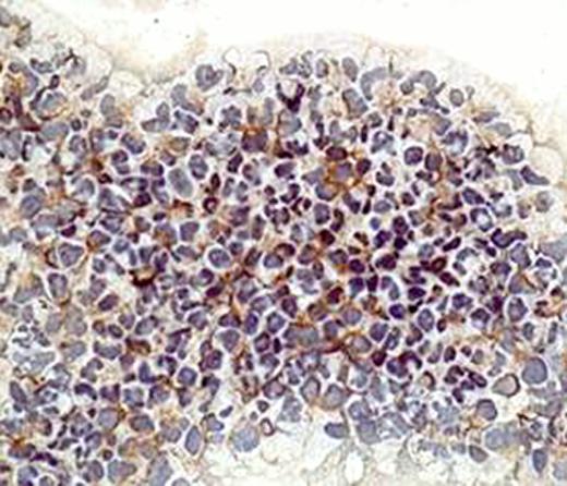
For the 98 MDS patients studied, 78% were older than 60 years; by IPSS score, 17 (17.3%) low risk, 35 (35.7%) INT-1, 24 (24.5%) INT-2, 10 (10.2%) high risk, 12 (12.2%) not available. By cytogenetics, diploid 44 (45%), 20q- 7 (7%), -5/5q- 7 (7%), -7/7q- 7 (7%), -5/5q- and -7/7q- 6 (6%), 8+ 6 (6%), IM 6 (6%), MISC 14 (14%). By real-time PCR, we observed FAK overexpression in 28% of MDS CD34+ cells (fold 2.2–26). We then analyzed FAK protein expression in 10 MDS CD34+ cell cytospin with either higher or lower RNA expression by QPCR using immunohistochemistry. The protein expression patterns were 100% consistent with RNA expression level. This result suggests that FAK expression is upregulated in MDS CD34+ cells. We then performed analysis of clinical associations between FAK expression and clinical characteristics. No association was observed in particular between FAK expression and survival. Because of the potential role of FAK as a therapeutic target, we then studied the effects of FAK inhibition in cell lines. We studied FAK expression level in leukemia cell lines of AML origin and found high protein expression level of FAK in AML leukemia cell line OCI-AML3 and KG1. We treated these cells with FAK inhibitor 14 and detected a dose dependent anti-proliferative effect on both cell lines. Using Annexin V - FITC analysis by flow cytometry, we found the FAK inhibition could induce apoptosis in both cell lines at concentrations of 10uM both at 24 hours and 48 hours.
This study shows that FAK is aberrantly expressed in MDS CD34+ cells, together with the anti-proliferative and apoptosis induction effect of FAK inhibitor, our study demonstrates that FAK may be a potential therapeutic target in MDS and AML patients.
No relevant conflicts of interest to declare.
Author notes
Asterisk with author names denotes non-ASH members.


This feature is available to Subscribers Only
Sign In or Create an Account Close Modal