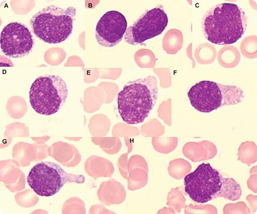
An 81-year-old man had multiple skin lesions (brown-red plaques on the face, abdomen, and back) and asthenia. There was no hepatosplenomegaly or adenopathy. Despite normal blood count values, the peripheral smear contained blasts (7%). Bone marrow examination showed 50% of blasts and mild erythroid dysplasia. The abnormal cells were medium-sized with blastic round (panels A-B) or convoluted (panels C-D,H) nuclei, fine to slightly condensed chromatin, and variable nucleoli. Some blasts had a pseudolymphoid appearance (panel B). The cytoplasm had faint heterogeneous basophilia, no granulation, and peripherally located vacuoles (panels A-F). Occasional cells with pseudopodia were present (panels F-H). Myeloperoxidase and butyrate esterase was negative. Flow cytometry showed a CD45low population expressing CD4, CD56, CD303, CD304, and high levels of CD123 and TCL1. There was no T-cell, B-cell or myeloid expression except for CD33 and CD2, and no expression of CD34 or Tdt. Karyotype was normal. A diagnosis of blastic plasmacytoid dendritic cell neoplasm (BPDCN) was made. A course of chemotherapy led to a short cutaneous remission, but he relapsed and died 7 months later.
BPDCN is an aggressive hematologic malignancy that involves precursors of plasmacytoid dendritic cells. The cells involve the skin and disseminate rapidly. Most patients are elderly with a male predominance. Despite responses to chemotherapy, the medial survival is short.
For additional images, visit the ASH IMAGE BANK, a reference and teaching tool that is continually updated with new atlas and case study images. For more information visit http://imagebank.hematology.org.


This feature is available to Subscribers Only
Sign In or Create an Account Close Modal