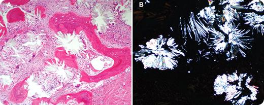
A 28-year-old woman presented with pancytopenia. Her past medical history was significant for primary hyperoxaluria. She had recurrent renal stones and urinary tract infections since the age of 5 years. Subsequently she developed uremic symptoms with end-stage renal disease. For the past 5 years she required regular hemodialysis and transfusions. There was no history of joint pain, cardiac problems, or brain sequelae of oxalosis. On physical examination she had massive hepatosplenomegaly but no lymphadenopathy. Liver enzymes and bilirubin were normal and studies for viral hepatitis were negative. Liver biopsy documented secondary hemochromatosis (transfusion dependent). Laboratory studies showed pancytopenia with hemoglobin of 63g/L, a white blood cell count of 2.6 × 10⁹/L, and a platelet count of 106 × 109/L. Peripheral blood film showed leukoerythroblastic picture with teardrop poikilocytosis. The anemia persisted with little, if any, response to erythropoietin, despite the use of high-dose erythropoietin (100 000 units 4 times per week). Bone marrow biopsy revealed replacement by extensive gray-white crystal deposition (panel A). The crystals were birefringent under polarized light (panel B).
Bone marrow oxalate deposition has been associated with variable degrees of cytopenias, leukoerythroblastic picture, and hepatosplenomegaly. In some patients, as in this case, bone marrow oxalosis can result in resistance to erythropoietin.
For additional images, visit the ASH IMAGE BANK, a reference and teaching tool that is continually updated with new atlas and case study images. For more information visit http://imagebank. hematology.org.


This feature is available to Subscribers Only
Sign In or Create an Account Close Modal