Abstract
Nodal marginal zone lymphoma (NMZL) is a small B-cell neoplasm whose molecular pathogenesis is still essentially unknown and whose differentiation from other small B-cell lymphomas is hampered by the lack of specific markers. We have analyzed gene expression, miRNA profile, and copy number data from 15 NMZL cases. For comparison, 16 follicular lymphomas (FLs), 9 extranodal marginal zone lymphomas, and 8 reactive lymph nodes and B-cell subtypes were included. The results were validated by quantitative RT-PCR in an independent series, including 61 paraffin-embedded NMZLs. NMZL signature showed an enriched expression of gene sets identifying interleukins, integrins, CD40, PI3K, NF-κB, and TGF-β, and included genes expressed by normal marginal zone cells and memory B cells. The most highly overexpressed genes were SYK, TACI, CD74, CD82, and CDC42EP5. Genes linked to G2/M and germinal center were down-regulated. Comparison of the gene expression profiles of NMZL and FL showed enriched expression of CHIT1, TGFB1, and TACI in NMZL, and BCL6, LMO2, and CD10 in FL. NMZL displayed increased expression of miR-221, miR-223, and let-7f, whereas FL strongly expressed miR-494. Our study identifies new candidate diagnostic molecules for NMZL and reveals survival pathways activated in NMZL.
Introduction
The term marginal zone lymphoma (MZL) encompasses 3 rather unrelated lymphoma subtypes: the extranodal marginal zone lymphoma of mucosa-associated lymphoid tissue (MALT lymphoma), the nodal marginal zone lymphoma (NMZL), and splenic B-cell marginal zone lymphoma (SMZL). In the World Health Organization Classification of Tumors,1 all 3 types of MZL are considered distinct clinicopathologic entities. NMZL is an uncommon form of small B-cell neoplasm originating in the lymph node, whose morphology resembles lymph nodes involved in MZL of the extranodal or splenic types, but without evidence of extranodal or splenic disease. The relative rarity of NMZLs, which account for fewer than 2% of all lymphoid neoplasms, is a significant obstacle to their molecular investigation.1 Genomic alterations, including translocations and genomic copy number alterations, are important events in lymphomagenesis,1 providing diagnostic markers and potential therapeutic targets. However, no characteristic translocations or chromosome imbalances have been described in NMZL. Alterations reported in other MZLs, such as the t(11;18) in MALT-type2 and the loss of 7q in SMZL,1,3 have not been found in NMZL cases. Only a few cytogenetic alterations of NMZL have been reported, including trisomy 3 in 50% to 70% of cases. More recently, inactivating mutations encoding truncated A20 proteins have been found in various types of MZL, including 3 of 9 NMZL cases.4 A20 is the protein coded by TNFAIP3 gene, a negative regulator of NF-κB, located in a frequently lost chromosomal region (6q21-q25).4-6 Other alterations described to date are rearrangements involving 1p/q, +3/3q, +12, and +18.1 The absence of molecular or phenotypic markers hinders accurate diagnosis and the differential diagnosis of NMZL from other types of B-cell lymphomas, specifically follicular lymphoma (FL) and reactive lymphoid hyperplasia. Thirteen NMZLs and 8 FLs in this series of cases were included in a previous study by our group that identified MNDA as a marker of potential diagnostic value in NMZL.7 Some cases of NMZL were classified in the past as monocytoid B-cell lymphomas and linked with the marginal zone.8 The normal marginal zone is present in the secondary lymphoid follicles as a concentric area surrounding the mantle follicle and is composed basically of B lymphocytes, macrophages, granulocytes, and dendritic cells that are specialized to capture blood-borne antigens and present them to the resident marginal zone B cells.9 The majority of these MZ cells have an IgMhigh, IgDlow, and CD27+ phenotype and exhibit somatic hypermutation in their Ig-variable genes, according to the data showing that this zone contains mainly memory B cells.9 MZ B cells are involved in early immune response, and the gene expression profile of normal MZ B cells exhibits strong expression of genes, such as CARD11, CXCL12, CXCR6, TACI, MMP12, APRIL, LTB, IFNGR1, COL3A1, AKAP13, and IL2R.10
Gene expression profiles (GEPs) have been obtained for other low-grade B-cell lymphomas, such as FL,11 SMZL,12 and MALT lymphoma.13 These gene expression studies have enabled the identification of genes and pathways related to pathogenesis and of subgroups with distinct pathologic and clinical features and have led to the proposal of new therapeutic targets. However, molecular pathogenesis is essentially unknown in NMZL, and the gene expression profile has yet to be fully described.
miRNAs are 21- to 23-nt-long RNA molecules that regulate the expression of protein-coding genes. More than 700 miRNAs have been identified in mammals and are known to play a role in multiple biologic functions. B-cell differentiation is tightly regulated by miRNAs, and the expression of characteristic sets of miRNAs distinguishes specific stages of B-cell differentiation and B-cell lymphoma main tumor types.14,15 The miRNA profile in NMZL has not been described so far, but memory B cells have been shown to display an increased expression of miR-223, miR-146b, and miR-150, and members of the miR-29 and miR-181 families and let-7 cluster, among others.15,16
The aim of this study was to improve our knowledge of the molecular mechanisms involved in NMZL, to find new diagnostic markers of use for its differential diagnosis from other small B-cell lymphomas and reactive therapeutic targets.
Methods
Patients and tissue samples
The series included 15 patients with NMZL. Clinical information about NMZL cases was retrieved from medical records, surgical pathology reports, and the referring clinicians (Table 1). For comparison purposes, we included B-cell subtypes and a set of 33 lymph node samples: 8 lymph nodes with reactive lymphoid hyperplasia (RLN), 5 lymph nodes infiltrated by MALT lymphoma (MZL-MALT), 4 lymph nodes with SMZL, and 16 lymph nodes infiltrated by FL. The criterion for inclusion was the availability of frozen tissue from the diagnostic lymph node specimens in each case. Morphologic examination and CD20 immunostaining revealed the percentage of tumoral cells exceeding 75% in all cases in this study.
Clinical characteristics of NMZL patient series
| Case no. . | Age, y/sex . | Clinical stage . | HCV . | IGHV gene . | ID, % homology . | Treatment . | Outcome (mo) . |
|---|---|---|---|---|---|---|---|
| 1 | 60/F | III | Negative | VH3-33 | 98.2 | CHT | CR (120) |
| 2 | 80/M | II | ND | VH4-39 | 94 | RT | AWD (64) |
| 3 | 74/F | I | ND | VH3-72 | 93 | ND | AWD (30) |
| 4 | 76/F | III | ND | NR | — | CHT | CR (24) |
| 5 | 73/F | III | Negative | NR | — | CHT | DOD (18) |
| 6 | 62/F | ND | ND | NR | — | ND | ND |
| 7 | 65/F | ND | ND | VH4-34/VH3-7 | 95.9/89.6 | ND | ND |
| 8 | 82/F | III | Negative | VH4-39 | 92.3 | CHT | DXT (54) |
| 9 | 71/M | IV | Positive | VH3-74 | 97.3 | CHT | AWD (19) |
| 10 | 76/M | I | ND | VH3-21 | 93.8 | ND | ND |
| 11 | 55/F | IV | Positive | NR | — | CHT | AWD (34) |
| 12 | 69/F | III | Negative | NR | — | ND | CR (25) |
| 13 | 62/M | I | ND | NR | — | RT | CR (46) |
| 14 | 80/M | IV | ND | VH3-23/VH4b | 89.3/91.1 | CHT | DXT (13) |
| 15 | 53/M | IV | ND | VH3-11 | 89.7 | CHT | DOD (7) |
NMZL patients had a median age of 68.5 y (range, 53-82 y); 64% of them were women.
ID indicates identity frequency; CHT, chemotherapy; CR, complete remission; ND, not determined; RT, radiotherapy; AWD, alive with disease; NR, no rearrangement; —, not applicable; DOD, dead of disease; and DXT, dead, nonlymphoma-related.
To validate the levels of gene expression and miRNA profiles found by microarray analysis, a quantitative RT-PCR experiment was performed with formalin-fixed, paraffin-embedded (FFPE) tissues from an independent series, including 125 cases: 61 NMZL, 57 FL, and 7 RLN.
All cases were selected from the routine and consultation files of the Pathology and Genetics Laboratories of the Virgen de la Salud Hospital (HVS, Toledo, Spain), the Spanish National Cancer Research Center (Madrid, Spain), and the Spanish Tumor Bank Network. Cases were diagnosed on the basis of morphology, immunophenotype, and molecular findings according to the World Health Organization classification criteria.1 Research was performed under the supervision of the Institutional Review Board of the HVS, Toledo, Spain.
Selection of B-cell subsets
B-cell populations were obtained by magnetic cell separation from 6 patients undergoing routine tonsillectomy. The germinal center (GC) B cells were recognized by CD38high IgD−, CD27+, and CD10+ expression, whereas memory B cells were isolated by stainings with CD38low, CD27+, IgDlow, and CD10−. The complete procedure for isolating B-cell subsets is described in supplemental Material 1 (available on the Blood Web site; see the Supplemental Materials link at the top of the online article).
RNA and DNA isolation
For GEP and miRNA hybridization, total RNA was isolated from each B-cell subset, 40 frozen tumoral blocks and 8 control samples from RLN by TRIzol Reagent (Invitrogen) following the manufacturer's recommendations. The quality of the RNA produced was checked by 1% agarose electrophoresis. Cases with poor-quality RNA were discarded.
Copy number alteration (CNA) was assayed with the DNA from 15 NMZL of frozen-tissue cases, extracted using the standard phenol-chloroform protocol.
Microarray procedures: GEP, miRNA, and CNA hybridization
RNA for GEP was hybridized on a Whole Human Genome Agilent 4 × 44K Oligonucleotide Microarray (Agilent Technologies) as reported.17
The miRNA microarray experiments were done using the Agilent Human miRNA Microarray (V1), 8 × 15K (Agilent Technologies). For each tissue sample, 100 ng total RNA was hybridized with the miRNA array and further processed as previously described.18
For CNA, the DNA was hybridized on an Agilent Human Genome CGH Microarray Kit 4 × 44K (Agilent Technologies) as described.19 Human female- and male-pooled gDNA (Promega) was used to normalize the comparative genomic hybridization (CGH) results. Results were considered valuable in 14 cases. The samples (1 μg DNA) were labeled with Cy5, and the DNA donor pool was labeled with Cy3. The commonly affected regions were compared with the Database of Genomic Variants (http://projects.tcag.ca/variation): regions with an overlap of more than 80% between probes and known copy number variations were considered bona fide copy number variations and excluded from further analysis.
Microarray data analysis: GEP, miRNA, B-cell subsets
The background subtraction of microarray data was carried out using GEPAS Version 4.0 (http://gepas.bioinfo.cipf.es). The dataset was normalized by lowess within-array normalization and quantile between-array normalization, and then preprocessed.
We used ANOVA and t tests (http://pomelo2.bioinfo.cnio.es/) to compare the expression of the NMZL gene and miRNA signatures with that of the RLN, with the other MZL subtypes (SMZL and MALT lymphoma) and FL. In all these comparisons, the genes and the miRNAs with false discovery rate (FDR) < 0.05 were considered significant.
The t statistic in gene set enrichment analysis (GSEA; http://www.broad.mit.edu/gsea/) was calculated to identify the pathways and functional groups enriched in the NMZL signature. The GSEA gene sets used were selected from a curated version of Biocarta, KEGG, and CCG pathway databases, as previously described.20 The gene sets with values of P < .05 and FDR < 0.25 were considered to be enriched and potentially relevant in each case.
CNA was normalized using CAPweb Version 2.021 from the Curie Institute (http://bioinfo-out.curie.fr/CAPweb).
miRNA target prediction and miRNA target correlation
We followed the previously described procedure22 to identify associations between differentially expressed miRNAs (FDR < 0.05) and gene expression signatures (FDR < 0.05). The GSEA (Pearson correlation) was used to test the enrichment of gene sets related to each corresponding miRNA. Those with values of P < .05 and FDR < 0.25 were considered to be enriched in each case.
Quantitative real-time RT-PCR analysis
To validate GEP and miRNA microarray data, we carried out a quantitative RT-PCR assay. Total RNA was extracted from FFPE sections of an independent patient group following the manufacturer's instructions using an miRNeasy FFPE Kit (QIAGEN).
Quantitative RT-PCR experiments were performed on selected genes, on the tested miRNAs, and on each B-cell subset using TaqMan probes (Applied Biosystems) as previously described.12,22 The relative degree of change for each gene and miRNA was calculated using the RQ = 2−ΔCt method (for GEP ΔCt = (Ctgene − CtGAPDH) and for miRNAs ΔCt =(CtmiRNA − CtU6)) with GAPDH as the GEP endogenous control and RNU6B as the miRNA endogenous control. Ct values of at least 36 were considered beyond the limit of detection.
The t test and one-way ANOVA (SPSS Version 17.0) were used, and the genes and miRNAs with values of P < .05 were considered significant.
Immunostaining techniques
A series of FFPE were analyzed using IHC by staining 2- to 4-μm-thick sections, following the Dako EnVision FLEX procedure (Dako Denmark). Some of the most relevant genes in the assay, such as CD82, TACI, CD44, CHIT1, TOM1, and LASS4, were studied by IHC. The antibodies used are detailed in (supplemental Table 8).
For immunohistochemical evaluation, cases were scored semiquantitatively, with respect to the number of positive cells and the intensity of the expression. Antibodies producing inconsistent or unreliable results were not quantified. Cases were considered as CD44-positive and LASS4-positive if these markers were expressed in more than 50% of tumoral cells at an intermediate-high intensity, and for CHIT1 when immunostaining was present in more than 10% of macrophage cells.
Results
Nodal marginal zone signature
We have characterized the protein-coding genes and miRNA signature for NMZL and selected B-cell populations. The expression of selected protein-coding genes and miRNAs was validated in a large independent FFPE series by quantitative RT-PCR. Putative targets for the miRNAs were also identified.
Gene expression profiling
Unsupervised hierarchical clustering revealed a relatively homogeneous profile for the entire series, whereby most NMZL, FL, and RLN cases were clustered in well-separated groups (supplemental Figure 1). The expression profiles of lymph nodes infiltrated by NMZL, MZL-MALT, and SMZL were compared statistically revealing no differentially expressed genes, although the analysis of these data should take into consideration the small number of lymph nodes involved in MZL-MALT and SMZL available for study.
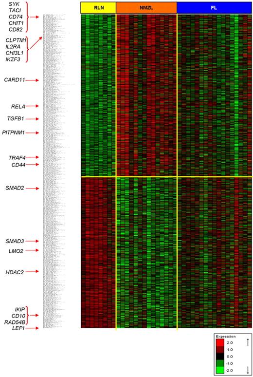
Supervised hierarchical clustering identified an NMZL signature containing 264 up-regulated genes and 184 down-regulated genes (Figure 1). The most relevant significantly deregulated genes in the NMZL signature are shown in Table 2 and Figure 1 (complete NMZL signature in supplemental Table 1). The 10 most up-regulated genes were SYK, TACI, CD74, CD82, CDC42EP5, TFEB, LYN, UCP2, ACP5, and HLA-DMA. Among the most underexpressed genes were those associated with proliferation and cell cycle (PBK, CD2AP, and CDC7), DNA repair (RAD54B, PSMC3IP, MSH2), GC (CD10), meiosis (MND1, MNS1), chromatin modification (HDAC2), cell apoptosis (BNIP3, IKIP), and extracellular matrix and cell adhesion (ANXA1, LMO7).
NMZL gene expression signature. Hierarchical clustering of genes with FDR < 0.10 in NMZL versus RLN t test comparison. Some relevant genes of the signature are marked with red arrows. Red and green represent high- and low-level expression, respectively.
NMZL gene expression signature. Hierarchical clustering of genes with FDR < 0.10 in NMZL versus RLN t test comparison. Some relevant genes of the signature are marked with red arrows. Red and green represent high- and low-level expression, respectively.
NMZL gene expression signature: most relevant NMZL signature genes
| Gene . | NMZL vs RLN comparison . | NMZL vs FL comparison . | Memory vs GC comparison . | Cytoband . | Description . | |||
|---|---|---|---|---|---|---|---|---|
| Fold change . | FDR . | Fold change . | FDR . | Fold change . | P . | |||
| TACI* | 2.075 | 0.032 | 1.987 | 0.042 | 0.925 | < .001 | 17p11.2 | TNF receptor, B-cell stimulation. NF-κB activator |
| CHIT1* | 2.044 | NS (0.072) | 2.527 | 0.020 | −0.234 | NS | 1q31-q32 | Microenvironment involved |
| CD82* | 2.045 | 0.012 | 1.727 | 0.027 | 0.399 | NS | 11p11.2 | Coactivator for the BCR pathway |
| CHI3L1 | 1.588 | 0.028 | 1.635 | 0.029 | 0.133 | ND | 1q32.1 | Microenvironment involved |
| CLPTM1 | 1.652 | 0.009 | 1.334 | 0.049 | 0.249 | ND | 19q13.32 | Lymphocyte activation |
| PTPN1 | 1.487 | 0.008 | 1.243 | 0.026 | 0.245 | ND | 20q13.13 | Signal transduction |
| TRAF4 | 1.036 | 0.033 | 1.139 | 0.022 | −0.110 | ND | 17q11.2 | TNF receptor-associated factor; NF-κB activation |
| TGF-β1* | 1.175 | 0.010 | 1.009 | 0.025 | 0.371 | NS | 19q13.2 | Cell proliferation and differentiation |
| CD44* | 0.874 | 0.029 | 1.285 | 0.006 | 1.131 | NS | 11p13 | Adhesion molecule related to marginal zone |
| SMAD2 | −0.837 | 0.010 | −0.712 | 0.030 | 0.000 | ND | 18q21.1 | TGF-β signaling pathway |
| HDAC2 | −1.258 | 0.007 | −1.016 | 0.013 | −0.171 | ND | 6q22.1 | Chromatin modification |
| IKIP* | −1.428 | 0.029 | −1.270 | 0.045 | −0.179 | ND | 12q23.1 | Apoptosis |
| RAD54B* | −1.775 | 0.007 | −1.291 | 0.026 | −0.241 | NS | 8q22.1 | DNA repair |
| SMAD3 | −2.447 | 0.029 | −1.226 | 0.044 | 0.335 | ND | 6q23.1 | TGF-β signaling pathway |
| LMO2* | −2.533 | 0.006 | −1.732 | 0.014 | −0.545 | NS | 11p13 | GC marker |
| LEF1 | −2.828 | 0.009 | −1.768 | 0.021 | 1.682 | ND | 4q25 | Implicated in gene expression transcription |
| CD10 | −4.269 | 0.006 | −2.172 | 0.041 | −0.612 | ND | 3q32.31 | GC marker |
Fold changes corresponding to the log2 difference between the NMZL and RLN, NMZL, and FL, and memory B-cell and GC B-cell averages, respectively. FDR and P values are from the t test (http://pomelo2.bioinfo.cnio.es/).
NS indicates not significant; and ND, not determined.
To validate the microarray data, the gene was included in the quantitative RT-PCR assay.
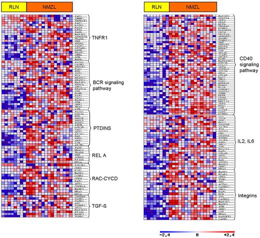
GSEA revealed enriched pathways in NMZL compared with RLN, including IL6, integrins, IL2RB, CD40, RAC-CYCD, TGFβ, PTDINS (PIK3C2A), IL2, RELA, and TNFR1 gene sets (Figure 2; Table 3).
Gene set and pathways enriched in NMZL. The expression of genes representing different B-cell pathways by GSEA analysis (t test). Red and blue represent higher and lower expression, respectively.
Gene set and pathways enriched in NMZL. The expression of genes representing different B-cell pathways by GSEA analysis (t test). Red and blue represent higher and lower expression, respectively.
GSEA for NMZL gene expression profile
| Gene sets . | P . | FDR . | Up-regulated genes . |
|---|---|---|---|
| IL6 pathway | .004 | 0.177 | SHC1, JAK1, CSNK2A1, RAF1, STAT3, JUN, HRAS, MAPK3, GRB2 |
| Integrin pathway | .006 | 0.135 | BCR, RAF1, ZYX, JUN, CAPNS1, CSK, MAP2K2, HRAS, MAPK3, GRB2, RAPGEF1, CAPN1 |
| IL2RB pathway | .004 | 0.152 | SHC1, PTPN6, IL2RA, JAK1, SYK, RAF1, CFLAR, SOCS1, BAD, AKT1, BCL2L1, HRAS, MAPK3, IKZF3, GRB2, CSNK2A1, JUN |
| CD40 signaling during GC development | .028 | 0.202 | KCNN4, BTG2, CAPG, LYN, LITAF, IRF5, DUSP2, JUNB, CD44, IRF4, CD74, NF-KB2, IFI30, ADAM8, TNF |
| RAC and CYCD pathway | .021 | 0.210 | RELA, HRAS, MAPK3, CDKN1A, AKT1, RAF1, CDK4 |
| TGF-β pathway | .023 | 0.210 | MAP3K7IP1, EP300, MAPK3, TGF-β1 |
| PTD INS pathway | .028 | 0.195 | PFKM, GSK3B, VAV2, AP2A1, BAD, AKT1, PFKL, ARF1, AP2M1, LYN |
| IL2 pathway | .030 | 0.197 | SHC1, PTPN6, IL2RA, JAK1, SYK, RAF1, CFLAR, SOCS1, BAD, AKT1, BCL2L1, HRAS, MAPK3, IKZF3, GRB2, CSNK2A1, JUN |
| RELA pathway | .042 | 0.203 | RELA, EP300, IKBKG, TNF, HDAC3, FADD, TRADD |
| TNFR1 pathway | .023 | 0.214 | MADD, JUN, LMNA, DFFB, SPTAN1, CASP2, PRKDC, TNF, TRADD, ARHGDIB, MAP3K1, FADD |
The gene sets were considered up-regulated values of P < .05 and FDR < 0.250 in GSEA analysis using a t test.
miRNA profiling
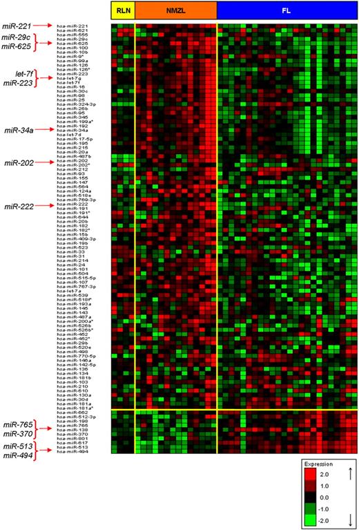
The t test showed 4 miRNAs to be significantly deregulated in NMZL compared with RLN: 3 of them were up-regulated (miR-221, miR-555, and miR-29c) and one was down-regulated (miR-532-5p; Table 4; Figure 3; supplemental Figure 3). The prediction targets for miR-221 and miR-555 included the repressed genes LMO2 and CD10, whereas miR-532-5p showed targets in the up-regulated SYK, LYN, and RELA genes.
NMZL miRNA signature
| Gene . | NMZL vs RLN comparison . | NMZL vs FL comparison . | Memory vs GC comparison . | Cytoband . | CNA* . | Putative prediction targets . | |||
|---|---|---|---|---|---|---|---|---|---|
| Fold change . | FDR . | Fold change . | FDR . | Fold change . | P . | ||||
| miR-223 | 0.298 | NS | 0.978 | 0.009 | 1.023 | NS (.054) | Xq12 | — | LMO2, MYBL1 |
| miR-29c | 0.809 | 0.031 | 0.188 | NS | 1.081 | < .001 | 1q32.2 | — | PTEN, PLAG1, GNB4, MEST |
| miR-221 | 0.562 | 0.002 | 0.430 | 0.009 | 1.057 | NS | Xp11.3 | Gain | CD10, LMO2 |
| miR-34a | 0.432 | NS | 0.494 | 0.024 | 0.992 | ND | 1p36.22 | Gain | LEF1, LASS6, GRSF1, E2F3 |
| let-7f | 0.109 | NS | 0.809 | 0.008 | 1.020 | < .05 | 9q22.32 | — | SMAD2, LBR, CCDC100 |
| miR-625 | 0.396 | 0.092 | 0.447 | 0.002 | 1.010 | ND | 14q23.3 | — | PAG1, SFRS1, BAT3, ABCF3 |
| miR-222 | 0.330 | NS | 0.052 | NS | 1.025 | ND | Xp11.3 | Gain | MYO10 |
| miR-202 | 0.320 | NS | −0.036 | NS | 1.045 | ND | 10q26.3 | Gain | MYCBP, LEPROTL1, STX17 |
| miR-765 | −0.207 | NS | −1.491 | 0.000 | 0.990 | ND | 1q23.1 | — | TGF-β1 |
| miR-370 | −0.221 | NS | −1.961 | 0.000 | 0.960 | ND | 14q32.2 | — | TRAF4 |
| miR-513 | −0.230 | NS | −2.226 | 0.000 | 0.939 | ND | Xq27.3 | — | DRAP1, SMARCAD1, HMGB1 |
| miR-494 | −0.937 | NS | −2.969 | 0.000 | 0.989 | < .001 | 14q32.31 | — | CCND2, ASL, PMPCA, BCL6 |
Most relevant miRNAs with significantly differential expression in comparisons of NMZL and RLN, NMZL and FL, and of memory B cells versus GC B cells. Fold changes correspond to the log2 difference between averages. FDR and P values are from the t test (http://pomelo2.bioinfo.cnio.es/). To validate the microarray data, all miRNAs in this table were included in the quantitative RT-PCR assay. Only 5 miRNAs had valuable data in the quantitative RT-PCR assay (Ct value > 36): miR-223, miR-29c, miR-221, let-7f, and miR-494.
NS indicates not significant; —, not applicable; and ND, not determined.
Chromosomal sites with alterations in the CGH microarray data.
NMZL miRNA signature. Hierarchical clustering of miRNAs with FDR < 0.05 in ANOVA (NMZL vs RLN vs FL) and t test comparison. Some important miRNAs in the signature are marked with red arrows. Red and green represent high- and low-level expression, respectively.
NMZL miRNA signature. Hierarchical clustering of miRNAs with FDR < 0.05 in ANOVA (NMZL vs RLN vs FL) and t test comparison. Some important miRNAs in the signature are marked with red arrows. Red and green represent high- and low-level expression, respectively.
The results detailing the relationship between gene sets and miRNAs are shown in supplemental Material 2.
NMZL versus FL
The differential diagnosis of NMZL and FL is not always clear. The microarray data from frozen tissues in 15 samples of NMZL and 16 of FL were used to compare NMZL and FL gene expression and miRNA profiles. The correlation between miRNAs and gene targets was also investigated.
Supervised hierarchical clustering highlighted a list of interesting deregulated genes. NMZL cases had increased expression of CHIT1, TACI, TRAF4, TGFB1, CD82, PTPN1, and CD44. Conversely, FL cases were characterized by the increased expression of a series of GC markers, including CD10 (MME), BCL6, GCET1 (SERPINA9), and LMO2, whereas genes that are normal marginal zone-related, such as TACI and CD44, were up-regulated in NMZL. Furthermore, NF-κB-related and binding genes (TRAF4, CD82, CLIC1, CSNK2B, and VARS) were up-regulated in NMZL. In the same way, IL32, histones (some isoforms of HIST1H and HIST2H), TNF family members (TACI, TNFRSF14), and regulatory genes involved in lymphocyte activation, such as CLPTM1 and TGFB1, were more highly expressed in NMZL than in FL (Table 2; Figure 1; supplemental Table 2).
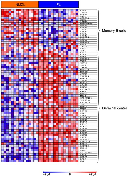
GSEA analysis revealed enriched pathways related to memory B cells (IgM+ IgD− CD27+) in NMZL compared with FL. We also found the IL10 pathway to be strongly represented in NMZL. Genes linked to the GC were up-regulated in FL (Figure 4).
B cell signature expression in NMZL and FL. The GSEA assay revealed up-regulation of genes related to the germinal center were overexpressed in FL cases. Red and blue indicate higher and lower expression, respectively.
B cell signature expression in NMZL and FL. The GSEA assay revealed up-regulation of genes related to the germinal center were overexpressed in FL cases. Red and blue indicate higher and lower expression, respectively.
miRNAs differentially expressed between NMZL and FL were also investigated, revealing 61 deregulated miRNAs: 24 were up-regulated and 37 were repressed in NMZL (Table 4; Figure 3; supplemental Table 3). Some of the up-regulated miRNAs were miR-223 and let-7f, whose putative targets are LMO2 and cell cycle-related genes, respectively. The functional relationship between miR-223 and LMO2 in B cells has already been demonstrated.16 The let-7 cluster, including let-7f miRNA, is involved in cell cycle regulation and cell division.23 The miRNAs down-regulated in NMZL included miR-494, miR-765, miR-370, miR-30d, miR-181a, and miR-29b. The miRNAs miR-370 and miR-765 have the TRAF4 and TGFB1 genes as their respective potential targets. CCND2 is a putative target of miR-494 according to the TargetScan algorithm.
NMZL homology with memory B cells
To determine whether NMZL signature has homology with the gene expression patterns of the normal memory B-cell subpopulation, we identified the GEP and miRNA profile of memory B cells and GC B cells. The comparison of the GEP and miRNAs of memory and GC B cells is presented in supplemental Tables 6 and 7.
The most relevant genes and miRNAs that are up-regulated in NMZL cells, including TACI, CD44, let-7 family members, and miR-223, have been previously described in memory B cells,10,13,15,24 whereas the GC B cells showed overexpression of GC markers, such as CD10, BCL6, and LMO2 and the miRNAs miR-28 and miR-17-5p, among others. Thus, protein-coding genes and miRNA profiles of NMZL both reproduced the findings obtained for memory B cells, which is consistent with the data showing that memory B cells are the predominant cell population in the human normal marginal zone.24
Validation of microarray data in an independent FFPE series
Microarray results were validated by quantitative RT-PCR and IHC assays in an independent FFPE series. The set of genes selected for validation by quantitative RT-PCR was a combination of those differentially expressed genes that were common in NMZL against RLN and opposite FL comparisons, including TACI, CHIT1, CD82, TGFB1, CD44, IKIP, RAD54B, and LMO2 (Table 2). The selected miRNAs for validation were those with higher differential expression between NMZL versus RLN and NMZL versus FL (Table 4). The genes with higher differential expression between NMZL versus RLN were selected for confirmation by IHC assay (Table 2).
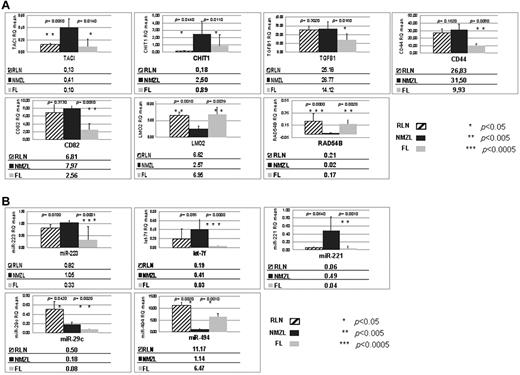
The quantitative RT-PCR analysis showed higher expression levels in NMZL versus FL and versus RLN of TACI and CHIT1 and a lower level of LMO2 and RAD54B, with significant P values for all genes. The complete results are shown in Figure 5A. The data for IKIP could not be evaluated because the TaqMan probe was not amplified. The differential expression of miR-221, miR-223, let-7f, and miR-494 was also confirmed (Figure 5B). Quantitative RT-PCR failed to confirm the results concerning miR-29c. Data for the miRNAs not shown in Figure 5B could not be evaluated. The results of the ANOVA for the most relevant genes and the miRNAs in the assay are shown in supplemental Table 4. The lack of validation for some genes and miRNAs may be the result of the relatively few cases of RLN in the FFPE series.
Quantitative RT-PCR assay. (A) Quantitative RT-PCR assay for GEP. (B) Quantitative RT-PCR study for miRNAs. Expression data (RQ mean) of the validated quantitative RT-PCR genes and miRNAs included in the NMZL signature compared with RLN and FL. RQ values are shown below the corresponding graph, and the t test (P) values are shown on the bars for each comparison. The genes and miRNAs that appear in Tables 2 and 4 (and that are not shown in Figure 5) had no valuable data in the quantitative RT-PCR assay (Ct values > 36): IKIP gene and miR-34a, miR-625, miR-222, miR-202, miR-765, miR-370, and miR-513. Oblique striped represent RLN cases; black, NMZL cases; and light gray, FL cases.
Quantitative RT-PCR assay. (A) Quantitative RT-PCR assay for GEP. (B) Quantitative RT-PCR study for miRNAs. Expression data (RQ mean) of the validated quantitative RT-PCR genes and miRNAs included in the NMZL signature compared with RLN and FL. RQ values are shown below the corresponding graph, and the t test (P) values are shown on the bars for each comparison. The genes and miRNAs that appear in Tables 2 and 4 (and that are not shown in Figure 5) had no valuable data in the quantitative RT-PCR assay (Ct values > 36): IKIP gene and miR-34a, miR-625, miR-222, miR-202, miR-765, miR-370, and miR-513. Oblique striped represent RLN cases; black, NMZL cases; and light gray, FL cases.
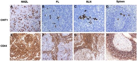
CD44 protein expression was significantly higher in the NMZL cases than in FL (P = .0157); thus, CD44 was positive in 90% of the NMZL cases but was only expressed in 69% of FL cases. CHIT1 expression, present in macrophages but not in the tumoral cells, was stronger in NMZLs (48%) than in FLs (38%), although this difference was not statistically significant (Figure 6; supplemental Table 8).
IHC tissue staining. NMZL, FL, and RLN differential expression data revealed by IHC. Examples of cases immunostained for CHIT1 and CD44. All images were acquired using an Olympus A×80 microscope (Olympus) with a magnification 400×, captured with an Olympus DP72 3.0 camera and processed with Cell A software (Olympus Soft Imaging Solutions, Version 3.29).
IHC tissue staining. NMZL, FL, and RLN differential expression data revealed by IHC. Examples of cases immunostained for CHIT1 and CD44. All images were acquired using an Olympus A×80 microscope (Olympus) with a magnification 400×, captured with an Olympus DP72 3.0 camera and processed with Cell A software (Olympus Soft Imaging Solutions, Version 3.29).
Thus, the validation of the NMZL versus RLN comparison was corroborated in 4 of 7 genes (TACI, CHIT1, LMO2, and RAD54B) and 1 of 2 miRNAs (miR-221). The NMZL versus FL assay was validated in all genes (7 of 7) and all miRNAs (4 of 4): miR-223, let-7f, miR-221, and miR-494. Finally, CD44 was not validated in the NMZL versus RLN comparison by RT-PCR, although it was using IHC.
CNAs
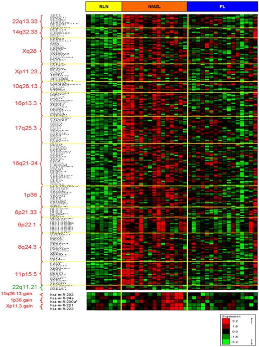
Details of the CNA analysis are shown in supplemental Table 5 and supplemental Figure 3. This assay revealed gains at various chromosomal sites where we studied the relationship between the CNA and GEP data (Table 5; Figure 7). We found miRNAs belonging to the NMZL signature in several of these gained bands. We also observed a very low frequency of CNA losses.
Chromosomal band gains and losses by CGH microarray
| Band . | Imbalance . | Frequency, no. (%) . | Genes involved* . |
|---|---|---|---|
| 22q13.32-33 | Gain | 8/14 (67) | CHKB, SBF1 |
| 14q32.33 | Gain | 5/14 (36) | — |
| Xq28 | Gain | 5/14 (36) | TAZ |
| 10q26.13 | Gain | 4/14 (29) | — |
| 16p13.3 | Gain | 4/14 (29) | RHOT2, RAB40C |
| 17q25 | Gain | 4/14 (29) | SYNGR2, WBP2 |
| 16q21-24 | Gain | 3/14 (21) | PLCG2 |
| 1p36 | Gain | 2/14 (14) | MIB2, SDF4 |
| 6p21.33 | Gain | 2/14 (14) | HLA-DMA, HLA-E, HLA-H, HLA-A, HLA-B |
| 6p22.1 | Gain | 2/14 (14) | HIST1H2 cluster |
| 8q24.3 | Gain | 2/14 (14) | — |
| Xp11.23 | Gain | 2/14 (14) | OTUD5 |
| 11p15.5 | Gain | 2/14 (14) | IFITM family genes |
| 22q11.21 | Loss | 2/14 (14) | PRAME; described as a polymorphism resulting from CNV_53983, CNV_35989, and CNV_53720 |
—indicates not applicable.
Data are the deregulated genes located in the corresponding cytoband. The deregulated genes belong to the NMZL signature (FDR < 0.05 in the NMZL vs RLN comparison, all NMZL cases included).
CNA, GEP and miRNA integration data. Hierarchical clustering of signature genes located in the chromosomal bands with copy number aberrations. Red and green represent high- and low-level expression, respectively.
CNA, GEP and miRNA integration data. Hierarchical clustering of signature genes located in the chromosomal bands with copy number aberrations. Red and green represent high- and low-level expression, respectively.
Discussion
The diagnosis of NMZL is still hampered by the lack of consistent, specific markers. Here we have analyzed the gene expression profile and copy number in a series of cases selected using very conservative diagnostic criteria that excluded all cases with intermediate features or insufficient clinical data. This reduced the number of cases that could be analyzed but improved the chance of identifying specific immunohistochemical or molecular markers. The gene expression profiles were very similar throughout the NMZL series, which guarantees the homogeneity of data and suggests that NMZL is a unique entity.
The NMZL gene expression profile identified in this way encompasses pathways and genes of the normal marginal zone and memory B cells, the cell subpopulation that occupies the normal marginal zone.24 These findings confirm that NMZL is the tumoral counterpart of marginal zone cells. This corroborates previous observations mainly based on the morphologic analysis of MZLs and suggests that typical MZL cases could be more homogeneous than previously thought.
The gene expression profile of all MZL subtypes described to date12,13,25,26 suggests that chronic antigenic stimulation, potentially originating from pathogenic organisms or arising from autoimmune disorders, has an important role in the ontogeny of these tumors. The inflammatory microenvironment, composed of proinflammatory cytokines and cells including antigen-presenting cells andT cells, seems to be crucial in these different lymphoma types. Remarkably, in this NMZL series, the GSEA analysis found up-regulation of the IL6 and IL2 cytokine pathways, and CD40 signaling, which are involved in B-cell survival. Other up-regulated cytokines, such as IFN-γ and CD70 (CD27L), also appear in the NMZL signature. Accordingly, inflammatory cytokines present in the microenvironment might contribute to the onset and progression of lymphoma or at least contribute to the survival of tumor cells. The overexpression of genes functionally related to processes of antigen presentation, such as class I and class II HLAs, and other structurally related genes, such as CD81 and CD74, suggests an important role of the immune response in this lymphoma. CD74 is critical to MHC class II antigen processing and has been proposed as a candidate target for the immunotherapy of B-cell neoplasms by Stein et al.27 This chaperone was shown to be directly involved in the maturation of B cells through a pathway involving NF-κB.28
TACI (TNFRSF13B), a transmembrane activator and CAML interactor, is a TNF family receptor involved in the TI-2 response for the efficient differentiation from marginal zone B cells to plasma cells.29 TACI has multiple functions in the B cell, apoptosis regulation, and NF-κB canonical and noncanonical (through recruitment of TRAF2, TRAF5, and TRAF6) activation.30 Therefore, TACI should be considered an important gene in the molecular mechanism of NMZL and could be a new candidate for diagnosis and as a therapeutic target. The splenic marginal zone is essential for an appropriate TI-2 immune response, with absence or dysfunction of the spleen, resulting in an increased risk of infection by pathogens with polysaccharide capsule.31 Transforming growth factor β-1 (TGFB1) is a member of the TGF-β superfamily, which plays a crucial role in regulating the balance between proliferation and differentiation in hematopoietic cells.32 TGFB1 has been ascribed context-dependent33 contrary functions in hematopoietic cells, with respect to growth inhibition and tumor progression. Increased TGFB1 signaling, as seen in NMZL cases, is an indication of the role that other cell subpopulations, macrophages, regulatory T cells, and other cells could play in NMZL pathogenesis.33
Antigen stimulation and selection of neoplastic cells are thought to occur in gastric MALT lymphoma and SMZL.12,13 At present, the role of antigen stimulation in NMZL is unclear, although association with hepatitis C virus (HCV) has been described in a highly variable proportion of cases.34 The small number (2) of HCV-positive cases in our series prevents any firm conclusion being drawn, but the up-regulation of surface molecules and genes functionally related to antigen-presenting processes suggests that antigen stimulation may play a crucial role in the pathogenesis of this lymphoma. CD81 has been described as being an HCV receptor35 and was up-regulated in the HCV-positive cases.
In addition, the data obtained here seem to identify signaling from BCR and coregulated receptors as being essential for NMZL survival. SYK, LYN, BLK, and BLNK are tyrosine kinases involved in the BCR signaling pathway, which is also known to be overexpressed in other MZLs.12,13
In the molecular signature of NMZL, we found a large number of overexpressed genes associated with NF-κB signaling pathway, such as CD74, CD81, CD82, RELA, and TRAF4, thus extending and confirming the role of this transcription factor in lymphoma pathogenesis.7,36 Neither copy number nor expression profiling data in this study confirmed previous findings concerning the frequent loss of TNFAIP3 in NMZL.7
The NMZL cases overexpressed marginal zone-like genes, such as CD44 and TACI, genes of diagnostic value for NMZL, such as MNDA, and other relevant genes, including CHIT1. Expression of MNDA protein was previously evaluated by our group and found to be significantly higher in NMZL than in FL. The FL samples strongly expressed known GC markers, such as CD10, BCL6, and LMO2. CD44 is an adhesion molecule expressed in MZLs and other non-Hodgkin lymphomas and is an integral member of the CD74 receptor complex.25,37 The increased expression of chiotrisidase-1 or chitinase-1 (CHIT1), a marker of macrophages, in the NMZL cases highlights the essential role of specific subpopulations of accompanying cells in the normal and neoplastic marginal zone.38 A previous independent study found TACI protein expression to be higher in NMZL than in FL.39 TACI, CHIT1, and TGFB1 showed stronger expression, with microarrays and RT-PCR, in NMZL than FL and could be candidates for novel diagnostic markers in NMZL.
Data obtained suggest that the differential expression of miR-221 in NMZL against RLN, and miR-223, let-7f, and miR-494 in NMZL versus FL could be of potential diagnostic value. FL and GC cells are distinguished by an increased expression of LMO2, and a diminished expression of miR-223. LMO2 targeting by miR-223 is essential to the regulation of B-cell development.16 The data here obtained showing LMO2 down-regulation and increased miR-223 expression suggest that this relation could also play a role in NMZL. Let-7 cluster is implicated in multiple molecular processes as cell cycle, apoptosis, and cell proliferation, so this miRNA has been proposed as a putative therapeutic target.23 CCND2 is an miR-494 putative target and is essential for the control of the cell cycle at the G1 to S transition.40
Although the results of this study warrant functional validation, our findings suggest that the miRNA signature is related to the gene expression profile in NMZL in several ways. For instance, the up-regulation of miR-223 and miR-221, which target the GC-related genes LMO2 and CD10, could be partially responsible for the expression of a marginal zone signature. Similarly, the most up-regulated miRNAs in the signature showed a significant positive correlation (Pearson GSEA analysis) with important B-cell pathways, such as BCR, IL2, IL6, CD40, NF-κB, TGFB, and memory B cells. These pathways were also found in the GSEA analysis (NMZL vs RLN comparison), suggesting a significant role for these pathways in NMZL.
Even though deletions of the chromosomal band 6q21-q25 involving the NF-KB negative regulator TNFAIP3 (A20) have been described as characteristic cytogenetic abnormalities for MZLs,4-6 we were unable to confirm this finding in our series (Figure 6; supplemental Table 5; supplemental Figure 3). This could be a consequence of the small number of cases studied or the resolution of the CGH arrays used for each study.
In conclusion, this analysis shows that the NMZL gene expression profile reproduces the signature of normal marginal zone and memory B cells, identifies BCR signaling, interleukins (IL2, IL6, IL10), integrins (CD40), and survival pathways (MAPKs, TNF, TGFB, NF-κB) as the most significant pathways in NMZL pathogenesis, and allows new specific markers (TACI, CHIT1, CD44, CD82, TGFB1, miR-223, let-7f, and miR-221) to be proposed as a means of distinguishing this lymphoma type from FL. Our study also identifies some possible therapeutic targets, such as TACI and CD74.
The online version of this article contains a data supplement.
The publication costs of this article were defrayed in part by page charge payment. Therefore, and solely to indicate this fact, this article is hereby marked “advertisement” in accordance with 18 USC section 1734.
Acknowledgments
The authors thank R. Diaz de Otazu (Vitoria), C. Lobo (San Sebastián), S. Nieto (Madrid), P. Domínguez (Madrid), J. San Juan (Valencia), P. Navarro (Valencia), J. Escobar Jarrio (Asturias), and Laura Cereceda (Spanish National Cancer Research Center Tumor Bank) for kindly providing the cases included in this series and S. Opazo, Y. Ruano, and B. Meléndez (HVS) for their excellent technical help.
This work was supported by the Ministerio de Sanidad y Consumo (RETICS, FIS PI052742, PI081666, PI052800, and INT09/276), Ministerio de Ciencia e Innovación (SAF2008-03871), the Servicio de Salud de Castilla la Mancha (FISCAM PI2008/31), and the Asociación Española Contra el Cancer.
Authorship
Contribution: A.J.A. analyzed microarray data, performed research, and wrote the paper; Y.C.-M. and P.A. analyzed data and performed research; C.G.-A. analyzed CGH data and performed research; M.S.-B. performed research and contributed analytical tools; M.S.R.-P. performed research and reviewed cases; S.M.-M. and J.A.-F. performed research and contributed samples; and M.A.P. and M.M. designed the study.
Conflict-of-interest disclosure: The authors declare no competing financial interests.
Correspondence: Manuela Mollejo, Avda Barber 30, CP 45004, Toledo, Spain; e-mail: mmollejov@sescam.jccm.es.

