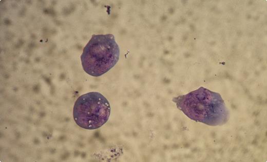
A 76-year-old woman presented with diffuse large B-cell non-Hodgkin lymphoma (NHL), stage III A, low-intermediate International Prognostic Index. The disease involved the retroperitoneum, laterocervical, and supraclavicular regions. Bone marrow and other organ sites were negative. She received 5 of 6 planned cycles with rituximab, cyclophosphamide, doxorubicin, vincristine, and prednisone. One month after the fifth cycle, while in complete remission, she developed flaccid quadriplegia along with left hemiparesis, gaze palsy, dysphonia, and dysphagia. The clinical picture was suggestive of Guillain-Barrè syndrome but there was no response to high-dose intravenous immunoglobulins. Brain CT showed diffuse meningeal enhancement. The cerebrospinal fluid showed mononucleated cells 12 000/mm3, protein 151 mg/dL, and glucose 5 mg/dL. Microscopic examination of the fluid showed large mononucleated cells with multiple prominent nucleoli (see figure). The cells were positive for CD19 and CD20, as were the original malignant B cells.
A diagnosis of leptomeningeal lymphoma was made. After intrathecal chemotherapy with methotrexate, cytosine arabinoside, and dexamethasone, she showed improvement but died of bacterial sepsis. This case illustrates an example of meningeal lymphoma arising in a patient with NHL in presumed complete remission. The neurologic abnormalities and abnormal cerebrospinal fluid and the microscopic appearance of lymphoma cells established the diagnosis.
For additional images, visit the ASH IMAGE BANK, a reference and teaching tool that is continually updated with new atlas and case study images. For more information visit http://imagebank.hematology.org.


This feature is available to Subscribers Only
Sign In or Create an Account Close Modal