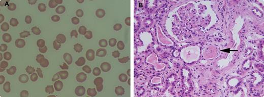
A 59-year-old female who had been diagnosed with multiple myeloma received treatment with bortezomib and dexamethasone. Subsequently, she underwent autologous peripheral stem cell transplantation. Two months later she was admitted with a urinary tract infection complicated by Klebsiella bacteremia. Her complete blood count showed anemia (8.5 g/dL), thrombocytopenia (22 K/uL), increased reticulocytes, and normal white blood cells (8.9 K/uL). The peripheral blood smear (panel A) revealed moderate schistocytosis. The LDH was 2920 IU/L, haptoglobin was undetectable, D-dimer was 2020, fibrinogen was 375, and ADAMTS13 activity was low.
Acute renal failure ensued that required hemodialysis. A kidney biopsy showed thrombotic microangiopathic nephropathy (panel B; arrow shows a thrombus). A diagnosis of thrombotic thrombocytopenic purpura (TTP) was made and plasmapheresis was started with resolution of TTP over the next few weeks. Although schistocytes are a clue to disseminated intravascular coagulopathy, malignancy, anatomic vascular abnormalities, hypertension, and connective tissue disease among others, they were indicative of TTP in this patient.
For additional images, visit the ASH IMAGE BANK, a reference and teaching tool that is continually updated with new atlas and case study images. For more information visit http://imagebank.hematology.org.


This feature is available to Subscribers Only
Sign In or Create an Account Close Modal