Abstract
Patients with Glanzmann thrombasthenia or Leukocyte Adhesion Deficiency-III syndrome (LAD-III or LAD-1/variant) present with increased bleeding tendency because of the lack or dysfunction of the fibrinogen receptor GPIIb/IIIa (integrin αIIbβ3), respectively. Although the bleeding disorder is more severe in LAD-III patients, classic aggregometry or perfusion of Glanzmann or LAD-III platelets over collagen-coated slides under physiologic shear rate does not discriminate between these 2 conditions. However, in a novel flow cytometry-based aggregation assay, Glanzmann platelets were still capable of forming small aggregates upon collagen stimulation, whereas LAD-III platelets were not. These aggregates required functional GPIa/IIa (integrin α2β1) instead of integrin αIIbβ3, thus explaining the clinically more severe bleeding manifestations in LAD-III patients, in which all platelet integrins are functionally defective. These findings provide genetic evidence for the differential requirements of platelet integrins in thrombus formation and demonstrate that correct integrin function assessment can be achieved with a combination of diagnostic methods.
Introduction
Damage to blood vessels exposes collagen to platelets, which bind to it through several receptors, supporting adhesion and stimulating platelet activation. Fibrinogen, present in plasma and released by activated platelets, forms a bridge between platelets in blood clots. Collagen is bound by GPIa/IIa (integrin α2β1) and GPVI, and activation of α2β1 enhances platelet adhesion to collagen.1–5 The notion of thrombus formation thus far suggests that activation of the fibrinogen receptor GPIIb/IIIa (integrin αIIbβ3), induced by GPVI, is required for further α2β1 activation and blood clot formation.5–7 GPIb (VWF receptor) and GPIV have also been described as functional collagen receptors.8
Defective integrin expression or function results in bleeding tendency, as observed in Glanzmann thrombasthenia or Leukocyte Adhesion Deficiency type III (LAD-III or LAD-1/variant).9–13 Glanzmann thrombasthenia is a rare congenital disorder characterized by mutations in the ITGA2B (αIIb) or ITGB3 (β3) genes, causing qualitative or quantitative abnormalities of integrin αIIbβ3,9,10 leading to mucocutaneous bleeding with variable clinical manifestations.14
LAD-III is an autosomal recessive disorder characterized by integrin signaling dysfunction in leukocytes and platelets, whereas integrin expression is normal.11,12 We and others have identified mutations in the FERMT3 gene encoding the hematopoietic specific integrin-activating cytoplasmic protein kindlin-315 as the primary cause of the disease.13,16,17 Kindlin-3 knockout mice phenocopy the disorder.18
We performed a comparative study of the adhesion and aggregation characteristics of Glanzmann and LAD-III platelets by combining several assays. We show that integrin α2β1 in Glanzmann—not in LAD-III—platelets still binds collagen, allowing formation of small aggregates with functional relevance, as it explains that the bleeding tendency in LAD-III patients is clinically more severe than in Glanzmann patients.13,14,19
Methods
The study was approved by the Academic Medical Centre Institutional Medical Ethics Committee in accordance with the 1964 Declaration of Helsinki.
Light transmission aggregometry
Activation of platelet-rich plasma with 10 μg/mL type I collagen (Horm, Nycomed Arzneimittel) was traced for 10 minutes at 800 rpm in a standard aggregometer (model 490, Chronolog).13
Flow chamber perfusion
Slides (μ-Slide-I 0.1 Luer, Ibidi) were coated with 100 μg/mL collagen. Platelet-rich plasma (250-500 × 106 platelets/mL) was perfused 5 minutes at 1500 second−1 shear rate. After washing 2 minutes with PBS, images were taken at 600× magnification with an EVOS microscope (AMG).
Flow cytometry aggregation assay
CFSE- and PKH26-labeled platelets were mixed 1:1 and preincubated with or without 0.5 μg/mL tirofiban (Aggrastat, Merck), 10 μg/mL mAb LIA1/2.1 (LIA),20 or 9O1221 as antagonists, 15 minutes at 37°C. As agonists, we used 100 ng/mL phorbol myristate acetate (PMA; Sigma-Aldrich), 1.5 mg/mL ristocetin (Biopool; Trinity Biotech), 10 μg/mL collagen, or 2 μg/mL cross-linked collagen-related peptide (CRP)22 in the presence of 3mM CaCl2. Time samples fixed in 0.5% formaldehyde/PBS were measured on an LSRII + HTS flow cytometer and analyzed for double-colored events by FACSDiva Version 6.1 software (both BD Biosciences; I.M.D.C., M. Meinders, E.v.d.V., D. de Korte, L. Porcelijn, M. de Haas, T.W.K., A.J.V., A Novel flowcytometry-based platelet aggregation assay, manuscript submitted).
Integrin activation phenotyping
Platelets were stimulated 5 minutes with 100 ng/mL PMA, 10 μg/mL collagen, 2 μg/mL CRP, or 1.5 mg/mL ristocetin. Total expression of β1 and β3 integrins was measured with HUTS-21-PE (BD Biosciences) and C17-FITC (Sanquin Reagents), respectively. The high-affinity conformation of integrin β1 was measured with HUTS-4 (Millipore) and goat anti–mouse-FITC (Invitrogen), and of integrin β3 with PAC-1–FITC (BD Biosciences). The extent of activated integrin was determined relative to total integrin expression after background correction with isotype controls and normalizing ratios of unstimulated platelets to 1.
Results and discussion
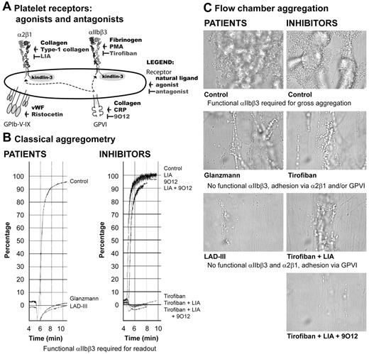
We investigated the aggregation response of platelets from Glanzmann thrombasthenia (type I, < 5% αIIbβ3 protein expression) and LAD-III patients because of their differences in integrin expression and function.9–11,19 In parallel, we studied platelets from healthy donors, preincubated with the antagonists tirofiban (an integrin αIIbβ3 inhibitor), LIA20 (an integrin α2β1-blocking mAb), or 9O1221 (a GPVI-blocking mAb; Figure 1A).
Classic aggregometry and flow chamber perfusion assay. (A) Scheme representing the integrin and nonintegrin receptors studied, with their natural ligand, agonist, and antagonist indicated. (B) Aggregation of control, Glanzmann, and LAD-III platelets (left) or control platelets preincubated with different antagonists (right) upon stimulation with 10 μg/mL collagen measured by light transmission aggregometry. (C) Binding of control, Glanzmann, and LAD-III platelets (left) or control platelets preincubated with different antagonists (right) to collagen-coated slides. Pictures were taken using an EVOS fl (fluorescence) digital inverted microscope by Advanced Microscopy Group at 600× magnification using an 60×/1.35 oil objective. The imaging medium was PBS. The EVOS fl contains a CCD camera and EVOS fl software was used for acquiring images. Adobe Suite CS 5 Photoshop was used to adjust brightness and contrast.
Classic aggregometry and flow chamber perfusion assay. (A) Scheme representing the integrin and nonintegrin receptors studied, with their natural ligand, agonist, and antagonist indicated. (B) Aggregation of control, Glanzmann, and LAD-III platelets (left) or control platelets preincubated with different antagonists (right) upon stimulation with 10 μg/mL collagen measured by light transmission aggregometry. (C) Binding of control, Glanzmann, and LAD-III platelets (left) or control platelets preincubated with different antagonists (right) to collagen-coated slides. Pictures were taken using an EVOS fl (fluorescence) digital inverted microscope by Advanced Microscopy Group at 600× magnification using an 60×/1.35 oil objective. The imaging medium was PBS. The EVOS fl contains a CCD camera and EVOS fl software was used for acquiring images. Adobe Suite CS 5 Photoshop was used to adjust brightness and contrast.
Platelet aggregation is routinely measured by light transmission aggregometry, in which the increase in light transmission upon platelet aggregation is dependent on αIIbβ3.23 Collagen-induced light transmission aggregometry of normal platelets was completely inhibited by tirofiban, whereas inhibition of integrin α2β1, GPVI, or both had no effect. Neither Glanzmann nor LAD-III platelets aggregated because of absent or dysfunctional αIIbβ3 integrin (Figure 1B).9–11,14,19
Visualization with wide-field microscopy of platelet binding to collagen in flow chambers under physiologic shear rate confirmed that large aggregate formation is αIIbβ3-dependent because tirofiban inhibited this process in control platelets. In contrast, both Glanzmann and LAD-III platelets showed only direct platelet-collagen interactions (Figure 1C), through collagen receptors that were not inhibited by tirofiban. Thus, these assays do not discriminate Glanzmann from LAD-III platelets.3,24
Next, we measured platelet aggregation in a novel flow cytometric assay (Figure 2A), which detects the formation of small double-colored aggregates over time, thereby rendering it more sensitive for receptor/integrin function dissection (I.M.D.C., M. Meinders, E.v.d.V., D. de Korte, L. Porcelijn, M. de Haas, T.W.K., A.J.V., A Novel flowcytometry-based platelet aggregation assay, manuscript submitted).
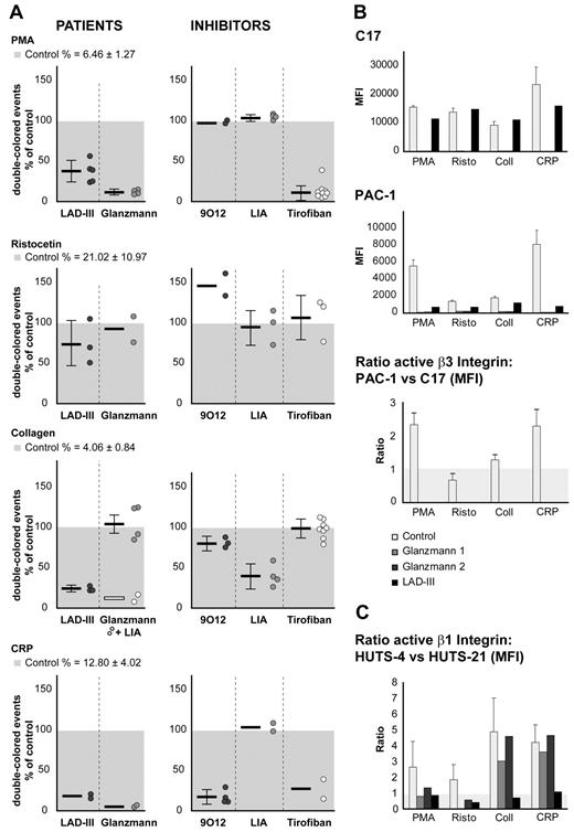
Flow cytometric aggregation assay and characterization of integrin activation. (A) Aggregation of control, Glanzmann, and LAD-III platelets upon stimulation with PMA, ristocetin, collagen (Student t test LAD-III vs Glanzmann, P < .001), or CRP. Preincubation with 9O12, mAb LIA, or tirofiban blocks ligand binding to GPVI, integrin α2β1, and αIIbβ3, respectively. Aggregation of the control platelets was set to 100% for each separate experiment (n = 4-6). Combinations of inhibitors did not add to the effects noticed when used as a single blocking agent (data not shown). (B) Characterization of αIIbβ3 integrin by flow cytometry upon stimulation with PMA, ristocetin, collagen, or CRP in control, Glanzmann, and LAD-III platelets. αIIbβ3 total expression was measured with C17 (top panel), and active αIIbβ3 was measured with PAC-1 antibodies (middle panel). The ratio of active integrin versus total integrin expression was calculated from the respective mean fluorescence intensity (MFI) after subtraction of isotype controls and normalizing unstimulated platelets to 1 (bottom panel). (C) Characterization of α2β1 integrin by flow cytometry upon stimulation with PMA, ristocetin, collagen, or CRP in control, Glanzmann, and LAD-III platelets. α2β1 total expression was measured with HUTS-21, and active α2β1 was measured with HUTS-4. The ratio of active integrin versus total integrin expression was calculated from the respective mean fluorescence intensity (MFI) after subtraction of isotype controls and normalizing unstimulated platelets to 1.
Flow cytometric aggregation assay and characterization of integrin activation. (A) Aggregation of control, Glanzmann, and LAD-III platelets upon stimulation with PMA, ristocetin, collagen (Student t test LAD-III vs Glanzmann, P < .001), or CRP. Preincubation with 9O12, mAb LIA, or tirofiban blocks ligand binding to GPVI, integrin α2β1, and αIIbβ3, respectively. Aggregation of the control platelets was set to 100% for each separate experiment (n = 4-6). Combinations of inhibitors did not add to the effects noticed when used as a single blocking agent (data not shown). (B) Characterization of αIIbβ3 integrin by flow cytometry upon stimulation with PMA, ristocetin, collagen, or CRP in control, Glanzmann, and LAD-III platelets. αIIbβ3 total expression was measured with C17 (top panel), and active αIIbβ3 was measured with PAC-1 antibodies (middle panel). The ratio of active integrin versus total integrin expression was calculated from the respective mean fluorescence intensity (MFI) after subtraction of isotype controls and normalizing unstimulated platelets to 1 (bottom panel). (C) Characterization of α2β1 integrin by flow cytometry upon stimulation with PMA, ristocetin, collagen, or CRP in control, Glanzmann, and LAD-III platelets. α2β1 total expression was measured with HUTS-21, and active α2β1 was measured with HUTS-4. The ratio of active integrin versus total integrin expression was calculated from the respective mean fluorescence intensity (MFI) after subtraction of isotype controls and normalizing unstimulated platelets to 1.
When stimulated with PMA, aggregation was absent in platelets from Glanzmann and LAD-III patients. This response is αIIbβ3-dependent, as corroborated by the inhibition by tirofiban in control platelets. As expected, phenotyping with antibody C17 showed absence of β3 integrin in Glanzmann, whereas LAD-III platelets had normal expression, although dysfunctional (Figure 2B). PMA stimulation induced major activation of β3 and minor activation of β1 integrin, probably mediated by β3 activation, as this is not detected in Glanzmann platelets (Figure 2B-C).
When stimulated with ristocetin, control platelets aggregated normally irrespective of the antagonist added, showing that GPIb-mediated platelet activation does not require functional integrins. Aggregate formation was equal to controls in Glanzmann and LAD-III platelets, revealing a normal GPIb response in these patients. Ristocetin did not induce significant changes in either β1 or β3 integrin conformation (Figure 2B-C).
In control platelets, collagen-induced aggregation was mainly integrin α2β1-dependent as judged by the decrease in aggregation in the presence of LIA. Remarkably, we measured αIIbβ3-independent aggregation because tirofiban exerted no effects. GPVI had only a minor contribution, as shown by the slight decrease in aggregation in the presence of 9O12. The aggregation of LAD-III platelets was strongly reduced to approximately 25% of control platelets, whereas, as shown previously, Glanzmann platelets aggregated normally25 (Figure 2A). In control and Glanzmann platelets, but not in LAD-III patients, collagen induced the activation of β1 (Figure 2C) and aggregation was specifically inhibited by LIA (Figure 2A). Thus, functional α2β1 explains the collagen response and the milder bleeding tendency of Glanzmann compared with LAD-III patients.
We next used CRP, which bears the GPVI but not the integrin α2β1-binding site.3,22 The aggregation of normal platelets in response to CRP was inhibited by tirofiban and 9O12 but not by LIA, reflecting aggregate formation essentially dependent on αIIbβ3 activation without contribution of α2β1 integrin. Platelets from both Glanzmann and LAD-III patients19 were unable to form aggregates upon CRP stimulation. As described,1 CRP induced activation of β1 and β3 integrins in control and Glanzmann platelets (Figure 2B-C).
In conclusion, functional integrin α2β1 may explain the relatively milder bleeding phenotype in Glanzmann disease compared with LAD-III because αIIbβ3-independent α2β1 activation and collagen-induced aggregate formation25 in vivo may be sufficient to limit spontaneous bleeding. We highlight the synergistic role of collagen receptors and integrin function in platelet adhesion and aggregation and the requirement of distinct methods for correct assessment of integrin function.
The publication costs of this article were defrayed in part by page charge payment. Therefore, and solely to indicate this fact, this article is hereby marked “advertisement” in accordance with 18 USC section 1734.
Acknowledgments
The authors thank Dr F. Sánchez-Madrid (Universidad Autónoma de Madrid, Madrid, Spain) for providing mAb LIA1/2.1, Dr M. Jandrot-Perrus (Inserm U698, Hôpital Bichat, Paris, France) for providing mAb 9O12, Prof R. W. Farndale (University of Cambridge, Cambridge, United Kingdom) for providing cross-linked CRP, Dr P. W. Kamphuisen and Dr M. Peters (both Academic Medical Center, Amsterdam, The Netherlands) and Dr Ö. Sanal and Dr M. Çetin (both Hacepette University, Ankara, Turkey) for patient material, and Prof Dirk Roos for critically reading the manuscript.
T.W.K. and E.v.d.V. were supported by the Landsteiner Foundation for Blood Transfusion Research (LSBR 0619). L.G. was supported by The Netherlands Organization for Scientific Research (NWO 863.09.012).
Authorship
Contribution: E.v.d.V. and I.M.D.C. designed and performed experiments and wrote the paper; A.J.G. performed experiments; A.J.V., L.G., T.K.v.d.B., and T.W.K. designed experiments and wrote the paper; and K.S. provided patient material and helped with discussions.
Conflict-of-interest disclosure: The authors declare no competing financial interests.
The current affiliation for A.J.V. is Department of Medical Biochemistry, Academic Medical Centre, University of Amsterdam, Amsterdam, The Netherlands.
Correspondence: Edith van de Vijver, Department of Blood Cell Research, Sanquin Research and Landsteiner Laboratory, Plesmanlaan 125, 1066 CX Amsterdam, The Netherlands; e-mail: e.vijver@sanquin.nl.
References
Author notes
E.v.d.V. and I.M.D.C. contributed equally to this study.


This feature is available to Subscribers Only
Sign In or Create an Account Close Modal