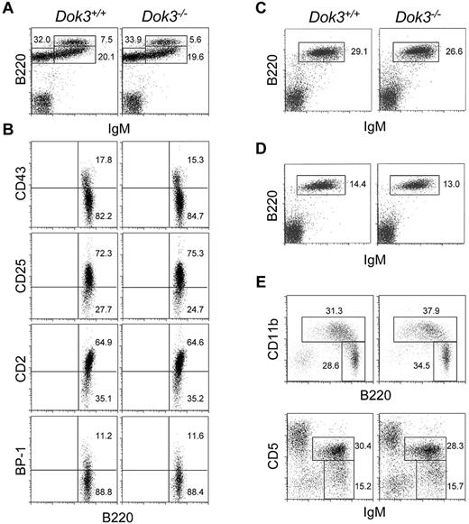
On page 261 of the 1 July 2007 issue, there are errors in Figure 2. Figure 2C and D depicted a representative flow cytometry analysis of IgM+B220+ B-cell population in spleen and lymph nodes of Dok-3+/+ and Dok-3−/− mice. Because of an oversight, the data files for Dok-3+/+ were duplicated. The duplication does not affect any other data or conclusions of the paper. The corrected figure shown was prepared from the printout of the original FACS data.
Normal B cell development in Dok-3−/− mice. Flow cytometry analyses of B cell populations in the bone marrow (A and B), spleen (C), lymph nodes (D) and peritoneal cavity (E) of wild-type and Dok-3−/− mice. Bone marrow B220 +IgM− cells were further analyzed for their expression of CD43, CD2, CD25 and BP-1 (B). Numbers indicate percent of cells in the lymphocyte gate for (A, C, D and E) and percent of B220 +IgM− cells for (B). Data shown are representative of 3 independent experiments.
Normal B cell development in Dok-3−/− mice. Flow cytometry analyses of B cell populations in the bone marrow (A and B), spleen (C), lymph nodes (D) and peritoneal cavity (E) of wild-type and Dok-3−/− mice. Bone marrow B220 +IgM− cells were further analyzed for their expression of CD43, CD2, CD25 and BP-1 (B). Numbers indicate percent of cells in the lymphocyte gate for (A, C, D and E) and percent of B220 +IgM− cells for (B). Data shown are representative of 3 independent experiments.


This feature is available to Subscribers Only
Sign In or Create an Account Close Modal