Abstract
Patients with anti–IFN-γ autoantibodies have impaired IFN-γ signaling, leading to severe disseminated infections with intracellular pathogens, especially nontuberculous mycobacteria. Disease may be severe and progressive, despite aggressive treatment. To address the underlying pathogenic IFN-γ autoantibodies we used the therapeutic monoclonal rituximab (anti-CD20) to target patient B cells. All subjects received between 8 and 12 doses of rituximab within the first year to maintain disease remission. Subsequent doses were given for relapsed infection. We report 4 patients with refractory disease treated with rituximab who had clinical and laboratory evidence of therapeutic response as determined by clearance of infection, resolution of inflammation, reduction of anti–IFN-γ autoantibody levels, and improved IFN-γ signaling.
Introduction
The IFN-γ–IL-12 axis is critical for control of mycobacteria as well as other opportunistic pathogens.1 High-titer neutralizing anti–IFN-γ autoantibodies affect the same pathway and cause a syndrome of disseminated nontuberculous mycobacterial and other opportunistic infections.2-5 These patients can have severe and progressive disease despite prolonged antimicrobial therapy, which can result in drug toxicity and therapeutic failure.
Rituximab is a chimeric mAb directed against human CD20 on mature B cells and plasmablasts that causes rapid and sustained depletion of circulating and tissue-based B cells.6 Diseases for which rituximab is approved by the Food and Drug Administration include B-cell lymphoma, dosed at 375 mg/m2 for 4 or 8 doses,6,7 or rheumatoid arthritis, dosed at 1000 mg for 2 doses on day 1 and day 15.8 Rituximab has also been used off-label for the treatment of diseases caused by pathogenic autoantibodies, including the anti–desmoglein 3 autoantibodies of pemphigus vulgaris,9 the anti-acetylcholine or anti-muscle–specific kinase autoantibodies of myasthenia gravis,10,11 the anti–GM-CSF autoantibodies of pulmonary alveolar proteinosis,12 and the anti-erythropoietin autoantibodies of pure red cell aplasia.13
We used rituximab in 4 patients with high-titer anti–IFN-γ autoantibodies who had progressive refractory nontuberculous mycobacterial disease despite aggressive anti-infective treatment. Our initial approach was to treat according to a lymphoma regimen, given our aim of depleting B cells. Additional doses of rituximab were given for signs and symptoms consistent with infection persistence or relapse.
Methods
Subjects
The 4 patients were seen at the National Institutes of Health and consented to evaluation and treatment of disseminated nontuberculous mycobacterial infection under protocol 01-I-0202. Control plasma and PBMCs were obtained though the National Institutes of Health Blood Bank under appropriate protocols.
Clinical monitoring
Patients had routine laboratories, including HIV testing, complete blood count with differential, serum electrolytes, renal and hepatic function chemistries, inflammatory markers, quantitative immunoglobulin levels, and lymphocyte markers that included total T cells (CD3+; BD PharMingen), CD4+ T cells (Immunotech), CD8+ T cells (Immunotech), CD20+ B cells (BD PharMingen); and CD16+ or CD56+ natural killer cells (BD PharMingen). Disease activity was determined by evidence of active infection on computed tomographic scan, bone scan, culture, or smear, as indicated. Treatment and clinical data were collected by review of electronic chart records. Salient clinical features of disease activity were compiled retrospectively into a scoring system.
Plasma collection
Plasma from each subject was collected and stored at −80°C before, during (2-9 months after start), and after treatment (1-4 years after start) with rituximab. All samples were tested at first thaw. For each patient, the same 3 plasma collection dates for before, during, and after rituximab treatment were used across experiments. For patient 2, plasma and clinical data had been collected and was studied at a time of relapse followed by retreatment.
Determination of IFN-γ autoantibody titers
Relative titers of IFN-γ autoantibody were determined by performing serial 10-fold dilutions of plasma and measuring IFN-γ autoantibody levels by a particle-based technique as previously described.14 Briefly, magnetic beads (Bio-Rad) conjugated to recombinant human IFN-γ (R&D Systems) were incubated 1 hour with subject or control plasma, washed, and incubated with mouse anti–human IgG conjugated to PE (eBioscience). The beads were run on the Bio-plex (Bio-Rad) instrument, and the fluorescence intensity was plotted at each dilution to generate titration curves for each patient (GraphPad Prism Version 5.0c). For all patients, a dilution factor of 1:5000 yielded a fluorescence intensity within the dynamic range of the assay and was chosen as a standard dilution factor to test Ab levels. First-thaw aliquots of plasma for each patient from each date were tested ≥ 3 times at 1:5000 dilution, with error bars indicating the range of results obtained.
Cell culture and stimulation
PBMCs were obtained by density gradient centrifugation as previously described,15 and cultured at 106 cells/mL in complete medium consisting of RPMI 1640 (Gibco BRL), 2mM glutamine, 20mM HEPES, 0.01 mg/mL penicillin/streptomycin with 10% plasma from patients or from healthy blood bank donors.
Detection of phosphoSTAT-1 by flow cytometry
For detection of phosphorylated STAT-1 (pSTAT-1), PBMCs (5 × 10⁵ cells) from healthy donors were cultured in complete RPMI media containing control or patient plasma (10%) and left unstimulated or stimulated with IFN-γ (1000 U/mL; Intermune Inc) or IFN-α2b (1000 U/mL; Merck) for 15 minutes at 37°C. Monocytes were identified by CD14 (BD PharMingen) before being fixed and permeabilized for intracellular staining with phosphoSTAT-1 (Y701) Ab (BD PharMingen) as previously described.5 Data were collected with FACSCalibur (BD Biosciences) and analyzed with FlowJo Version 9.4.10 (TreeStar).
Detection of phosphoSTAT-1 by immunoblot
For immunoblot analysis, PBMCs (3 × 10⁶ cells/well) were cultured in RPMI medium in the presence of 10% normal or patient plasma and were left unstimulated or stimulated with IFN-α or IFN-γ (1000 U/mL) for 30 minutes. Cell lysates were collected and analyzed for pSTAT-1 production as described previously.16
Real-time quantitative PCR
For evaluation of gene expression, PBMCs isolated from healthy donors were cultured in complete RPMI 1640 medium containing 10% normal or patient plasma at 37°C and were unstimulated or treated with IFN-γ at specified concentrations or IFN-α (1000 U/mL) for 3 hours, at which time cells were harvested for RNA isolation. Total RNA was extracted with the RNeasy kit according to the manufacturer's protocols (QIAGEN). RNA (1 μg) was used for reverse transcription by oligo-dT primer (Invitrogen), and the resulting cDNA was amplified by PCR with the use of the ABI 7500 Sequence detector (Applied Biosystems). Amplification was performed with Taqman expression assays (Applied Biosystems). GAPDH was used as a normalization control, and results are expressed as fold-induction over unstimulated. Because normal PBMCs across individual donors can show large differences in IFN-γ responsiveness, a difference in fold-induction of IFN-γ–induced RNA might reflect differences between normal values and not the actual inhibitory capacity of the plasma. To avoid this confounder, the IFN-γ–induced RNA fold-induction for normal PBMCs with normal plasma was set at 100%, and the IFN-γ–induced RNA fold-induction for the same PBMCs with patient plasma was calculated as a percentage of normal.
Results
Patients
All subjects were HIV negative, had proven anti–IFN-γ autoantibodies as described previously,5 and, although modest abnormalities were noted, none had quantitative deficiencies in lymphocyte subsets sufficient to explain their infections (Table 1). Patient 2 was noted to have a mildly decreased CD4 count (291/μL) immediately before treatment, but CD4 counts obtained previously, during ongoing infection, were normal at 471/μL and 530/μL, respectively.
Baseline immunophenotyping
| Parameter (absolute) . | Patient 1 . | Patient 2 . | Patient 3 . | Patient 4 . | Normal range . |
|---|---|---|---|---|---|
| CD3, cells/μL | 1593 | 694 | 1041 | 1285 | 650-2108 |
| CD4/CD3, cells/μL | 513 | 289 | 637 | 460 | 358-1259 |
| CD8/CD3, cells/μL | 1021 | 391 | 342 | 698 | 194-836 |
| T4/T8 ratio | 0.50 | 0.74 | 1.87 | 0.66 | 0.74-3.58 |
| CD20, cells/μL | 204 | 40 | 100 | 50 | 49-424 |
| CD16+ or CD56+/CD3−, cells/μL | 481 | 227 | 549 | 535 | 87-505 |
| IFNγR-α on CD14, % | 100 | 100 | 100 | 100 | 100 |
Disseminated nontuberculous mycobacterial infection was shown by culture from multiple noncontiguous sterile sites of Mycobacterium abscessus, M avium, and M intracellulare as listed in Table 2. All subjects had demonstrated progressive mycobacterial infection over 1-5 years in the context of treatments with ≥ 3 antimycobacterial agents before rituximab treatment (Table 2).
Clinical and laboratory features
| Parameter (normal range) . | Patient 1 . | Patient 2 . | Patient 3 . | Patient 4 . |
|---|---|---|---|---|
| Age, y/sex/ethnicity | 46/F/Filipino* | 69/F/Filipino† | 50/F/Laotian | 60/F/Vietnamese |
| Treatment duration before rituximab, y | 5 | 7‡ | > 1 | 1 |
| Directed therapy prior to rituximab‡ | IFN-γ | IFN-γ | IFN-γ | Amikacin |
| Clarithromycin | Intravenous immunoglobulin | Clarithromycin | Azithromycin | |
| Ethambutol | Plasmapheresis | Ethambutol | Clarithromycin | |
| Isoniazid | Amikacin | Moxifloxacin | Ethambutol | |
| Linezolid | Amoxicillin/clavulanate | Isoniazid | ||
| Moxifloxacin | Azithromycin | Levofloxacin | ||
| Tigecycline | Ciprofloxacin | Moxifloxacin | ||
| Ertapenem | Pyrazinamide | |||
| Ethambutol | Rifampin | |||
| Isoniazid | ||||
| Linezolid | ||||
| Meropenem | ||||
| Pyrazinamide | ||||
| Rifampin | ||||
| Tigecycline | ||||
| Total no. of rituximab doses received | 15 over 3 y | 18 over 5 y | 11 over 1 y | 9 over 2 y |
| Follow-up after start of rituximab, y | 5 | 6 | 4 | 2 |
| Mycobacterial species | M abscessus | M abscessus | M avium | M intracellulare |
| M avium complex | ||||
| Sites of infection (culture proven) | Lymph nodes, blood, urine, pelvic abscess, skin | Lymph nodes, blood, bone | Lymph nodes, bone, muscle | Bone, muscle, skin |
| Clinical course after rituximab | Clearance of bacteremia; resolution and closure of draining lymph nodes and pelvic abscess | Clearance of bacteremia; resolution of vertebral osteomyelitis and cord compression; normalization of liver enzymes | Improved lytic and blastic bony disease; resolution of muscle abscess, resolution and closure of draining lymph nodes | Improved lytic and blastic bony disease; resolution of multiple draining sinuses (sternum, clavicle, scapula) |
| Weight gain, kg | ND | ND | 15 | 17 |
| Culture | Bacteremia cleared | Bacteremia cleared | Sinus tracts closed, culture negative | Sinus tracts closed, culture negative |
| Imaging | Resolution of pelvic abscess | Healing of osteomyelitis after decompression and stabilization of spine | Resolution of pelvic abscesses; healing of osteomyelitis | Healing of osteomyelitis |
| Laboratory | ||||
| B lymphocytes; normal, 81-493/μL (time to full recovery after last rituximab, mo) | 165 (14) | 345 (12) | 260 (13) | 166 (7) |
| IgG; normal, 642-1730 mg/dL | Decreased from 2140 to 1280 | Decreased from to 3390§ to 1160 | Decreased from 2560 to 1050 | Decreased from 1940 to 1150 |
| ESR; normal, < 42.0 mm/h | Decreased from 81 to 59 | Decreased from 64 to 34 | Decreased from 67 to 51 | Decreased from > 140 to 111 |
| CRP; normal, < 0.8 mg/dL | Increased from 1.06 to 1.08 | ND | Decreased from 0.7 to 0.5 | Decreased from 8.8 to 4.6 |
| Anti–IFN-γ autoantibody levels | 80% decrease from baseline | 73.7% decrease from baseline | 65% decrease from baseline | 58% decrease from baseline |
| Comments | Relapsed twice and retreated with clinical improvement | Relapsed twice and retreated with clinical improvement | Relapsed twice and retreated with clinical improvement | 6 mo of rituximab before clinical improvement |
ND indicates not done; ESR, erythrocyte sedimentation rate; and CRP, C-reactive protein.
Patient 2 in prior report.5
Patient 5 in prior report.5
Three or more antimycobacterials given at any time for all patients; IFN-γ treatment given before rituximab and continued while receiving rituximab for patients 2 and 3.
Value drawn 6 months before treatment.
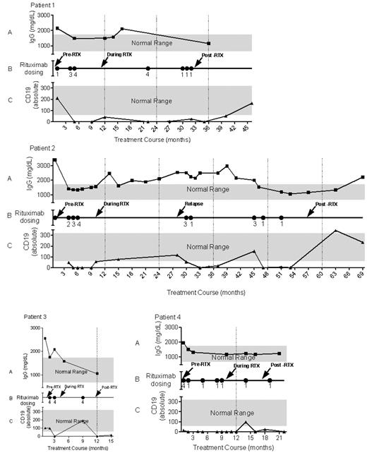
Rituximab was given at 375mg/m2 weekly for ≥ 4 doses and then at wider intervals (Figure 1). All patients received between 8 and 12 doses over the first year with subsequent additional doses determined by recurrence of infection. All patients had transient complete elimination of CD19+ B cells in the peripheral blood after rituximab. B cells recovered to the normal range in all patients at ∼ 1 year after rituximab, with B-cell levels often detectable before that, similar to previous reports17 (Figure 1; Table 2). All patients had improvement in inflammatory markers such as hypergammaglobulinemia with total IgG decreasing to normal, as well (Figure 1).
Timeline for rituximab therapy. Each timeline represents the treatment course for 1 patient. Panel A reflects IgG levels with normal range in gray, panel B has dots indicating months in which rituximab was given and a number indicating how many doses at 375mg/m2 were given in that month. Panel C reflects total absolute B-cell numbers by CD19 staining with normal range in gray. Black arrows are the times at which plasma was collected for Ab titers and functional studies for Figures 2 and 3.
Timeline for rituximab therapy. Each timeline represents the treatment course for 1 patient. Panel A reflects IgG levels with normal range in gray, panel B has dots indicating months in which rituximab was given and a number indicating how many doses at 375mg/m2 were given in that month. Panel C reflects total absolute B-cell numbers by CD19 staining with normal range in gray. Black arrows are the times at which plasma was collected for Ab titers and functional studies for Figures 2 and 3.
All patients had objective evidence of progressive improvement initially and remission or cure of infection after rituximab, as determined by clearance of cultures from previously infected sites, radiographic improvement of sites of soft tissue and bone infections, and weight gain (Table 2). These were the features that prompted initial use and subsequent retreatment with rituximab. With the use of these clinical indicators of systemic infection we developed a scoring system retrospectively to quantify the degree of disease activity (Table 3).
Retrospective clinical scoring before and after rituximab administration
| Parameter (values assigned) . | Patient 1 . | Patient 2 . | Patient 3 . | Patient 4 . | ||||
|---|---|---|---|---|---|---|---|---|
| Before . | After . | Before . | After . | Before . | After . | Before . | After . | |
| Constitutional (fever, weight loss, malaise: 1 point each) | 0 (unknown) | 0 | 2 (fever, malaise) | 0 | 2 (fever, weight loss) | 0 | 2 (weight loss, malaise) | 0 |
| Clinical infection (0, healing of sinus tract; 1, stable lesion; 2, development of sinus tract) | 2 | 0 | 0 | 0 | 2 | 0 | 2 | 0 |
| Positive blood cultures (0, negative; 2, positive) | 2 | 0 | 2 | 0 | 0 | 0 | 0 | 0 |
| Radiographic change (0, resolved; 1, improvement; 2, stable; 3, progression) | 3 | 1 | 3 | 1 | 3 | 1 | 3 | 1 |
| Hypergammaglobulinemia (0, normal; 1, elevated) | 1 | 0 | 1 | 0 | 1 | 0 | 1 | 0 |
| Total score | 8 | 1 | 8 | 1 | 8 | 1 | 8 | 1 |
Anti–IFN-γ autoantibody titers
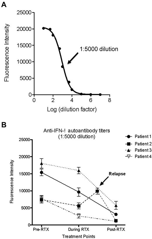
Anti–IFN-γ autoantibodies were measured by a particle-based technology.14 Titration curves using serial dilutions of subject plasma generated sigmoidal curves at each time point to determine a dilution factor within the dynamic range of the assay (Figure 2A). Relative Ab titers were determined at 1:5000 dilution for each subject. Ab titers decreased by between 1 and 2 logs from baseline in all subjects (Figure 2B).
Anti–IFN-γ autoantibody titers in relation to treatment with rituximab. (A) IFN-γ labeled beads were incubated with subject plasma at 10-fold serial dilutions to generate an Ab titration curve, as a function of fluorescence intensity, and determine a common dilution factor within the dynamic range of the assay. A representative example of the titration curve for patient 4 during rituximab therapy with the 1:5000 dilution indicated (black arrow) that lies within the linear phase of the dilution curve. (B) Each patient's Ab titer over the course of rituximab therapy was plotted at 1:5000 dilution.
Anti–IFN-γ autoantibody titers in relation to treatment with rituximab. (A) IFN-γ labeled beads were incubated with subject plasma at 10-fold serial dilutions to generate an Ab titration curve, as a function of fluorescence intensity, and determine a common dilution factor within the dynamic range of the assay. A representative example of the titration curve for patient 4 during rituximab therapy with the 1:5000 dilution indicated (black arrow) that lies within the linear phase of the dilution curve. (B) Each patient's Ab titer over the course of rituximab therapy was plotted at 1:5000 dilution.
Plasma-mediated inhibition of IFN-γ–stimulated STAT-1 phosphorylation
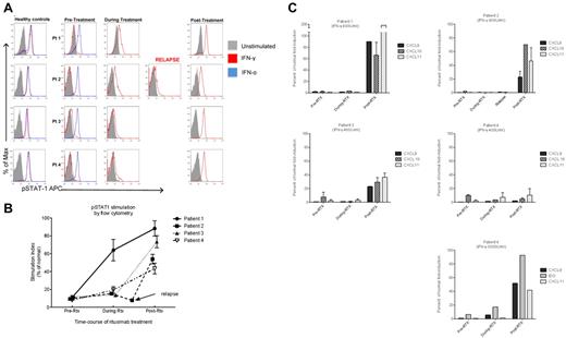
Plasma inhibition of IFN-γ signaling was followed by flow cytometry (Figure 3) and corroborated by immunoblot analysis (not shown). Stimulated normal PBMCs were assayed for pSTAT-1 production in the presence of control or subject plasma after stimulation with IFN-α or IFN-γ. In all patients, IFN-α–induced pSTAT-1 generation was preserved at all time points (shown for baseline time point; Figure 3A). In contrast, all baseline plasma samples completely inhibited IFN-γ–induced pSTAT-1 production. After treatment with rituximab, plasma from all subjects allowed IFN-γ–induced STAT-1 phosphorylation (Figure 3A). The degree of IFN-γ stimulation permitted by patient plasma was calculated across time points as a percentage of total stimulation observed by normal PBMCs in the presence of normal plasma (IFN-γ–induced pSTAT-1 mean fluorescence intensity in CD14+ cells, ratio of stimulated/unstimulated; Figure 3B).
Patient plasma inhibition of IFN-γ–induced pSTAT-1 production and RNA expression. Normal PBMC incubated in 10% control or patient plasma at time points specified in Figure 1 were left unstimulated or stimulated with IFN-α or IFN-γ for 15 minutes. CD14+ cells were assayed for intracellular pSTAT-1 by flow cytometry. (A) pSTAT-1 in CD14+ cells, solid gray, unstimulated; red line, IFN-γ stimulated; blue line, IFN-α stimulated. Representative example of 1 of 3 experiments performed for each patient. (B) To determine the relative inhibitory effect of plasma on IFN-γ induced pSTAT-1 production, a stimulation index for each plasma (ratio of the geometric mean channel for stimulated to unstimulated) was calculated and then graphed as a percentage of the stimulation index seen in the presence of normal plasma. Error bars represent the SEM seen for 3 separate experiments performed for each series of patient plasma. (C) PBMC obtained from healthy donors (n = 4 experiments per series of patient plasmas) were cultured in the presence of normal or patient plasma (10%) and stimulated with IFN-γ (400U/mL) for 3 hours. Target gene expression was evaluated by real time PCR and values are mean fold induction (± SD) relative to the unstimulated cells. GAPDH was used as normalization control. Fold-induction of IFN-γ–induced gene expression in presence of subject plasma was calculated as a percentage of the fold-induction expression seen for the same PBMCs in the presence of normal plasma. Plasma from patient 4 inhibited IFN-γ induced gene expression when stimulated with IFN-γ 400U/mL at all time points, and so the experiment was repeated using IFN-γ at a higher concentration of 3000U/mL.
Patient plasma inhibition of IFN-γ–induced pSTAT-1 production and RNA expression. Normal PBMC incubated in 10% control or patient plasma at time points specified in Figure 1 were left unstimulated or stimulated with IFN-α or IFN-γ for 15 minutes. CD14+ cells were assayed for intracellular pSTAT-1 by flow cytometry. (A) pSTAT-1 in CD14+ cells, solid gray, unstimulated; red line, IFN-γ stimulated; blue line, IFN-α stimulated. Representative example of 1 of 3 experiments performed for each patient. (B) To determine the relative inhibitory effect of plasma on IFN-γ induced pSTAT-1 production, a stimulation index for each plasma (ratio of the geometric mean channel for stimulated to unstimulated) was calculated and then graphed as a percentage of the stimulation index seen in the presence of normal plasma. Error bars represent the SEM seen for 3 separate experiments performed for each series of patient plasma. (C) PBMC obtained from healthy donors (n = 4 experiments per series of patient plasmas) were cultured in the presence of normal or patient plasma (10%) and stimulated with IFN-γ (400U/mL) for 3 hours. Target gene expression was evaluated by real time PCR and values are mean fold induction (± SD) relative to the unstimulated cells. GAPDH was used as normalization control. Fold-induction of IFN-γ–induced gene expression in presence of subject plasma was calculated as a percentage of the fold-induction expression seen for the same PBMCs in the presence of normal plasma. Plasma from patient 4 inhibited IFN-γ induced gene expression when stimulated with IFN-γ 400U/mL at all time points, and so the experiment was repeated using IFN-γ at a higher concentration of 3000U/mL.
Rituximab and IFN-γ–responsive gene expression
Plasma inhibition of IFN-γ–induced gene transcription was studied with he use of normal PBMCs incubated with 10% patient plasma unstimulated or stimulated with IFN-γ for 3 hours before collecting RNA for gene expression. Baseline plasma from all patients inhibited IFN-γ–induced transcription of CXCL9, CXCL10, and CXCL11. After treatment with rituximab, plasma from all patients showed partial reconstitution of IFN-γ–induced chemokine expression. Interestingly, plasma from patient 4 did not allow gene transcription after IFN-γ 400 U/mL for any time point. Therefore, we increased the dose of IFN-γ to overcome plasma inhibition, and at 3000 U/mL her plasma showed a progressive alleviation of IFN-γ signal inhibition after treatment with rituximab, akin to the other 3 patients (Figure 3C).
Discussion
Our patients were all previously healthy Asian women with adult onset of disseminated nontuberculous mycobacteria and high-titer anti–IFN-γ autoantibodies, similar to previous reports.2,3,5,18-22 Patient 2 had a slightly decreased CD4 lymphocyte count but not low enough to cause disseminated nontuberculous mycobacterial disease. All patients had been healthy into adulthood, whereas primary immunodeficiencies tend to present early in life. All patients had extensive disease with nontuberculous mycobacteria that continued to progress over years of extensive antimycobacterial and other adjunctive therapy, including plasmapheresis (1 patient) and exogenous IFN-γ (3 patients). Within 2-6 months after initiation of rituximab treatment, all patients had marked clinical, radiologic, and laboratory improvement. All patients had improvement of inflammatory markers, although some did not completely normalize. This is not surprising, given the chronic nature of this illness, potential for relapse, and ongoing need for antimicrobials. In conjunction with clinical improvement, we found convincing reduction in autoantibody titers in all 4 patients, with concordant relief of plasma-mediated inhibition of IFN-γ signaling.
Our patients received modestly differing amounts of rituximab at different dosing schedules, but all patients received between 8 and 12 doses of rituximab in the first year. Subsequent doses were given for signs and symptoms of persistent or relapsed infection. All patients had been on long-duration, best-tolerated, optimized anti-infective therapy. Despite aggressive antimicrobials, all had progressive infection. Therefore, changes of anti-infectives are unlikely explanations of the striking clinical responses, decline of Ab titers, and amelioration of plasma-mediated IFN-γ signaling inhibition.
Rituximab is approved by the Food and Drug Administration for treatment of lymphoma at 375/m2 weekly for 4-8 doses6,7 and rheumatoid arthritis (RA), which is given as two 1000-mg doses at day 1 and day 15.8 Our patients received 375/m2 (roughly 500 mg) for 4 doses over a month, which is a similar dose intensity to both regimens. In contrast to patients with RA, our patients required a longer duration of therapy, between 8 and 12 doses within the first year to avoid disease progression. For example, although patient 3 had improvement over the initial 2 months of treatment, by 3 months she had recrudescence of fever, inflammatory markers, and development of abscesses. In concordance with this, her titers only slightly declined (Figure 2B) and remained neutralizing (Figure 3), possibly accounting for her persistent mycobacterial infection. The Ab titers of patients 2 and 4 during treatment showed only modest initial declines with continued in vitro inhibitory activity, possibly due in part to the long half-life of IgG. Although patient 1 showed a greater autoantibody decline at her first time point, the collection date was 1 year after initiation of rituximab (rather than 2-8 months for the others). At the time of clinical relapse for patient 2, her plasma showed an increase in titer and in vitro inhibitory activity, which improved after rituximab was readministered (Figures 2–3). The need for prolonged rituximab therapy in our patients and the tendency for early relapse may reflect persistence of autoantibody-producing cells in privileged areas, despite immediate B-cell depletion in the periphery, as reported in RA.23,24
To date none of our patients has had infectious complications because of administration of rituximab. Side effects have been minimal and limited to those previously reported, such as mild infusion reactions and 2 cases of transient neutropenia (1030 neutrophils/μL for patient 2; 510 neutrophils/μL for patient 3). None of these complications of rituximab precluded continuation of therapy. All patients showed the ability to recover B cells, even after extensive treatment (Table 2).
All patients showed ongoing reduction of Ab titers and improved signaling over the years during and after rituximab treatment, suggesting that the benefits of CD20 depletion may accrue over time, perhaps because of depletion of plasmablasts that would replace autoantibody-producing plasma cells. Interestingly, we saw a lag between clinical improvement and in vitro improvement of IFN-γ signaling (Figure 3). Patient 4 had a protracted clinical course with less robust reconstitution of in vitro IFN-γ signaling. Nevertheless, her autoantibody titers did eventually decline concomitant with increased responsiveness to IFN-γ. In vivo, plasma constitutes a far greater percentage of blood than the 10% plasma concentration we used in our experiments. Although the stringency of our experimental conditions underscores the potency of these autoantibodies, it may overlook subtle alterations in Ab activity only apparent in vivo. Other factors not reflected in our measurement of Ab titer include changes in Ab avidity. The proportion of biologically active epitopes can also affect disease activity, as has been shown in pemphigus vulgaris.25 These differences in avidity and activity may account for the differences in Ab titer between subjects, indicating that titer alone is probably not adequate as the sole correlate of biologic activity.
In severe nontuberculous mycobacterial disease, antimycobacterials alone are often ineffective without intact IFN-γ signaling, as evidenced by the exceedingly poor survival in patients with complete IFN-γ receptor deficiency.1 This in part reflects the intrinsic difficulties of treating disseminated nontuberculous mycobacterial infections, but the fact that survival is excellent in those with partial defects in IFN-γ signaling indicates that even small amounts of IFN-γ signaling are able to effectively control mycobacterial infection.1 Thus, small improvements in IFN-γ signal transduction during treatment with rituximab could account for improved immune function, resulting in control of infection, even before this effect was detectable in vitro. These patients had dramatic improvement in IFN-γ signaling from pretreatment levels that matched decreasing Ab titers and clinical improvement.
Rituximab has been effective in other diseases caused by neutralizing IgG autoantibodies,9-13 although plasma cells do not express CD20. Possible mechanisms of action include modulation of other cell surface receptors, modulation of B-cell help to T cells, and elimination of plasmablasts with slow attrition of plasma cells that produce pathologic Abs.26,27 The prolonged time to clinical improvement in this disease and the protracted fall in autoantibody levels fits this hypothesis.
Not all cases of anti–IFN-γ autoantibody-mediated disseminated mycobacterial disease require rituximab therapy. Of the 11 patients we follow with this syndrome, so far only these 4 have required rituximab; the other 7 patients responded to antimycobacterial therapy alone. The cause of the difference between these 2 groups will be important to identify. It will be valuable to determine whether those who recover without rituximab do so because of spontaneous reduction of Ab titer or because their Abs were less intrinsically neutralizing in the first place. However, for those patients with persistent, progressive, severe anti–IFN-γ autoantibody–associated infection, rituximab may be an important adjunct.
The publication costs of this article were defrayed in part by page charge payment. Therefore, and solely to indicate this fact, this article is hereby marked “advertisement” in accordance with 18 USC section 1734.
Acknowledgments
This work was supported by the Division of Intramural Research, National Institute of Allergy and Infectious Diseases, National Institutes of Health, and the National Cancer Institute, National Institutes of Health (contract HHSN261200800001E).
The content of this publication does not necessarily reflect the views or policies of the Department of Health and Human Services, nor does mention of trade names, commercial products, or organizations imply endorsement by the US government.
National Institutes of Health
Authorship
Contribution: S.K.B., R.Z., E.P.S., K.J., L.B.R., L.D., M.J.P., L.M.Y., and D.L.P. performed experiments; S.K.B, R.Z., E.P.S., and L.B.R. analyzed results and made figures; S.K.B., G.U., A.F.F., C.E.H., R.B., S.H.C., and S.M.H. participated in clinical care; and S.K.B. and S.M.H. designed research and wrote the paper.
Conflict-of-interest disclosure: The authors declare no competing financial interests.
Correspondence: Sarah K. Browne, CRC B3-4141, MSC 1684, Bethesda, MD 20892-1684; e-mail: brownesa@niaid.nih.gov.


This feature is available to Subscribers Only
Sign In or Create an Account Close Modal