Abstract
Vascular endothelial growth factors (VEGFs) regulate blood and lymph vessel development by activating 3 receptor tyrosine kinases (RTKs), VEGFR-1, -2, and -3, and by binding to coreceptors such as neuropilin-1 (NRP-1). We investigated how different VEGF-A isoforms, in particular VEGF-A165a and VEGF-A165b, control the balance between VEGFR-2 recycling, degradation, and signaling. Stimulation of cells with the NRP-1–binding VEGF-A165a led to sequential NRP-1–mediated VEGFR-2 recycling through Rab5, Rab4, and Rab11 vesicles. Recycling was accompanied by dephosphorylation of VEGFR-2 between Rab4 and Rab11 vesicles and quantitatively and qualitatively altered signal output. In cells stimulated with VEGF-A165b, an isoform unable to bind NRP-1, VEGFR-2 bypassed Rab11 vesicles and was routed to the degradative pathway specified by Rab7 vesicles. Deletion of the GIPC (synectin) binding motif of NRP-1 prevented transition of VEGFR-2 through Rab11 vesicles and attenuated signaling. Coreceptor engagement was specific for VEGFR-2 because EGFR recycled through Rab11 vesicles in the absence of known coreceptors. Our data establish a distinct role of NRP-1 in VEGFR-2 signaling and reveal a general mechanism for the function of coreceptors in modulating RTK signal output.
Introduction
Activation of receptor tyrosine kinases (RTKs) is initiated on ligand-mediated dimerization of membrane-associated receptors. Receptors are subsequently internalized by endocytosis and either recycled to the plasma membrane or degraded.1 RTKs elicit signal output at the plasma membrane immediately after ligand binding and during subsequent transfer of receptors through endocytic vesicles such as early and late endosomes.2 The use of Rab family GTPases as markers for intracellular vesicular traffic allows the assignment of transport routes of membrane proteins such as RTKs.1,3 Rab5 is involved in the initial internalization step, the transfer from the plasma membrane to early endosomes from where receptors are further sorted to specific vesicular compartments.4 Rab4 and Rab11 define 2 distinct markers for recycling pathways, also known as fast (Rab4) and slow (Rab11) recycling. Rab7 specifies vesicles destined to target proteins to the degradative pathway.
Vascular endothelial growth factor (VEGF) family proteins (VEGF-A, -B, -C, and -D, and placenta growth factor [PlGF]) are encoded by distinct genes and expressed in multiple isoforms on mRNA splicing and posttranslational modification, in particular proteolytic processing.5 VEGFs promote endothelial cell survival, migration, proliferation, and differentiation and are indispensible for blood and lymph vessel formation. In addition, VEGFs regulate endothelial cell permeability and vessel contraction. Each VEGF isoform activates distinct signaling pathways eliciting specific biologic output. The molecular basis for ligand-specificity of VEGFR signaling is poorly understood. One possibility is that VEGF receptors, by associating with distinct coreceptors such as neuropilins (NRPs), integrins, semaphorins, or heparan sulfate glycosaminoglycans (HSPGs), engage distinct signaling molecules giving rise to specific signal output. Alternatively, ligand-specific signaling may result from receptor trafficking to specific cellular compartments where receptors encounter distinct signaling molecules.6-8
VEGFR-2 is the major mediator of angiogenic signaling in endothelial cells and is required for de novo vessel formation, vasculogenesis, and for angiogenesis, the formation of vessels from pre-existing vasculature.9 The pathways leading to VEGFR internalization and the role of receptor degradation in VEGF signaling remain controversial and differ for VEGFR-1, -2, and -3. VEGFRs generate signal output at the plasma membrane and on their way to degradation through endocytic vesicles.10 In unstimulated cells, VEGFR-2 was shown to be predominantly located in recycling endosomes identified by Rab411 and/or Rab5.12 On ligand stimulation, VEGFR-2 enters late endosomes through Rab7 vesicles and is subsequently degraded in lysosomes or proteasomes.13,14 VEGFR internalization is clathrin-mediated and transport is further directed by the endosomal sorting complex required for transport (ESCRT) proteins.15 VEGFR signaling is also regulated by ubiquitination, not only of the receptor itself, but also of receptor-associated signaling molecules such as phospholipase Cγ-1 (PLCγ-1).13 Specific VEGFR trafficking regulates biologic output as for instance shown for arterial morphogenesis.16
Neuropilins were first discovered as receptors of class-3 semaphorins.17,18 NRP-1 was also shown to act as a coreceptor for VEGF family proteins such as VEGF-A165, PlGF-2, VEGF-C, and some pox virus VEGF homologues collectively called VEGF-E. 5 Studies with knockout mice revealed that Nrp-1 plays an important role in axon guidance, vasculogenesis, and angiogenesis.19,20 The most prominent VEGF-A isoform, VEGF-A165, exists in 2 isoforms generated by alternative splicing, VEGF-A165a and VEGF-A165b, respectively.21,22 NRP-1 binding of VEGF-A165a is mediated by the carboxyterminal 8 amino acids encoded by exon 8a and by the HSPG binding domain encoded by exon 7, which also promotes extracellular matrix association. VEGF-A165b, on the other hand, shows reduced binding to HSPGs and does not bind NRP-1.23 Multimeric receptor/coreceptor complexes are stabilized by ligand bound to the extracellular domains of VEGFR-2 and NRP-1, and by intracellular proteins such as the scaffolding protein GIPC, also called synectin, interacting with the intracellular domains of these receptors.23-25 We showed earlier that VEGF-A isoforms lacking a NRP-1 binding site do not promote vessel sprouting, apparently because of a deficiency in activation of the p38 MAP kinase.7 On the other hand, Pan et al investigated NRP-1 binding of several VEGF-A isoforms and showed that the presence of an HSPG binding domain is not required for this interaction.26 These data are also in agreement with an earlier study showing that VEGF-A121, lacking the HSPG binding domain, is more potent in activating VEGFR-2 in the presence of NRP-1.27 Taken together, these data suggest that NRP-1 either modulates signaling by VEGFR-2 on formation of coreceptor complexes, or by activating additional independent pathways converging with VEGFR-2 signals.
Here we performed a comprehensive analysis of the role of NRP-1 signaling by VEGFR-2. We were particularly interested in how NRP-1 influences VEGFR-2 internalization, trafficking, degradation, and signaling. The data presented show that VEGFR-2 is localized in Rab5 and Rab4 vesicles in unstimulated cells, whereas NRP-1 is localized in Rab5, Rab4, and Rab11 vesicles. In the absence of NRP-1, VEGF-A165a stimulation led to the activation and phosphorylation of VEGFR-2 and translocation to Rab7 vesicles. In cells expressing VEGFR-2 together with NRP-1, VEGF-A165a linked both receptors on the extracellular side and directed VEGFR-2 together with NRP-1 to Rab11 vesicles. This led to enhanced activation of downstream signaling molecules. Most interestingly, our data show that association of VEGFR-2 with NRP-1 promotes increased signal output by p38 MAP kinase that is only weakly activated in the absence of NRP-1.
Methods
Plasmids
The coding regions of the Rab proteins were cloned carboxy-terminally of fluorescent proteins (mTFP1, EGFP, mCitrine, or mCherry) and expressed under the control of the CMV promoter. All constructs were sequenced before use. The construction of the mCitrine-Rab5-mCherry-Rab4-mTFP1-Rab11 and mCitrine-Rab5-mTFP1-Rab4-mCherry-Rab7 vectors was based on MultiLabel technology that allows the assembly of several expression cassettes in a single plasmid.28
Cell culture
Human umbilical vein endothelial cells (HUVECs) were obtained from Lonza and cultured in supplemented EGM-2 medium according to the manufacturer's recommendations. The porcine aortic endothelial cell (PAEC) lines stably expressing VEGFR-2, NRP-1, or both, which were used for immunofluorescence analysis, were derived from previously described cell lines.29 For Western blot analysis we engineered 2 additional PAEC lines expressing equal levels of VEGFR-2 together with mutated NRP-1 variants. The hygromycin resistant PAEC expressing VEGFR-2 was stably transfected with AA5-NRP-1, AA5-NRP-1-887HA (NRP-1 ends at amino acid 887, followed by an HA-tag; depicted as NRP-1-ΔC in the figures), and AA5-NRP-1-S921X. Selection was performed with Zeocin. PAECs were maintained in 10% FBS/HamF12/Penicillin/Streptomycin. For transfection, cells were plated at a density of approximately 30% on glass coverslips and then transfected with Fugene HD (Roche Diagnostics) according to the manufacturer's recommendations. Stimulation with different VEGFs was done 45 hours after transfection for the indicated time periods at a concentration of 50 ng/mL. The experiments in Figure 4 were performed in the presence of 10 μm cycloheximide. Cells were fixed by transferring the cover slips into 4% formaldehyde/PBS for 10 minutes and then washed 3 times with PBS. Spheroids consisting of 500 cells were generated by the hanging drop method in 0.25% Methocel/10% FBS/HamF12/Penicillin/Streptomycin. They were then embedded in a collagen matrix and cultured in the presence or absence of VEGF-A165a for 24 hours and then fixed.30
Immunostaining
Cells were permeabilized with 1% NP40 in PBS for 10 minutes and then incubated with the following antibodies, diluted in PBS: Goat anti–NRP-1 (Santa Cruz Biotechnology; sc7239, 1:200), mouse anti–VEGFR-2 (Sigma-Aldrich; V9134, 1:200), rabbit anti–VEGFR-2 (Cell Signaling; #2479, 1:500), rabbit anti–VEGFR-2 pY1175 (Cell Signaling; #2478, 1:100), rabbit anti–VEGFR-2 pY1212 (Cell Signaling, #2477, 1:100), rabbit anti-Rab4 (StressMarq; #SPC141, 1:150), rabbit anti-Rab5 (StressMarq; #SPC168, 1:150), rabbit anti-Rab7 (Cell Signaling; #2094, 1:150), rabbit anti-Rab11 (Cell Signaling; #3539, 1:150). Secondary antibodies were labeled with Alexa488, Cy3, or Cy5 (Invitrogen; Jackson ImmunoResearch Laboratories). Samples were mounted in Citifluor AF1 (Citifluor) and analyzed on a Leica SP5 confocal microscope. Single sections are shown in the figures, levels and γ settings were adjusted in Adobe Photoshop CS3 extended. All operations were applied to the entire image.
Western blot analysis
Cells were plated in 3.5 cm dishes and starved overnight in 1% BSA/DMEM before stimulation with VEGF. Cells were lysed in 0.5% Triton-X100/100mM NaCl/50 mM Tris-HCl pH 7.5 at the indicated time points. The supernatant was used for Western blot analysis after sonification and centrifugation. The following antibodies were used: rabbit anti-phospho PLCγ-1 (Cell Signaling, #2821), rabbit anti–PLCγ-1 (Cell Signaling; #2822), rabbit anti-phospho–p38 (Cell Signaling; #4631), rabbit anti-p38 (Sigma-Aldrich; M0800), rabbit anti–VEGFR-2 (Cell Signaling, #2479), rabbit anti-phospho–VEGFR-2 (pY1175; Cell Signaling; #2478), mouse anti-actin (Sigma-Aldrich; A5316), goat anti–NRP-1 (Santa Cruz Biotechnology; sc7239; 1:2000), mouse anti–HA-tag (12CA5; 1:100), and mouse anti-tubulin (Sigma-Aldrich; T5168; 1:4000). All antibodies were diluted 1:1000 (if not stated otherwise) in 3% BSA/TBST. As secondary antibodies, alkaline phosphatase-coupled goat anti–mouse, anti–goat, and anti–rabbit antibodies were used, followed by chemiluminescence detection with a CCD camera. Lysates were loaded from the parental PAE-VEGFR-2 cell line together with a PAEC line expressing VEGFR-2 and NRP-1. All data were normalized to VEGFR-2 activation in the PAE-VEGFR-2 cell line after 10 minutes. Quantification was performed with NIH ImageJ 1.43μ. The experiment was repeated 3 times for each cell line. Two cell lines were tested for each NRP-1 mutant.
Results
Localization of VEGFR-2 and NRP-1 in HUVECs
HUVECs are primary endothelial cells endogenously expressing VEGFR-2 and NRP-1. Rab5, Rab4, Rab11, and Rab7 define vesicular compartments for receptor trafficking and degradation.1 We initially determined whether Rab11 defines an independent recycling pathway or whether it acts downstream of Rab4 as previously described for transferrin recycling in A431 cells.31 We confirmed the described sequential recycling path in HUVECs and PAECs by transient coexpression of mCitrine-Rab5, mCherry-Rab4, and mTFP1-Rab11. Rab5 vesicles are linked to Rab4 vesicles and Rab4 vesicles are then linked to Rab11 vesicles. The direct Rab5 to Rab11 transition seems to play a minor role in these 2 cell types (supplemental Figure 1A-C; available on the Blood Web site; see the Supplemental Materials link at the top of the online article). A similar experiment with a mCitrine-Rab5, mTFP1-Rab4, and mCherry-Rab7 construct revealed that Rab7 vesicles were linked to the Rab4 compartment (supplemental Figure 1B-D). The connection of the different Rab vesicle compartments is summarized in supplemental Figure 1E.
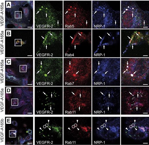
HUVECs were starved for 5 hours in basal medium without VEGF followed by stimulation for 30 minutes with VEGF-A165a or VEGF-A165b. VEGFR-2 and NRP-1 were detected in Rab5, Rab4, Rab11, and Rab7 vesicles after stimulation with VEGF-A165a (Figure 1A-D). On stimulation with VEGF-A165b, NRP-1 was detected in all 4 Rab compartments whereas VEGFR-2 was only detected in Rab5, Rab4, and Rab7 and was absent from Rab11 vesicles (Figure 1E and data not shown).
Localization of VEGFR-2 and NRP-1 in HUVECs. (A-D) HUVECs were stimulated with VEGF-A165a for 30 minutes and then stained for VEGFR-2, NRP-1, and the indicated Rab GTPase. VEGFR-2 and NRP-1 colocalized with Rab5, Rab4, Rab7, and Rab11 (arrows). (E) When the cells were stimulated with VEGF-A165b, no colocalization of VEGFR-2 with Rab11 was found whereas NRP-1 was still able to traffic through the Rab11 compartment (circles). Arrowheads indicate vesicles positive for NRP-1 and VEGFR-2. Scale bar: 20 μm (insets: 5 μm).
Localization of VEGFR-2 and NRP-1 in HUVECs. (A-D) HUVECs were stimulated with VEGF-A165a for 30 minutes and then stained for VEGFR-2, NRP-1, and the indicated Rab GTPase. VEGFR-2 and NRP-1 colocalized with Rab5, Rab4, Rab7, and Rab11 (arrows). (E) When the cells were stimulated with VEGF-A165b, no colocalization of VEGFR-2 with Rab11 was found whereas NRP-1 was still able to traffic through the Rab11 compartment (circles). Arrowheads indicate vesicles positive for NRP-1 and VEGFR-2. Scale bar: 20 μm (insets: 5 μm).
The use of GFP-tagged, constitutively active Rab constructs was previously described as a method to monitor receptor tyrosine kinase or pathogen trafficking between the different intracellular vesicular compartments.32,33 We therefore transfected HUVECs with GFP-tagged Rab constructs and monitored VEGFR-2 localization in the different Rab compartments. VEGF-A165a stimulation led to the accumulation of VEGFR-2 in Rab11 vesicles whereas VEGF-A165b did not (supplemental Figure 2). These results show that the ability of VEGF-A165a to interact with NRP-1 determines VEGFR-2 trafficking through Rab11 vesicles.
VEGFR-2 and NRP-1 trafficking in unstimulated cells
The next series of experiments was performed in PAECs that lack endogenous VEGFR-2 and NRP-1 expression. The use of this endothelial cell line allowed studying membrane trafficking of VEGFR-2 and NRP-1 either separately, in singly transfected cell lines, or in cells coexpressing both receptors. In addition, transfection of fluorescently tagged, constitutively active Rab GTPases permitted to obtain quantitative data on receptor trafficking.
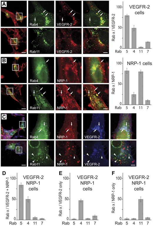
In the absence of VEGF, VEGFR-2 predominantly localized to the plasma membrane, Rab5, and Rab4 vesicles with a minor fraction colocalizing with Rab7 (Figure 2A, supplemental Figure 3A). NRP-1, on the other hand, was found on the plasma membrane and in Rab5 and Rab11 vesicles, with a small fraction colocalizing with Rab4 and Rab7 (Figure 2B, supplemental Figure 3B).
Localization of VEGFR-2 and NRP-1 in unstimulated PAEC. Cells stably expressing VEGFR-2 and/or NRP-1 were transiently transfected with constitutively active Rab constructs. Images for Rab5 and Rab7 are given in supplemental Figure 1. The quantification shows percentage of VEGFR-2 or NRP1-positive vesicles that were also positive for the indicated Rab. (A) VEGFR-2 predominantly localized to Rab4 and Rab5 vesicles in cells overexpressing VEGFR-2 only (arrows); no colocalization with Rab11 was observed. (B) NRP-1 was predominantly localized in Rab5 and Rab11 vesicles, with a minor population in Rab4 vesicles in cells expressing NRP-1 only (arrows). (C) In cells coexpressing VEGFR-2 and NRP-1, a fraction of VEGFR-2 and NRP-1 colocalized in Rab5 and Rab4 vesicles (Figure 1C arrows, quantified in Figure 1D) whereas the individual receptors were found in Rab4 and Rab11 vesicles, respectively (Figure 1C, quantified in Figure 1E-F). Scale bar: 20 μm (insets: 5 μm).
Localization of VEGFR-2 and NRP-1 in unstimulated PAEC. Cells stably expressing VEGFR-2 and/or NRP-1 were transiently transfected with constitutively active Rab constructs. Images for Rab5 and Rab7 are given in supplemental Figure 1. The quantification shows percentage of VEGFR-2 or NRP1-positive vesicles that were also positive for the indicated Rab. (A) VEGFR-2 predominantly localized to Rab4 and Rab5 vesicles in cells overexpressing VEGFR-2 only (arrows); no colocalization with Rab11 was observed. (B) NRP-1 was predominantly localized in Rab5 and Rab11 vesicles, with a minor population in Rab4 vesicles in cells expressing NRP-1 only (arrows). (C) In cells coexpressing VEGFR-2 and NRP-1, a fraction of VEGFR-2 and NRP-1 colocalized in Rab5 and Rab4 vesicles (Figure 1C arrows, quantified in Figure 1D) whereas the individual receptors were found in Rab4 and Rab11 vesicles, respectively (Figure 1C, quantified in Figure 1E-F). Scale bar: 20 μm (insets: 5 μm).
In unstimulated cells expressing both receptors, vesicles containing both VEGFR-2 and NRP-1 were positive for either Rab5 or Rab4 (Figure 2C, quantified in Figure 2D). Vesicles containing only VEGFR-2 were predominantly Rab4 positive (Figure 2C, supplemental Figure 3C, quantified in Figure 2E), whereas vesicles positive only for NRP-1 were almost exclusively Rab11 positive (Figure 2C, supplemental Figure 3C, quantified in Figure 2F). A very small fraction of receptors was found in Rab7 vesicles indicating that receptor degradation plays a minor role in the absence of ligands.
VEGF-A165 exon 8a or 8b variants promote different trafficking of VEGFR-2 and NRP-1
We applied 2 strategies to study the influence of NRP-1 on VEGFR-2 activation and trafficking. First, we used VEGF-A isoforms with different receptor binding specificities determined by sequences encoded by exon 8. The carboxyterminal 6 amino acids of VEGF-A165a and VEGF-A165b are encoded by either exon 8a or exon 8b. VEGF-A165a binds to VEGFR-1 (which is not the subject of this study), VEGFR-2, and NRP-1, whereas VEGF-A165b interacts with VEGFR-1 and VEGFR-2. Second, we studied receptor activation in PAECs expressing either VEGFR-2 alone or together with wild-type or mutant forms of NRP-1.
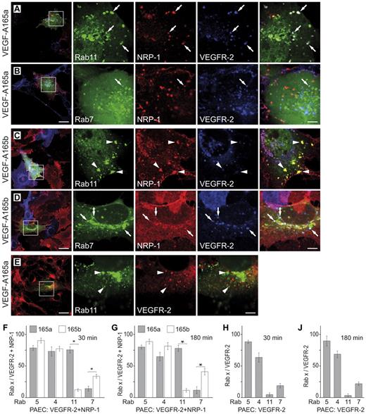
Receptor trafficking was remarkably different when PAECs expressing both receptors were stimulated with either VEGF-A165a or VEGF-A165b. Stimulation with VEGF-A165a promoted VEGFR-2 internalization and transport to Rab11 vesicles (Figure 3A-B, quantification in Figure 3F-G, supplemental Figure 4A-D). VEGF165b, on the other hand, promoted accumulation in Rab7 vesicles (Figure 3C-D, quantification in Figure 3F-G, supplemental Figure 4B). Receptor relocalization was visible shortly after stimulation and Rab7 localization became pronounced after 180 minutes (supplemental Figure 4). Most importantly, VEGFR-2 was hardly found in Rab11 vesicles in PAECs expressing VEGFR-2 alone (Figure 3E,H,J). These experiments show that VEGF-A165a, capable to simultaneously bind both receptors, relocalized VEGFR-2 to Rab11 vesicles. We hypothesize that this results from VEGF binding to the extracellular domains of VEGFR-2 and NRP-1 promoting stable receptor association as previously shown.23,24 Compared with cells expressing VEGFR-2 alone, this coreceptor complex is thus differently routed through intracellular membrane vesicle compartments. In particular, transport to Rab7 vesicles, indicative of receptor degradation, was significantly reduced in VEGF-A165a stimulated cells expressing both receptors.
VEGFR-2 is relocalized to Rab11 vesicles on interaction with NRP-1 in VEGF-A165a stimulated cells. PAECs expressing VEGFR-2 and NRP-1 (A-D) or VEGFR-2 alone (E) were transiently transfected with the indicated Rab constructs. Cells were stimulated with VEGF-A165a (A-B-E) or VEGF-A165b (C-D) for 180 minutes. Arrows indicate vesicles positive for the indicated Rab, VEGFR-2, and NRP-1. Arrowheads indicate Rab11 vesicles that are VEGFR-2 negative. Quantification of the data shows mean ± SD, n = 3; *P < .01 (paired Student t test). Images for Rab5 and Rab4 transfected and for cells stimulated for 30 minutes are given in supplemental Figure 2. Scale bar: 20 μm (insets: 5 μm).
VEGFR-2 is relocalized to Rab11 vesicles on interaction with NRP-1 in VEGF-A165a stimulated cells. PAECs expressing VEGFR-2 and NRP-1 (A-D) or VEGFR-2 alone (E) were transiently transfected with the indicated Rab constructs. Cells were stimulated with VEGF-A165a (A-B-E) or VEGF-A165b (C-D) for 180 minutes. Arrows indicate vesicles positive for the indicated Rab, VEGFR-2, and NRP-1. Arrowheads indicate Rab11 vesicles that are VEGFR-2 negative. Quantification of the data shows mean ± SD, n = 3; *P < .01 (paired Student t test). Images for Rab5 and Rab4 transfected and for cells stimulated for 30 minutes are given in supplemental Figure 2. Scale bar: 20 μm (insets: 5 μm).
The carboxyterminal SEA motif of NRP-1 is required for receptor trafficking to Rab11 vesicles
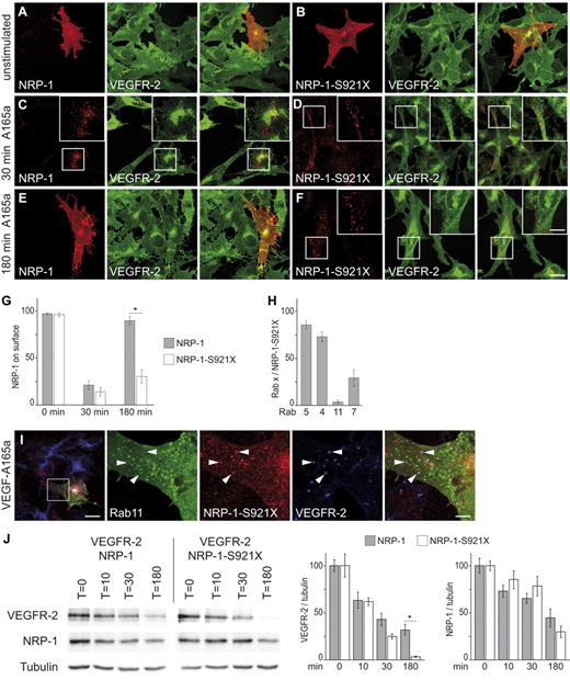
We next investigated the mechanism determining the sorting of NRP-1 to Rab11 vesicles. NRP-1 associates with a cytoplasmic adaptor protein that is essential for angiogenic signaling called GIPC or synectin.34,35 The last 3 amino acids of NRP-1, SEA, are required for PDZ domain-mediated GIPC binding.24 To test the influence of the GIPC binding motif on NRP-1 and VEGFR-2 localization, we expressed wild-type and a mutant NRP-1 (NRP-1-S921X) lacking the last 3 amino acids in PAECs stably expressing VEGFR-2. Both NRP-1 variants accumulated at the plasma membrane in unstimulated cells (Figure 4A-B). NRP-1 was cleared from the plasma membrane after VEGF stimulation and transferred to vesicles within 30 minutes (Figure 4C-D). Wild-type NRP-1 reappeared on the plasma membrane 180 minutes after stimulation with VEGF-A165a (Figure 4E), whereas NRP-1-S921X remained associated with intracellular vesicles (Figure 4F, quantified in Figure 4G). NRP-1-S921X was predominantly found in Rab5, Rab4, and Rab7 vesicles shortly after stimulation with VEGF-A165a (supplemental Figure 5) and was not localized in Rab11 vesicles (quantified in Figure 4H, Figure 4I). Localization was similar in control and ligand stimulated cells after 180 minutes (supplemental Figure 5). VEGFR-2 was translocated to Rab7 instead of Rab11 vesicles in NRP-1-S921X expressing cells (supplemental Figure 5) suggesting that NRP-1–mediated transfer of VEGFR-2 to Rab11 vesicles, and subsequently back to the plasma membrane, required the carboxyterminal PDZ binding motif of NRP-1.
The PDZ binding motif of NRP-1 is required for recycling of VEGFR-2/NRP-1 complexes to the plasma membrane through the Rab11 compartment. PAECs expressing VEGFR-2 were transiently transfected with either full-length NRP-1 (A,C,E) or NRP-1-S921X (B-D-F). Cells were analyzed before (A-B), 30 minutes (C-D), and 180 minutes after stimulation with VEGF-A165a (E-F). (G) Quantification of panels A through F (*P < .01; paired Student t test). (H-I) PAECs expressing VEGFR-2 were transiently transfected with NRP-1-S921X and fluorescently tagged Rab GTPase constructs. Cells were stimulated for 10 minutes with VEGF-A165a. No colocalization of NRP-1-S921X with Rab11 was found. Arrowheads indicate vesicles that are positive for NRP-1 and VEGFR-2, but negative for Rab11. Scale bar: 20 μm (insets: 5 μm). Quantification revealed that NRP-1-S921X is not able to enter the Rab11 compartment. Images of Rab11 transfected cells are shown in panel (I), images of all other Rab GTPases stimulated for various time points are shown in supplemental Figure 5. (J) Degradation of VEGFR-2 is slower in the presence of wild-type NRP-1. VEGFR-2 was nearly completely degraded in the presence of NRP-1-S921X after 180 minutes whereas > 30% of VEGFR-2 remained in the presence of NRP-1 (mean ± SD, n = 3; *P < .05; paired Student t test).
The PDZ binding motif of NRP-1 is required for recycling of VEGFR-2/NRP-1 complexes to the plasma membrane through the Rab11 compartment. PAECs expressing VEGFR-2 were transiently transfected with either full-length NRP-1 (A,C,E) or NRP-1-S921X (B-D-F). Cells were analyzed before (A-B), 30 minutes (C-D), and 180 minutes after stimulation with VEGF-A165a (E-F). (G) Quantification of panels A through F (*P < .01; paired Student t test). (H-I) PAECs expressing VEGFR-2 were transiently transfected with NRP-1-S921X and fluorescently tagged Rab GTPase constructs. Cells were stimulated for 10 minutes with VEGF-A165a. No colocalization of NRP-1-S921X with Rab11 was found. Arrowheads indicate vesicles that are positive for NRP-1 and VEGFR-2, but negative for Rab11. Scale bar: 20 μm (insets: 5 μm). Quantification revealed that NRP-1-S921X is not able to enter the Rab11 compartment. Images of Rab11 transfected cells are shown in panel (I), images of all other Rab GTPases stimulated for various time points are shown in supplemental Figure 5. (J) Degradation of VEGFR-2 is slower in the presence of wild-type NRP-1. VEGFR-2 was nearly completely degraded in the presence of NRP-1-S921X after 180 minutes whereas > 30% of VEGFR-2 remained in the presence of NRP-1 (mean ± SD, n = 3; *P < .05; paired Student t test).
We next quantified the effect of wild-type and S921X mutant NRP-1 on VEGFR-2 degradation by Western blot analysis. The degradation of VEGFR-2 was significantly slower in the presence of wild-type NRP-1, indicating that the receptor was partially recycled on coligation with NRP-1. Degradation of NRP-1 was independent of the SEA PDZ binding motif (Figure 4J).
Activity of VEGFR-2 in endocytic vesicles after VEGF-A stimulation
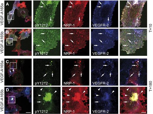
We next investigated VEGFR-2 activity in ligand stimulated cells using phospho-specific antibodies recognizing either phosphotyrosine pY1175 or pY1212. Phosphorylated VEGFR-2 was detectable at the cell membrane 10 minutes after stimulation with either VEGF-A165a or VEGF-A165b and was maintained in NRP-1–positive vesicles for 180 minutes. In cells stimulated with VEGF-A165a, most double-receptor positive vesicles were also positive for pY1175 and pY1212 (Figure 5A-C and supplemental Figure 6A-C). VEGF-A165b was less effective in activating VEGFR-2 but also promoted receptor phosphorylation and internalization (Figure 5B-D and supplemental Figure 6B-D). Interestingly, phosphorylated VEGFR-2 was always present in NRP-1 positive vesicles.
Tyrosine-phosphorylated VEGFR-2 always colocalizes with NRP-1 on stimulation with VEGF-A165a. PAECs stably expressing VEGFR-2 and NRP-1 were stimulated for 10 (A-B) or 180 (C-D) minutes with VEGF-A165a (A-C) or VEGF-A165b (B-D). Receptor phosphorylated at Y1212 accumulated at the plasma membrane 10 minutes after stimulation (A). In addition, vesicles with phosphorylated VEGFR-2 and NRP-1 are visible (A-B arrows). Phosphorylated VEGFR-2 disappeared from the plasma membrane 180 minutes after stimulation (C) and was now observed in NRP-1–positive vesicles (C arrows). Most VEGFR-2/NRP-1 positive vesicles showed a pY1212 signal after stimulation with VEGF-A165a (C). In contrast, many VEGFR-2/NRP-1 positive vesicles showed no pY1212 signal after stimulation with VEGF-A165b (D arrowheads). Similar data were observed for VEGFR-2 phosphorylation at Y1175 (supplemental Figure 4). Scale bar: 20 μm (insets: 5 μm).
Tyrosine-phosphorylated VEGFR-2 always colocalizes with NRP-1 on stimulation with VEGF-A165a. PAECs stably expressing VEGFR-2 and NRP-1 were stimulated for 10 (A-B) or 180 (C-D) minutes with VEGF-A165a (A-C) or VEGF-A165b (B-D). Receptor phosphorylated at Y1212 accumulated at the plasma membrane 10 minutes after stimulation (A). In addition, vesicles with phosphorylated VEGFR-2 and NRP-1 are visible (A-B arrows). Phosphorylated VEGFR-2 disappeared from the plasma membrane 180 minutes after stimulation (C) and was now observed in NRP-1–positive vesicles (C arrows). Most VEGFR-2/NRP-1 positive vesicles showed a pY1212 signal after stimulation with VEGF-A165a (C). In contrast, many VEGFR-2/NRP-1 positive vesicles showed no pY1212 signal after stimulation with VEGF-A165b (D arrowheads). Similar data were observed for VEGFR-2 phosphorylation at Y1175 (supplemental Figure 4). Scale bar: 20 μm (insets: 5 μm).
Phosphorylated VEGFR-2 is absent from Rab11 vesicles
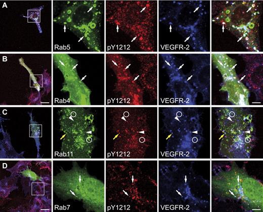
PAECs expressing VEGFR-2 and NRP-1 were transfected with GFP tagged Rab constructs and stained for phosphotyrosine and VEGFR-2. Cells were stimulated with VEGF-A165a or VEGF-A165b for 30 minutes followed by immunostaining with antibodies recognizing either pY1212 (Figure 6, supplemental Figure 7C) or pY1175 (supplemental Figure 7A-B). Stimulation of cells with exon 8a or 8b VEGF-A165 promoted phosphorylation and internalization of VEGFR-2, and the phosphorylated receptor was present in Rab5, Rab4, and Rab7 vesicles (Figure 6A,B,D, supplemental Figure 7). Interestingly, the fraction of VEGFR-2 present in Rab11 vesicles after activation with VEGF-A165a was not phosphorylated (Figure 6C, supplemental Figure 7A), but vesicles containing phosphorylated VEGFR-2 were often found in close proximity to Rab11 vesicles (Figure 6C yellow arrow) suggesting that VEGFR-2 was dephosphorylated before entering Rab11 vesicles and recycling to the plasma membrane. VEGFR-2 was also phosphorylated in VEGF-A165b stimulated cells but was never localized in Rab11 vesicles (supplemental Figure 7B-C). Taken together, these data show that stimulation of cells with VEGF-A165a led to VEGFR-2 association with NRP-1. Subsequently, receptor complexes were internalized, dephosphorylated, and recycled to the plasma membrane via Rab11 vesicles.
VEGFR-2 in Rab11 vesicles is dephosphorylated. PAECs expressing VEGFR-2 and NRP-1 were transiently transfected with the indicated Rab constructs and stimulated with VEGF-A165a for 30 minutes. Y1212-phosphorylated VEGFR-2 was present in Rab5, Rab4, and Rab7 vesicles (white arrows in panels A,B,D). Similar results were observed for Y1175 phosphorylation (supplemental Figure 5A). (C) No phosphotyrosine signal was observed for VEGFR-2 localized in Rab11 vesicles although the receptor localized to these vesicles (circles). Phosphorylated VEGFR-2 was found in these cells (arrowheads), sometimes in vesicles in close proximity to Rab11 vesicles (yellow arrow). Scale bar: 20 μm (insets: 5 μm).
VEGFR-2 in Rab11 vesicles is dephosphorylated. PAECs expressing VEGFR-2 and NRP-1 were transiently transfected with the indicated Rab constructs and stimulated with VEGF-A165a for 30 minutes. Y1212-phosphorylated VEGFR-2 was present in Rab5, Rab4, and Rab7 vesicles (white arrows in panels A,B,D). Similar results were observed for Y1175 phosphorylation (supplemental Figure 5A). (C) No phosphotyrosine signal was observed for VEGFR-2 localized in Rab11 vesicles although the receptor localized to these vesicles (circles). Phosphorylated VEGFR-2 was found in these cells (arrowheads), sometimes in vesicles in close proximity to Rab11 vesicles (yellow arrow). Scale bar: 20 μm (insets: 5 μm).
The cytoplasmic domain of NRP-1 modulates VEGFR-2 signaling
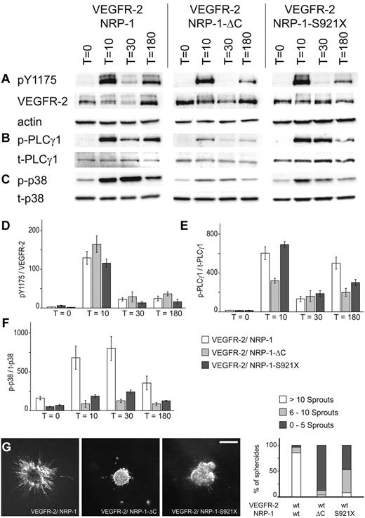
We next investigated how NRP-1 affects signal output by VEGFR-2. We generated a set of PAECs expressing VEGFR-2 together with either wild-type or mutant NRP-1 using our VEGFR-2 expressing PAEC line as a parent. All cell lines expressed similar levels of VEGFR-2, which is crucial for quantifying receptor phosphorylation and downstream signaling. NRP-1 and VEGFR-2 expressing cells were stimulated with VEGF-A165a and receptor activity was compared with cells expressing only VEGFR-2 (Figure 7). VEGFR-2 was phosphorylated at the major phosphorylation site, Y1175, 10 minutes after ligand stimulation and was dephosphorylated 20 minutes later. The relative level of VEGFR-2 phosphorylation was similar in all NRP-1 expressing lines and 20%-40% higher than in the parental VEGFR-2 expressing cell line. VEGFR-2 phosphorylation was biphasic with a second, weaker peak of activity observed 180 minutes after ligand addition (Figure 7A and D). Biphasic activation was observed in all cell lines including the parental line expressing only VEGFR-2 as described earlier.23
Modulation of VEGFR-2 signal output by the wild-type tail of NRP-1. PAEC expressing VEGFR-2 and NRP-1 were stimulated with VEGF-A165a and harvested at the indicated time points. Signaling was monitored with the indicated antibodies. Data were normalized to PAEC expressing only VEGFR-2 (not shown). (A-D) The relative VEGFR-2 phosphorylation was similar in all cell lines. (B-E) Full activation of PLCγ-1 was still observed by the mutant NRP-1-S921X. (C-F) p38 is only fully activated when the complete cytoplasmic tail is present. (G) Spheroids assembled of PAEC expressing VEGFR-2 and the indicated NRP-1 isoform were stimulated for 24 hours with VEGF-A165a and then analyzed. Only the full-length form of NRP-1 led to robust sprouting of spheres. Scale bar: 200 μm.
Modulation of VEGFR-2 signal output by the wild-type tail of NRP-1. PAEC expressing VEGFR-2 and NRP-1 were stimulated with VEGF-A165a and harvested at the indicated time points. Signaling was monitored with the indicated antibodies. Data were normalized to PAEC expressing only VEGFR-2 (not shown). (A-D) The relative VEGFR-2 phosphorylation was similar in all cell lines. (B-E) Full activation of PLCγ-1 was still observed by the mutant NRP-1-S921X. (C-F) p38 is only fully activated when the complete cytoplasmic tail is present. (G) Spheroids assembled of PAEC expressing VEGFR-2 and the indicated NRP-1 isoform were stimulated for 24 hours with VEGF-A165a and then analyzed. Only the full-length form of NRP-1 led to robust sprouting of spheres. Scale bar: 200 μm.
We next examined the phosphorylation of PLCγ-1, one of the signaling molecules directly activated by VEGFR-2. PLCγ-1 was phosphorylated to a higher degree in PAECs expressing VEGFR-2 together with wild-type NRP-1 or NRP-1-S921X than in cells expressing only VEGFR-2 or VEGFR-2 together with the NRP-1-ΔC mutant (Figure 7B-E). Therefore, the cytoplasmic domain lacking the carboxyterminal PDZ motif is sufficient for full activation of PLCγ-1. Phosphorylation of PLCγ-1 was biphasic as shown for VEGFR-2. We hypothesize that one or more of the putative phosphorylation sites in the cytoplasmic domain of NRP-1 play a regulatory role in PLCγ-1 activation.
We have shown earlier that p38 activity was required for vascular sprouting and was only activated when cells were treated with NRP-1–binding VEGF-A variants.7 We measured therefore p38 activity in VEGFR-2 cells coexpressing either wild-type NRP-1, NRP-1-ΔC, or NRP-1-S921X. Activity was highest in cells expressing VEGFR-2 together with wild-type NRP-1 indicating that the entire cytoplasmic part of NRP-1, including the last 3 amino acids, was required for full p38 activation (Figure 7C-F). The carboxyterminal SEA GIPC-binding motif is thus necessary for p38, but not for PLCγ-1 activation.
It has been shown before that VEGFR-2, NRP-1, and VEGF-A165a play essential roles in embryoid body sprouting.7 We generated spheroids with the 3 PAEC cell lines described in this section to test whether the cytoplasmic domain of NRP-1 was relevant for sprouting. Our data clearly show that wild-type NRP-1 coexpressed with VEGFR-2 was absolutely required for optimal sprouting of PAEC. Both mutations of NRP-1, S921X, and ΔC, severely attenuated VEGF-mediated sprouting (Figure 7G).
Not all RTKs require coreceptors for entering the Rab11 compartment
In the next set of experiments we investigated whether a related RTK such as the epidermal growth factor receptor (EGFR), relies on a similar strategy for ligand-mediated internalization and degradation as VEGFR-2. Microscopic analysis showed that EGFR was present in Rab5, Rab,4 and Rab11 vesicles in the absence of ligands (supplemental Figure 8A and data not shown). On stimulation with growth factor, the receptor was predominantly found in Rab7 vesicles and presumably subsequently degraded (supplemental Figure 8B-C).36 Interestingly, the EGFR was phosphorylated in all 4 Rab compartments, including Rab11, demonstrating that this receptor did not require dephosphorylation for trafficking through this vesicular compartment (supplemental Figure 8D and data not shown). This shows that the EGFR translocates to Rab11 vesicles independent from known coreceptors and suggests that different RTKs use distinct strategies for kinase activation and receptor recycling.
Discussion
RTK signaling is the result of a series of precisely orchestrated steps initiated at the plasma membrane after ligand binding. In this process, ligands not only bind to the extracellular domain of RTKs but in many cases interact with additional cell surface exposed molecules acting as coreceptors. Here we investigated the role of one of the coreceptors of VEGF-A, NRP-1, which emerged as a clinically relevant modulator of VEGF signaling.37 We specifically studied receptor internalization, trafficking, and signaling to downstream targets. In particular, we identified the vesicular compartments through which VEGFR-2 and NRP-1 were shuttled on their way from the plasma membrane to early and late endosomes using fluorescently tagged Rab proteins compartment markers.32,33 Based on our study we can now build a mechanistic concept for how VEGFR-2 signaling is influenced by coreceptors.
Two isoforms of VEGF-A165 generated by alternative splicing,21 and genetically engineered cell lines expressing VEGFR-2 together with either wild-type or mutant NRP-1, were used to dissect the function of NRP-1 in VEGFR-2 trafficking and signaling. VEGF-A165a binds NRP-1 and is prominently expressed in all tissues undergoing neovascularization. This isoform induced receptor internalization and recycling through Rab11 vesicles. On the other hand, VEGFR-2 activated by VEGF-A165b, lacking a NRP-1 binding site, bypassed Rab11 vesicles and rapidly accumulated in Rab7 vesicles indicative of receptor degradation. VEGF-A165b did not promote VEGFR-2 translocation to the Rab11 compartment and prevented receptor recycling to the plasma membrane in cells lacking NRP-1.
Trafficking of RTKs to distinct endocytic vesicular compartments identified by Rab proteins regulates receptor activation and signal. A number of recent studies addressed VEGFR-2 trafficking and degradation.13,14,16,38 In addition, cadherins regulating the formation of adherens junctions, seem to play a critical role in VEGFR-2 activation and turnover.8,39 Using our VEGFR-2 and NRP-1 expressing cell lines we found that the fraction of VEGFR-2 located in Rab11 vesicles was not phosphorylated suggesting that dephosphorylation by an as yet unidentified phosphatase is required for receptor recycling to the plasma membrane. Earlier studies identified PTP1b,16 HCPTPA,40 SH-PTP1,41 and DEP-18,42 as putative phosphatases inactivating VEGFR-2. Our data show that VEGFR-2 is dephosphorylated before entry into the Rab11 compartment and then targeted to the plasma membrane where it presumably initiates a new round of ligand binding and receptor activation thereby prolonging VEGF signaling to downstream targets. Receptor recycling through Rab11 was previously shown to direct membrane proteins such as transferrin receptors back to the plasma membrane, instead of lysosomes for degradation.43 We propose that VEGFR-2 destined for degradation remains phosphorylated and downstream signaling is only terminated after transfer of the receptor to multivesicular bodies as previously shown for the EGFR.44
A highly relevant question addressed in this study was how NRP-1 affects signaling by VEGF. We and others have shown earlier that p38 activation by VEGF-A165a is required for vascular sprouting.7,45 VEGF-A165b, which does not bind NRP-1, on the other hand, gave rise to attenuated signal output required for maintaining vessel integrity, but not for promoting new vessel formation.23 It was also shown that VEGF-A165a and VEGF-A165b induced different phosphorylation patterns of VEGFR-2.45 Here we show that NRP-1 does not only play a critical role in VEGFR-2 trafficking, but also in the regulation of downstream signaling. Engagement of NRP-1 by VEGF-A165a led to increased activation of signaling molecules by VEGFR-2. We also dissected the molecular interactions responsible for VEGFR-2 signaling to NRP-1–mediated receptor trafficking and signaling using a series of NRP-1 mutants. A mutant lacking the carboxyterminal PDZ domain-binding SEA motif was sufficient for fully activating PLCγ-1, whereas the entire cytoplasmic domain of NRP-1 was required for p38 activation. At present, the exact molecular mechanism responsible for enhancing VEGFR-2 signaling by NRP-1 is unknown. Based on our data we hypothesize that specific signaling molecules binding to the cytoplasmic domain of NRP-1 modulate VEGFR-2 activity. Alternatively, the cytoplasmic domain of NRP-1 may, either directly or indirectly, interact with and thereby structurally and functionally alter the intracellular kinase domain of VEGFR-2 as shown recently for GIPC.24 Together with altered receptor localization in intracellular vesicles this might affect VEGFR-2 phosphorylation giving rise to the specific signal output by VEGF-A165a compared with VEGF-A165b. The tyrosine phosphorylation site at position −4 located in the carboxyterminal domain of NRP-146 might thus fulfill a similar function as threonine at position −3 of PTEN that regulates binding to the PDZ domain.47 It is also tempting to speculate that phosphorylation of the carboxyterminal domain of NRP-1 by an unidentified kinase adjusts the balance between PLCγ-1 and p38 signal output.
At present GIPC is the only known molecule interacting with the SEA motif of NRP-1.35 The in vivo relevance of this interaction was confirmed by knocking down GIPC in zebrafish giving rise to similar vascular defects as observed for NRP-1 knockdowns.34 It was also shown previously that GIPC, in combination with APPL1, interacts with another RTK, TrkA.48 Members of the APPL family are thus important adaptors located in the endosomal compartment that regulate signal specificity.49 Future experiments will show whether an APPL family member is involved in the formation of the VEGFR-2/NRP-1/GIPC complex.
In summary, we present evidence that signaling by VEGFR-2 is modulated by the VEGF coreceptor NRP-1 and thereby indicating a specific biologic output. Using exon 8 VEGF-A165 variants differing in their ability to interact with NRP-1, and a set of cell lines expressing VEGFR-2 together with specific variants of NRP-1, we showed that coligation of VEGFR-2 with NRP-1 redirects receptors to Rab11 vesicles. This change in receptor trafficking clearly correlates with producing specific signal output documented by the activation of the p38 MAP kinase, an activity previously shown to be essential for sprouting angiogenesis.7
An Inside Blood analysis of this article appears at the front of this issue.
The online version of this article contains a data supplement.
The publication costs of this article were defrayed in part by page charge payment. Therefore, and solely to indicate this fact, this article is hereby marked “advertisement” in accordance with 18 USC section 1734.
Acknowledgments
The authors thank the Swiss National Science Foundation (grants 3100A-116507 and 31003A-118351, respectively) and Oncosuisse (grant OC2 01 200-08-2007) for continuous support of our work.
Authorship
Contribution: K.B.-H. and P.B. designed and performed experiments and wrote the paper; and A.E.A. and L.E.R. performed experiments.
Conflict-of-interest disclosure: The authors declare no competing financial interests.
Correspondence: Dr Philipp Berger, Paul Scherrer Institute, Biomolecular Research, Villigen, Switzerland CH-5232; e-mail: Philipp.Berger@psi.ch.

