Abstract
Abstract 4257
Interactions between leukemia stem cells and the microenvironment protect these cells from the effects of chemotherapy. Functional CXCR4 is necessary for the homing of leukemia cells to the marrow microenvironment. The CXCR4 inhibitor, AMD3100, has being tested in leukemia and multiple myeloma (MM) to disrupt the interaction of tumor cells with the microenvironment, mobilize leukemia cells into the peripheral blood and inhibit engraftment and enhance the sensitivity to therapy in xenograft model. We are interested in how CXCR4 antagonist affects a special type of childhood leukemia with t(4;11), which presents frequently in infant leukemia.
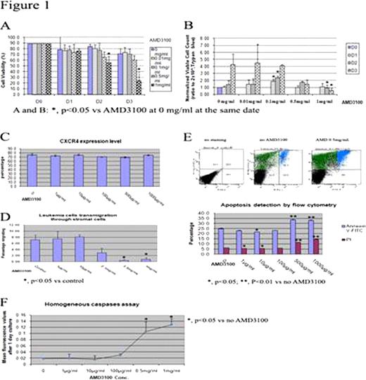
AMD3100 induces proliferation of t(4;11) leukemia cells at low concentration
AMD3100 above 500μ g/mL induces apoptosis of t(4;11) leukemia cells
When AMD3100 was included in the culture of t(4;11) leukemia cells, the cell viability percentage started to significantly decrease on day 2 at 1mg/mL (Figure 1A). After normalizing the cell count, it showed that AMD3100 decreased the cell number from concentration 0.5mg/ml and did so significantly at 1.0mg/ml (Figure 1B). Cell culture with AMD3100 did not change the expression level of its receptor CXCR4 (Figure 1C), but decreased the migration level through stromal cells (Figure 1D). To figure out if the reason of low transmigration was because of inhibiting migration or cell killing, we did two apoptosis related assay. First, AMD3100 at concentrations above 0.5 mg/mL significantly increased the apoptosis population showing with Annexin-V positive and cell death population with PI positive by flow cytometry (Figure 1E). Second, the homogneous caspases assay also showed that activated caspases level significantly increased after one day’s culture with AMD3100 at 0.5 mg/mL and 1 mg/mL (Figure 1F). These results confirm that AMD3100 induced apoptosis at higher concentrations.
Downstream kinases assay
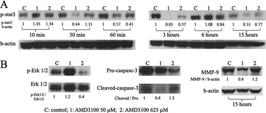
In summary, our study showed that AMD3100 at low concentrations increased proliferation and cell survival through activation of phosphorylation of Erk1/2. While at high concentrations, AMD3100 induced apoptosis through inhibiting the phophorylation of Stat3 and Erk1/2.
No relevant conflicts of interest to declare.
Author notes
Asterisk with author names denotes non-ASH members.


This feature is available to Subscribers Only
Sign In or Create an Account Close Modal