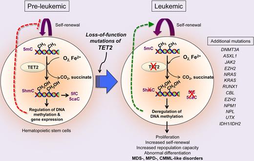
The TET-family enzymes TET1, TET2, and TET3 influence DNA methylation by modifying 5-methylcytosine.1-3 Somatic loss-of-function mutations in TET2 are frequently observed in myeloid neoplasms.2,4 In this issue of Blood, Li et al demonstrate that ablation of Tet2 alters the homeostasis and function of hematopoietic stem cells (HSCs) and induces leukemia in mice,5 as also reported by other groups.6-8
TET1, TET2, and TET3, members of the TET family of Fe2+ and α-ketoglutarate–dependent dioxygenases, successively oxidise 5-methylcytosine (5mC) to 5-hydroxymethylcytosine (5hmC), 5-formylcytosine (5fC), and 5-carboxycytosine (5caC; see figure).1-3 5caC can be excised by the base excision repair enzyme thymine-DNA glycosylase (TDG),9 suggesting that TET proteins cooperate with TDG to effect DNA demethylation. Thus, TET proteins have the potential to be important regulators of epigenetic status in the cell types in which they are expressed.
TET2 is frequently mutated in myeloid malignancies. Microdeletions and copy number–neutral loss of heterozygosity (also called uniparental disomy) are recurrently observed in the TET2 locus in diverse myeloid malignancies including myelodysplastic syndromes (MDS), myeloproliferative neoplasms (MPNs), chronic myelomonocytic leukemia (CMML), and acute myeloid leukemia (AML). TET2 is expressed, and 5hmC is readily detectable, in hematopoietic stem/progenitor cells and mature blood subsets.2,6 Moreover, leukemia-associated TET2 mutations have been shown to impair the catalytic activity of TET2 and diminish 5hmC levels in cells from patients with MDS/MPN/CMML and AML, and shRNA-mediated depletion of TET2 in hematopoietic stem cells (HSCs) resulted in skewing toward myeloid differentiation.2 Together, these results suggested a causal relation between loss-of-function mutations in TET2 and myeloid leukemias.
TET2 mutations in myeloid leukemogenesis. TET2 catalyzes the oxidation of 5mC to 5hmC, 5fC and 5caC in the genome and controls HSC self-renewal and function, presumably by regulating gene expression through effects on DNA methylation. Tet2 deficiency in mice impairs 5mC hydroxylation and leads to skewed differentiation and enhanced self-renewal and repopulating capacity of HSCs, promoting malignant transformation to cause disorders resembling MDS, MPD, or MDS/MPD overlap syndromes including CMML. TET2 mutations frequently coexist with other mutations in a wide spectrum of cancers including leukemias and lymphomas, suggesting that additional genetic alterations cooperate with TET2 mutations in different phases of tumorigenesis such as tumor initiation and progression.
TET2 mutations in myeloid leukemogenesis. TET2 catalyzes the oxidation of 5mC to 5hmC, 5fC and 5caC in the genome and controls HSC self-renewal and function, presumably by regulating gene expression through effects on DNA methylation. Tet2 deficiency in mice impairs 5mC hydroxylation and leads to skewed differentiation and enhanced self-renewal and repopulating capacity of HSCs, promoting malignant transformation to cause disorders resembling MDS, MPD, or MDS/MPD overlap syndromes including CMML. TET2 mutations frequently coexist with other mutations in a wide spectrum of cancers including leukemias and lymphomas, suggesting that additional genetic alterations cooperate with TET2 mutations in different phases of tumorigenesis such as tumor initiation and progression.
To address the function of TET2 in normal hematopoiesis and myeloid transformation, Li et al generated a gene-trap mouse strain in which a β-galactosidase-GFP cassette was inserted into exon 3 of the Tet2 locus, disrupting the endogenous start codon and inducing premature termination of transcription.5 They showed, as previously reported,2,6 that Tet2 was highly expressed in hematopoietic stem/progenitor cells and that ablation of Tet2 diminished levels of genomic 5hmC. Intriguingly, Tet2−/− mice developed diverse myeloid malignancies: ∼ 57% of deceased/moribund mice were found to have MDS with erythroid predominance and depletion of the hematopoietic stem/progenitor LSK (Lin−c-Kit+Sca-1+) population, and ∼ 20% displayed MPD- or CMML-like phenotypes with enlarged LSK and granulocyte/macrophage precursor (GMP) populations as assessed by increased monocytosis, splenomegaly, hepatomegaly, elevated WBC counts, and bone marrow hypercellularity. Tumors arising in Tet2−/− mice with myeloid but not erythroid infiltration were highly transplantable. Consistent with the high frequency of heterozygous TET2 mutations in myeloid malignancies in humans, Tet2+/− mice also developed MPD- or CMML-like leukemias. In preleukemic mice, Tet2 deficiency increased the size of the LSK compartment in which HSCs are enriched, and conferred a significant advantage on these cells in repopulating hematopoietic lineages in a cell-autonomous manner. Tet2−/− LSK cells were highly proliferative, displayed enhanced cloning efficiency, and were biased to differentiate into monocyte lineage in vivo and in vitro.
The phenotypes described by Li et al are consistent with recent reports using other Tet2−/− mouse models, from three independent groups.6-8 Moran-Crusio et al generated conditional Tet2-deficient mice targeting exon 3 in the Tet2 locus.6 Quivoron et al generated two different strains of Tet2-deficient mice: hypomorphic gene-trap mice in which β-galactosidase-neomycin was inserted into exon 9, and mice with a conditional deletion of exon 11.7 Ko et al generated Tet2-disrupted mice in which exons 8-10 were targeted.8 Exon 3 contains the start codon and exons 7-11 in the Tet2 locus encode the double-stranded β-helix domain, a core region of the catalytic domain1 ; thus, all mice with disruption of the Tet2 gene showed very similar loss-of-function phenotypes. Tet2-deficient LSK cells showed enhanced replating capacity but diminished differentiation in vitro. Loss of Tet2 resulted in expansion of the HSC compartment in a cell-intrinsic manner and enhanced HSC self-renewal, thereby conferring a competitive advantage relative to wild-type HSCs for reconstitution into all hematopoietic lineages in vivo. Cells obtained from serial methylcellulose cultures showed gene expression profiles similar to GMPs and had uniformly high levels of c-Kit (CD117) expression. Tet2 gene-trap or conditional exon 3–targeted mice were susceptible to CMML-like leukemia, and Tet2 haploinsufficiency sufficed to alter HSC properties and induce myeloid malignancies. Notably, Quivoron et al observed impaired lymphopoiesis in Tet2-disrupted mice.7 Furthermore, they observed rare somatic mutations of TET2 in B-cell (∼ 2%) and T-cell (∼ 11.9%) lymphoma samples and these mutations seemed to originate from CD34+ early progenitor cells with myeloid colony-forming potential, implicating TET2 loss-of-function in lymphoid malignancies as well.
In addition to TET2 loss-of-function mutations, many other recurrent mutations have been noted in MDS/MPN/CMML, some of which are presumed loss-of-function or possibly dominant interfering mutations (eg, DNMT3A, ASXL1, EZH2), whereas others are clearly neomorphic or gain-of-function (eg, JAK2, NRAS and KRAS, IDH1, and IDH2).4 Collectively, these findings suggest that oxidation of 5mC controlled by Tet2 and upstream regulators results in pleiotropic changes that protect HSCs from aberrant expansion and myeloid transformation. However, a molecular relation between TET2 and other mutations has been suggested only in the case of recurrent gain-of-function mutations in the metabolic enzymes isocitrate dehydrogenase (IDH) 1 and IDH2. Mutant IDH1/2 enzymes produce a novel “oncometabolite,“ 2-hydroxyglutarate, which competes with α-ketoglutarate to impair the catalytic activity of Fe2+/α-ketoglutarate–dependent dioxygenases including TET2.10 As a result, IDH1/2 mutations are rarely found together with TET2 mutations, but have similar effects as TET2 mutations on HSCs.10 Additional studies will be needed to clarify the mechanisms underlying the tumor-suppressor function of Tet2 in myeloid malignancies, as well as the mechanisms by which TET2 loss-of-function synergises with other mutations to promote malignant transformation.
How TET2 mutations affect the epigenetic landscape is still unclear. Because 5hmC can potentially be an intermediate in both passive and active demethylation,1,3,9 it is plausible that TET2 loss-of-function mutations impair the consumption of 5mC, leading to accumulation of 5mC at certain genomic locations. Indeed, Figueroa et al reported a correlation between TET2 mutations and DNA hypermethylation at HpaII sites in de novo AML.10 In contrast, Ko et al examined bone marrow samples from diverse myeloid malignancies and found that TET2 mutations (and more generally, low 5hmC levels) were associated with DNA hypomethylation at most differentially methylated CpG sites.2 This discrepancy may reflect differences in the cancer subtypes examined, the genomic CpG sites sampled, or the tools used for 5mC quantification and statistical analyses. It is noteworthy, however, that DNMT3A mutations are also associated with myeloid malignancies, and would also be predicted to be associated with DNA hypomethylation at certain CpG sites in the genome. Furthermore, 5hmC in embryonic stem cells was recently shown to be enriched in regions associated with specific chromatin modifications.11 Because of these associations, and because DNA methylation at promoters is often associated with transcriptional repression, TET2 mutations may alter gene expression programs that are critical for homeostasis and self-renewal of HSCs. Further studies will be necessary to resolve the epigenetic impact of TET2 mutations on regulated expression of genes that control the function, self-renewal, and differentiation of HSCs.
Conflict-of-interest disclosure: The authors declare no competing financial interests. ■


This feature is available to Subscribers Only
Sign In or Create an Account Close Modal