Merkel cell polyomavirus (MCPyV), a DNA tumor virus, has been found to be associated with Merkel cell carcinoma and chronic lymphocytic leukemia. MCPyV sequences have also been detected in various normal tissues in tumor-affected patients. Immunologic studies have detected MCPyV antibodies in as many as 80% of healthy blood donors. This high seroprevalence suggests that MCPyV infection is widespread in humans. In our study, buffy coats, which were examined for MCPyV DNA Tag sequences, showed a prevalence of 22%. Viral DNA load was revealed in blood samples from 10 to 100 molecules/100 000 cells. DNA sequencing confirmed that polymerase chain reaction amplicons belong to the MCPyV strain, MKL-1. To interpret the putative role of MCPyV in chronic lymphocytic leukemia, we may infer that, during a long period of viral persistence in blood cells, this DNA tumor virus may generate mutants, which are able to participate as cofactors in the multistep process of cell transformation.
Introduction
Merkel cell polyomavirus (MCPyV) is a small DNA tumor virus.1 A recent investigation has reported MCPyV sequences in 27.1% of purified malignant cells from human chronic lymphocytic leukemia (CLL) samples.2 CLL, the most common leukemia in the Western world, results from the expansion of a rare population of mature B-lymphocytes.3 In a previous study, MCPyV DNA was identified in Merkel cell carcinoma (MCC).4 This neoplasm is a rare but aggressive skin cancer of neuroendocrine origin.5 MCC has been detected in immunosuppressed patients who have undergone organ transplantation or are affected by HIV infection, with T-lymphocyte immunosuppression resulting from CLL.6 MCPyV DNA has been also identified in various tissue samples in patients affected by malignant or nonmalignant tumors.4,7,–9 Moreover, the presence of MCPyV in peripheral blood at low copy number has also been reported.4,10 Recent immunologic studies have detected MCPyV antibodies in 70% to 80% of adults.11,–13 Because human polyomavirus DNAs have been detected in tonsillar tissues, the high respiratory tract has been proposed as one of the entry portals for these viral agents.14,–16 However, the process by which polyomaviruses, and specifically MCPyV, gain access to and establish persistent infections in distal body compartments has not been well established.15
Methods
Samples and PCR techniques
Buffy coats (n = 60) were obtained from healthy blood donors as recently described.17 DNA was isolated as previously done, and its quality was confirmed by β-globin polymerase chain reaction (PCR).17 The pUC57MC1 recombinant plasmid, with MCC 350 strain sequences, was used as a positive control in MCPyV PCR amplification experiments.18 Two different MCPyV Tag regions, nucleotides (nt) 571 to 879 and nt 1709 to 1846, were investigated by single PCR analysis for 35 cycles using the primer sets, LT3F-LT3R and MCPyLT1709.F-MCPyLT1864.R, respectively. Amplicons of 308 bp and 138 bp are expected from these sPCR amplifications.4,18 However, no MCPyV Tag sequences were detected in our samples. Recently, similar data have been reported by other groups.19,20
To verify whether these negative data were the result of a low viral DNA load in our buffy coats, the same MCPyV sequences were investigated by PCR reamplifications, using 5 μL of the first PCR reaction, with the same technical conditions as the sPCR experiments.
To quantify the MCPyV DNA load, the samples found positive for nt 1709 to 1846 by PCR reamplification were further analyzed by quantitative real-time RT-PCR for Tag sequences, as previously described.17,21 Quantitative RT-PCR cycling conditions, primers, and probes for the Tag sequences were those reported by Goh et al.22 The positive control was a standard dilution of the recombinant plasmid pMCPyVLT.1, which contains 258 bp of the Tag (FJ472933).22 pMCPyVLT.1 sequences, from 10 to 107 copies, were run in duplicate in each experiment. To determine the human cell equivalents of each sample under analysis, quantitative RT-PCR assays were carried out simultaneously with the cellular RNase P gene.17,21
DNA sequence analysis
To verify the specificity of PCR-amplified products, 10 amplicons from the MCPyV Tag regions, obtained by PCR, were DNA sequenced (Macrogen Company South Korea). DNA sequence data were compared, using the National Center for Biotechnology Information Blast program, with the reference sequences from the National Center for Biotechnology Information Entrez Nucleotide database. Sequences for both nt 571 to 879 and nt 1709 to 1846 strands were identified as belonging to the MCPyV genome.
Results and discussion
PCR reamplifications, which were independently performed 5 times, indicated that 13 of 60 (22%) DNA samples tested were positive for MCPyV Tag coding sequences. A sample was considered positive when MCPyV Tag coding sequences were PCR amplified at least 3 of 5 times. A comparative analysis of the prevalence of MCPyV-positive samples indicated that a difference across 5 age cohorts (years 20-29, 30-39, 40-49, 50-59, and > 60) was not statistically significant (Fisher exact test).
A quantitative RT-PCR analysis, conducted on the 13 positive samples by PCR reamplification, confirmed the presence of Tag sequences in 8 of 13 samples (Table 1). Among the 13 MCPyV Tag samples that were found positive by PCR, 5 samples were negative by quantitative RT-PCR. We can infer that, in our quantitative RT-PCR experimental conditions, because of the low viral DNA load, MCPyV Tag sequences were not detectable in all samples. Indeed, it turned out that in the buffy coats under analysis the viral DNA copy numbers were very low, ranging from 10 to 100 copies/100 000 cells (Table 1), as demonstrated by the cycle threshold values (mean Tag = 37.7).
MCPyV DNA load, determined by quantitative RT-PCR in DNA, corresponding to 100 000 cells, from blood specimens found positive for the MCPyV-Tag region
| MCPyV DNA load expressed as viral copy number . | Tag+/sample analyzed, no. (%) . |
|---|---|
| 1-10 | 0/13 |
| 10-100 | 6/13 (46) |
| 100-1000 | 2/13 (15) |
| MCPyV DNA load expressed as viral copy number . | Tag+/sample analyzed, no. (%) . |
|---|---|
| 1-10 | 0/13 |
| 10-100 | 6/13 (46) |
| 100-1000 | 2/13 (15) |
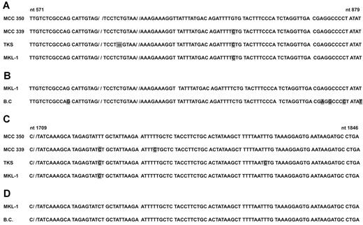
The sequencing result of the PCR products for both strands, nt 571 to 879 and nt 1709 to 1846, were identified as belonging to the MCPyV genome. A comparative analysis of the Tag sequences, from all 10 MCPyV-positive samples, with MCPyV sequences of isolates from the United States (MCC350, EU375803.1; MCC339, EU375804.1), Japan (TKS, FJ 464337), Sweden, and France (MKL-1, FJ173815) showed that they were very similar to those of the MKL-1 isolate from Sweden and France (Figure 1).12 Seven of 10 DNA sequences of buffy coats showed a single nucleotide substitution/deletion/insertion: specifically, in samples 1 to 3 at nt 582 G deletion, in sample 4 nt 868 A→G substitution, in sample 5 nt 870 C→G substitution, in sample 6 nt 874 C insertion, in sample 7 nt 879 A→T substitution.
MCPyV DNA sequence analysis and alignment of MCPyV Tag nucleotide sequences. (A,C) nt 571 to 879 and nt 1709 to 1846 from the MCC350 strain, respectively, compared with the MCC339, United States isolate, the TKS Japanese isolate, and the MKL-1 Swedish strain. Nucleotide substitutions/deletions (−) in different MCPyV strains are marked in gray. (B,D) nt 571 to 879 and 1709 to 1846 from the MKL-1 strain, respectively, compared with sequences obtained from buffy coats (B.C.). Seven of 10 DNA samples from B.C. contain a single nucleotide substitution, which is cumulatively marked in gray in the same sequence (B, B.C.).
MCPyV DNA sequence analysis and alignment of MCPyV Tag nucleotide sequences. (A,C) nt 571 to 879 and nt 1709 to 1846 from the MCC350 strain, respectively, compared with the MCC339, United States isolate, the TKS Japanese isolate, and the MKL-1 Swedish strain. Nucleotide substitutions/deletions (−) in different MCPyV strains are marked in gray. (B,D) nt 571 to 879 and 1709 to 1846 from the MKL-1 strain, respectively, compared with sequences obtained from buffy coats (B.C.). Seven of 10 DNA samples from B.C. contain a single nucleotide substitution, which is cumulatively marked in gray in the same sequence (B, B.C.).
In our investigation, MCPyV sequences were detected in buffy coats from healthy blood donors. The prevalence of 22%, which was determined in this study, is probably an underestimate because blood samples contain very low copies of MCPyV DNA. The discrepancy between our positive data and the negative results obtained in previous reports19,20 may be the result of the use of different technical approaches. Indeed, DNA isolation and purification, and/or different PCR protocols, could be responsible for the PCR-negative results in human sample target viral DNA, with a low molecular weight, when present at low copy numbers. Indeed, our quantitative PCR data revealed that MCPyV sequences are present at low copy numbers in blood specimens, ranging from 10 to 100 per 100 000 cells. In agreement with our results, previous studies have reported a similar low DNA viral load in nasopharyngeal aspirate samples22 as well as in other tissues.7 So far, high MCPyV DNA loads have only been detected in MCC specimens from oncologic patients.7,10 These data suggest that MCPyV is able to persist at a low viral copy in the peripheral blood mononuclear cells of immune-competent persons.
MCPyV Tag DNA sequencing data from 10 blood samples show high homology with previously published MCPyV sequences obtained from MCC specimens. The 10 MCPyV DNA showed little nucleotide variability and have a sequence that is highly homologous to the MKL-1 isolates from Sweden and France.12 This result, which was obtained using only 10 samples, is in agreement with other robust epidemiologic data, which indicate that MKL-1 isolate is very probably one of the main MCPyV strains circulating in Europe.12
MCPyV sequences detected in the buffy coats of healthy donors suggest that this viral agent may infect a specific blood leukocyte cell, where it remains in a latent/persistent state in the immune competent host. It has been reported that MCPyV-positive inflammatory monocytes are vehicles for the virus in the human host and are a source of MCPyV horizontal infection in the human population.23 Our results, together with data from recent reports,4,10 suggest that in a fraction of healthy persons MCPyV persists or remains latent in blood cells. In the long-term, viral persistent infection may allow MCPyV to generate mutants that can participate in the cell transformation process. Indeed, large T antigen MCPyV deletion mutants have been detected in CLL or integrated into MCC.2,24 This oncogenic process, together with the immune impairment of the host and other factors, is a well-known multistep cell transformation mechanism used by other DNA tumor viruses, such as human papilloma viruses,25 which are closely related to the Merkel cell polyomavirus.
An Inside Blood analysis of this article appears at the front of this issue.
The publication costs of this article were defrayed in part by page charge payment. Therefore, and solely to indicate this fact, this article is hereby marked “advertisement” in accordance with 18 USC section 1734.
Acknowledgments
The authors thank Prof Tobias Allander, Karolinska Institutet, Stockolm, Sweden, for his generous gift of the recombinant plasmid pMCPyVLT.1.
This work was supported in part by the University of Ferrara, the University Hospital of Ferrara, the Emilia Romagna Region, and Fondazione Cassa di Risparmio di Cento, Italy.
Authorship
Contribution: C.P., M.C., F.M., and M.T. designed the study; M.T. and F.M. collected the specimens; C.P., V.C., and S.M. were responsible for the DNA extractions and carried out qualitative and quantitative PCR analyses; C.P. and E.M. were responsible for the sequencing analysis data and the statistical results; C.P. and M.T. contributed to writing the report; M.T. edited the final version of the text; and all authors have seen and approved the manuscript.
Conflict-of-interest disclosure: The authors declare no competing financial interests.
Correspondence: Mauro Tognon, Department of Morphology and Embryology, Section of Cell Biology and Molecular Genetics, University of Ferrara, Via Fossato di Mortara 64/B, 44121 Ferrara, Italy; e-mail: tgm@unife.it.


This feature is available to Subscribers Only
Sign In or Create an Account Close Modal