Abstract
Survival in cytogenetically high-risk patients with acute myeloid leukemia or myelodysplastic syndromes is significantly worse in the presence of a monosomal karyotype (MK). The objective of the present study was to determine whether the same held true for primary myelofibrosis. Among 793 primary myelofibrosis patients seen at our institution, 62 displayed an unfavorable karyotype by way of complex karyotype (n = 41) or sole trisomy 8 (n = 21). Seventeen (41%) of the 41 patients with complex karyotype were classified as having an MK. Median survival was 6, 24, and 20 months in patients with MK, complex karyotype without monosomies, and sole trisomy 8, respectively (P < .0001). The corresponding 2-year leukemic transformation rates were 29.4%, 8.3%, and 0 (P < .0001); hazard ratios (95% confidence intervals) were 6.9 (1.3-37.3) and 14.8 (1.7-130.8). The prognostic relevance of MK was not accounted for by the Dynamic International Prognostic Scoring System. We conclude that MK in primary myelofibrosis is associated with extremely poor overall and leukemia-free survival.
Introduction
Prognosis in primary myelofibrosis (PMF) is assessed by the International Prognostic Scoring System (IPSS)1 or the dynamic IPSS (DIPSS),2 both of which use the following adverse risk factors: age > 65 years, hemoglobin < 10 g/dL, leukocyte count > 25 × 109/L, circulating blasts ≥ 1%, and constitutional symptoms. IPSS is applicable at the time of diagnosis, and DIPSS is applicable at any point during the disease course. Independent of IPSS, complex karyotype and sole trisomy 8 adversely affect both overall and leukemia-free survival in PMF.3,4
Two recent studies in acute myeloid leukemia (AML) have suggested that a monosomal karyotype (MK), which is defined as 2 or more autosomal monosomies or a single autosomal monosomy associated with at least 1 structural abnormality, identifies a subset of patients with unfavorable karyotype who are short-lived5,6 ; 4-year survival rates were 3%-4% in the presence of MK versus 13%-26% in its absence.6 We have recently shown that MK was equally detrimental in the setting of myelodysplastic syndromes (MDS)7 ; overall and leukemia-free survival rates in patients with the complex karyotype were negatively affected by the presence of MK (2-year survival rates were 6% and 23% in the presence or absence of MK, respectively). The objective of the present study was to determine whether MK has a similar adverse prognostic effect in PMF.
Methods
After receiving approval from the Mayo Clinic Institutional Review Board, we reviewed medical records of patients with PMF referred to the Mayo Clinic during 1970-2009. The study considered only those patients with available bone marrow and cytogenetic information at the time of their first referral to the Mayo Clinic. The diagnoses of PMF and leukemic transformation were according to World Health Organization criteria.8 Cytogenetic results were interpreted and reported according to the International System for Human Cytogenetic Nomenclature.9 The presence of fewer than 20 evaluable metaphases did not disqualify patients from study inclusion as long as ≥ 10 metaphases were examined in those patients with “normal” reports; patients with an insufficient number of metaphases were excluded. A complex karyotype was defined by the presence of 3 or more distinct numeric or structural cytogenetic abnormalities. MK was defined by the presence of 2 or more distinct autosomal monosomies or a single autosomal monosomy associated with at least 1 structural abnormality.5 An abnormality was considered clonal when at least 2 metaphases had the same aberration in case of a structural abnormality or an extra chromosome. For classification as a monosomy, the monosomy had to be present in at least 3 metaphases.
All statistical analyses considered parameters at time of first referral to the Mayo Clinic. Differences in the distribution of continuous variables between categories were analyzed by either Mann-Whitney (for comparison of 2 groups) or Kruskal-Wallis (comparison of 3 or more groups) test. Patient groups with nominal variables were compared by χ2 test. Overall survival analysis was considered from the date of first referral to our institution to date of death or last contact. Leukemia-free survival was calculated from the date of first referral to our institution to date of leukemic transformation or last contact/date of death. Overall and leukemia-free survival curves were prepared by the Kaplan-Meier method and compared by the log-rank test. Cox proportional hazard regression model was used for multivariable analysis. P values < .05 were considered significant. The Stat View (SAS Institute) statistical package was used for all calculations.
Results and discussion
The present study population of 793 patients with PMF was selected from a total of 923 consecutive patients with PMF seen at the Mayo Clinic between January 1970 and December 2009 and who had undergone bone marrow examination. A total of 130 patients were excluded because cytogenetic studies either were not performed (n = 65), resulted in insufficient mitotic figures (n = 40), or constituted normal karyotype with < 10 metaphases analyzed (n = 19). Six more patients were excluded because of inaccurate diagnosis. Among the 793 study patients, 341 (43%) displayed an abnormal karyotype, including 41 (12%) with complex karyotype and 21 (6%) with sole trisomy 8. Among the 41 patients with complex karyotype, 17 (41%) were classified as MK and 24 (59%) as “complex karyotype without monosomies.” To determine whether the presence of MK conferred additional prognostic significance, we compared the patient groups with MK, complex karyotype without monosomies, or sole trisomy 8. No significant differences were noted among the 3 groups with regard to age, hemoglobin level, leukocyte count, platelet count, peripheral blast count, DIPSS score, constitutional symptoms, spleen size, or JAK2V617F mutational status (Table 1). Among the 62 patients with MK, complex karyotype without monosomies, or sole trisomy 8, 32 (52%) were evaluated within 1 year of their initial diagnosis, and the median time from initial diagnosis in the remaining 30 patients was approximately 4 years (range 1.5-8 years). There was no difference in the interval between initial diagnosis and first bone marrow assessment at the Mayo Clinic between patients with MK and those with sole trisomy 8 (P = .7), whereas this interval was significantly longer in patients with complex karyotype without monosomies (P = .01).
Comparison of clinical characteristics of patients with PMF stratified by the presence of MK, complex karyotype without monosomies, and sole trisomy 8
| Variables . | MK (n = 17) . | Complex karyotype without monosomies (n = 24) . | Sole trisomy 8 (n = 21) . | P . |
|---|---|---|---|---|
| Median age, y (range) | 62 (30–83) | 60 (42–73) | 66 (28–85) | .2 |
| Age > 65 y; n (%) | 7 (47) | 6 (26) | 11 (52) | .2 |
| Males, n (%) | 9 (53) | 14 (58) | 11 (52) | .9 |
| Median hemoglobin, g/dL (range) | 9 (6–11) | 10 (5–16) | 10 (7–13) | .3 |
| Median leukocyte count × 109/L (range) | 6 (2–46) | 10 (2–157) | 12 (2–142) | .8 |
| Median platelet count × 109/L (range) | 50 (6–522) | 97 (8–981) | 133 (16–684) | .2 |
| DIPSS risk group, n (%) | ||||
| Low | 0 | 1 (5) | 0 | .7 |
| Intermediate-1 | 6 (35) | 5 (21) | 8 (38) | |
| Intermediate-2 | 8 (47) | 15 (62) | 10 (48) | |
| High | 3 (18) | 3 (12) | 3 (14) | |
| Constitutional symptoms, n (%) | 7 (41) | 10 (42) | 7 (33) | .8 |
| Circulating blasts ≥ 1%, n (%) | 13 (76) | 20 (87) | 15 (71) | .4 |
| Hemoglobin < 10 g/dL, n (%) | 11 (65) | 14 (58) | 12 (57) | .9 |
| Leucocytes > 25 × 109/L, n (%) | 3 (18) | 6 (25) | 3 (14) | .6 |
| Platelets < 100 × 109/L, n (%) | 12 (71) | 13 (54) | 9 (43) | .2 |
| Leucocytes < 4 × 10⁹/L, n (%) | 5 (29) | 5 (21) | 6 (29) | .8 |
| Palpable spleen > 10 cm, n (%) | 1 (33) | 9 (56) | 3 (23) | .2 |
| Splenectomy, n (%) | 3 (19) | 7 (30) | 4 (19) | .6 |
| JAK2V617F status, number tested (% positive) | 4 (100) | 6 (60) | 4 (67) | .3 |
| Transplanted, n (%) | 1 (6) | 0 | 1 (5) | .5 |
| Deaths, n (%) | 14 (82) | 21 (88) | 17 (81) | .8 |
| Leukemic transformations, n (%) | 5 (29) | 2 (8) | 1 (5) | .05 |
DIPSS indicates Dynamic International Prognostic Scoring System.2
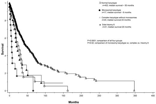
Overall survival was significantly worse for patients with MK than for those with either complex karyotype without monosomies or sole trisomy 8 (Figure 1). The corresponding median survival times were 6, 24, and 20 months (P < .0001; hazard ratio [95% confidence interval] 2.3 [1.1-4.8] and 2.4 [1.2-5.1], respectively). On multivariable analysis that included staging according to DIPSS, the independent prognostic significance of MK was sustained (supplemental Table, available on the Blood Web site; see the Supplemental Materials link at the top of the online article). The 2-year survival rates in the presence of complex karyotype without monosomies, sole trisomy 8, and MK were 51%, 45%, and 17%, respectively. Patients with MK also displayed significantly inferior leukemia-free survival (supplemental Figure). At 2 years, blast-phase PMF developed in 5 (29.4%) of 17 patients with MK compared with 2 (8.3%) of 24 with complex karyotype without monosomies and 0 of 21 with sole trisomy 8 (P < .0001). The respective hazard ratios (95% confidence intervals) were 6.9 (1.3-37.3) and 14.8 (1.7-130.8). Considering the previous categorization of certain cytogenetic abnormalities as being prognostically “unfavorable” in PMF,4,10 we re-ran the survival analysis comparing MK (n = 17) with unfavorable karyotype other than complex karyotype or sole trisomy 8 (n = 57). The corresponding median survivals were 6 and 11 months (P = .06), and leukemia-free survival was again inferior in patients with MK (P = .0002).
Overall survival of 62 patients with PMF and unfavorable cytogenetic findings further stratified into 3 groups by the presence of MK, complex karyotype without monosomies, or sole trisomy 8. For purposes of reference, the survival curve of 452 patients with PMF and normal karyotype is included. The latter group of patients was recruited from the same database used to identify the present study population.
Overall survival of 62 patients with PMF and unfavorable cytogenetic findings further stratified into 3 groups by the presence of MK, complex karyotype without monosomies, or sole trisomy 8. For purposes of reference, the survival curve of 452 patients with PMF and normal karyotype is included. The latter group of patients was recruited from the same database used to identify the present study population.
The results of the present study signify the across-the-board prognostic relevance of MK in myeloid malignancies. In an earlier study by Breems et al,5 MK+ AML was associated with a significantly shorter 4-year overall survival (4%) than MK− but cytogenetically high-risk AML (26%). Similar results were reported by another study in which patients with MK, compared with those with unfavorable cytogenetic findings without MK, displayed a significantly shorter 4-year survival (3% vs 13%).6 In this particular study, the inferior survival in MK+ patients was attributed in part to their lower complete remission rates (18% vs 34%), which was the case in all age groups above age 30 years. Furthermore, the negative prognostic impact of MK was less evident in patients with monosomy 7, inv(3)/t(3;3), del(5q), and t(9;22), as opposed to those with del(7q) or complex karyotype.6 In a more recent study,11 MK performed better than other “high-risk” cytogenetic categorization in predicting relapse incidence after allogeneic stem cell transplantation for AML.
We have recently demonstrated the adverse prognostic effect of MK in MDS.7 In that particular study,7 among 127 MDS patients with complex karyotype, the incidence of MK was 83%, which was substantially higher than the 42% seen in the present study. MK+ MDS patients' lives were significantly shorter than those with complex karyotype without MK, and the prognostic impact of MK was not accounted for by advanced age, bone marrow blast percentage, or the presence or absence of monosomy 7 or monosomy 5.7 We now show in the present study that MK is equally as bad in PMF in terms of both overall and leukemia-free survival. The presence of MK in PMF signified worse survival than that associated with either the complex karyotype without monosomies or sole trisomy 8, both of which were identified previously as being unfavorable cytogenetic findings in PMF.4 However, although statistical significance was demonstrated, the number of patients for each unfavorable cytogenetic category was too small to be certain about the difference in prognostic impact between MK and other unfavorable karyotype in PMF in terms of both survival and risk of leukemic transformation, and the present observations need to be validated in a larger group of informative patients. From a practical standpoint, these findings further underscore the importance of paying attention to cytogenetic findings in PMF and the prudence of early intervention with investigational drug therapy or allogeneic stem cell transplantation in MK+ PMF, although the value of such a treatment strategy in this particular patient population remains to be proven.
The online version of this article contains a data supplement.
The publication costs of this article were defrayed in part by page charge payment. Therefore, and solely to indicate this fact, this article is hereby marked “advertisement” in accordance with 18 USC section 1734.
Authorship
Contribution: R.V. collected data and participated in data analysis and the writing of the paper; D.C., K.H.B., and N.G. participated in data collection; D.L.V.D. reviewed cytogenetic information; C.H. reviewed histopathology; A.P. contributed patients and participated in data analysis; and A.T. designed the study, contributed patients, collected data, performed the statistical analysis, and wrote the paper. All authors approved the final draft of the paper.
Conflict-of-interest disclosure: The authors declare no competing financial interests.
Correspondence: Ayalew Tefferi, MD, Mayo Clinic, 200 First St SW, Rochester, MN 55905; e-mail: tefferi.ayalew@mayo.edu.


This feature is available to Subscribers Only
Sign In or Create an Account Close Modal