Abstract
Bortezomib is now widely used for the treatment of multiple myeloma (MM); however, its action mechanisms are not fully understood. Despite the initial results, recent investigations have indicated that bortezomib does not inactivate nuclear factor-κB activity in MM cells, suggesting the presence of other critical pathways leading to cytotoxicity. In this study, we show that histone deacetylases (HDACs) are critical targets of bortezomib, which specifically down-regulated the expression of class I HDACs (HDAC1, HDAC2, and HDAC3) in MM cell lines and primary MM cells at the transcriptional level, accompanied by reciprocal histone hyperacetylation. Transcriptional repression of HDACs was mediated by caspase-8–dependent degradation of Sp1 protein, the most potent transactivator of class I HDAC genes. Short-interfering RNA-mediated knockdown of HDAC1 enhanced bortezomib-induced apoptosis and histone hyperacetylation, whereas HDAC1 overexpression inhibited them. HDAC1 overexpression conferred resistance to bortezomib in MM cells, and administration of the HDAC inhibitor romidepsin restored sensitivity to bortezomib in HDAC1-overexpressing cells both in vitro and in vivo. These results suggest that bortezomib targets HDACs via distinct mechanisms from conventional HDAC inhibitors. Our findings provide a novel molecular basis and rationale for the use of bortezomib in MM treatment.
Introduction
Despite recent advances in treatment strategies using dose-intensified regimens and new molecular-targeted compounds, multiple myeloma (MM) remains incurable for most patients.1 To improve their prognosis, the development of molecular-targeted therapy, which involves therapeutic agents with distinct mechanisms of action and high specificity, is highly anticipated. Inhibitors of histone deacetylases (HDACs) and proteasome are promising candidates for these agents, and their clinical efficacy has been reported.2-4 Moreover, their combinations were proved to be additive in preclinical studies5,6 and are presently the focus of clinical trials.7
Aberrant transcriptional repression of genes regulating cell growth and differentiation is a hallmark of cancer.8 Recently, evidence has accumulated suggesting that altered activation of HDACs underlies transcriptional repression in malignancies.9 HDACs are a family of enzymes that catalyze the removal of acetyl groups from core histones, resulting in chromatin compaction and transcriptional repression.10 HDACs are divided into 5 groups: class I (HDAC1, HDAC2, HDAC3, and HDAC8), class IIa (HDAC4, HDAC5, HDAC7, and HDAC9), class IIb (HDAC6 and HDAC10), class III (SIRT family), and class IV (HDAC11). Class I HDACs are ubiquitously expressed and are generally involved in cell growth and differentiation,11 whereas class II HDACs have a more restricted pattern of expression (skeletal muscle, heart, and brain) and act in association with tissue-specific transcription factors. In leukemic cells, fusion proteins such as PML/RARα and AML-1/ETO form a complex with HDACs with higher affinities than their normal counterparts, and aberrantly suppress the expression of genes required for cell differentiation and growth control, leading to the transformation of hematopoietic progenitor cells.12,13 In addition, we have shown that class I HDACs are up-regulated in malignant melanoma and leukemias in association with histone hypoacetylation.14,15 Similarly, the aberrant expression of HDACs may contribute to the uncontrolled growth of myeloma cells.
Given the role of HDACs in tumor cells, the use of small compounds that inhibit HDAC activity, collectively referred to as HDAC inhibitors, is expected to become a novel strategy for the treatment of cancer.16 HDAC inhibitors are able to restore the expression of genes that are aberrantly suppressed in tumor cells, which may result in cell-cycle arrest, differentiation, and apoptosis.17
The proteasome inhibitor bortezomib (Velcade [Millennium Pharmaceuticals]; formerly known as PS-341) is now widely used for the treatment of MM.3,4 Bortezomib is a reversible inhibitor of the 26S proteasome complex, which catalyzes ubiquitin-dependent protein degradation. Inhibition of this complex ultimately leads to modulation of the abundance and functions of many intracellular proteins, which may be associated with cytotoxic effects on malignant cells.18 Recently, novel proteasome inhibitors, which target the ubiquitin-proteasome system in a manner distinct from bortezomib, have been developed and have shown strong activity in preclinical studies.19,20 These new inhibitors are thought to be promising candidates for MM treatment.
Recent genome-wide approaches have revealed that nuclear factor-κB (NF-κB) is frequently activated in MM cells.21,22 Because IκBα, which inactivates NF-κB, is a substrate of the proteasome complex, the initial rationale for the use of bortezomib was the inhibition of NF-κB activity.23 Despite several preclinical studies and clinical trials of MM,3,4 the inhibition of NF-κB activity has not been demonstrated in bortezomib-treated MM cells. In addition, Hideshima et al24 recently reported that bortezomib did not inactivate but rather activated the canonical NF-κB pathway in MM cells, suggesting that bortezomib-induced cytotoxicity could not be fully attributed to the inhibition of NF-κB activity. Taken together, there may be other critical pathways and target molecules of bortezomib that have not been fully investigated.
In this study, we found that bortezomib specifically down-regulated the expression of class I HDACs and induced histone hyperacetylation in MM cells. Gain- and loss-of-function analyses revealed that bortezomib-induced cytotoxicity depends on cellular HDAC activities both in vitro and in vivo. Based on these findings, we propose that HDACs are novel critical downstream targets of bortezomib. This finding may provide a novel molecular basis and rationale for the use of bortezomib in MM treatment.
Methods
Cells and cell culture
We used 3 bona fide human MM cell lines, KMS12-BM, RPMI8226, and U266, in this study.25 These cell lines were purchased from the Health Science Research Resources Bank and maintained in RPMI 1640 medium (Sigma-Aldrich) supplemented with 10% heat-inactivated fetal calf serum (Sigma-Aldrich) and antibiotics. Primary CD138+ MM cells were isolated from the bone marrow (BM) of patients at the time of diagnostic procedure using the magnetic-activated cell sorter (MACS) system (Miltenyi Biotec). Informed consent was obtained in accordance with the Declaration of Helsinki, and the protocol was approved by the institutional review board of Jichi Medical University.
Drugs
Bortezomib and romidepsin (formerly known as FK228 or depsipeptide) were obtained from Millennium Pharmaceuticals and Gloucester Pharmaceuticals, respectively. HDAC6-specific inhibitor tubacin and its inactive derivative niltubacin were provided by Dr Stuart L. Schreiber (Broad Institute of Harvard University and Massachusetts Institute of Technology). We used vincristine (Shionogi Co Ltd), doxorubicin (Meiji Co Ltd), and dexamethasone (Sigma-Aldrich) as conventional antimyeloma drugs. All drugs were dissolved in RPMI 1640 medium at appropriate concentrations and stored at −80°C until use.
Isobologram of Steel and Peckham
The cytotoxic interaction of bortezomib and romidepsin was evaluated at the point of IC80 by the isobologram of Steel and Peckham. IC80 was defined as the concentration of drugs that produced 80% inhibition of cell growth. The theoretical basis of the isobologram method has been previously described in detail.26
Assessment of cell death
Cells were washed with phosphate-buffered saline (PBS) and stained with allophycocyanin-conjugated annexin-V (annexin-V/APC; Biovision). Cell death/apoptosis was judged by annexin-V reactivity and BrdU/7-AAD double-staining using a FACSAria flow cytometer (Becton Dickinson) as described previously.27
Immunoblotting
Immunoblotting was carried out according to the standard method using the following antibodies: antiacetyl histone H3, antiacetyl histone H4 (Upstate Biotechnology/Millipore), antiacetyl histone H3–lysine18, antiacetyl histone H4–lysine12, antiacetyl–α-tubulin (Cell Signaling Technology), anti-HDAC1 (Sigma-Aldrich), anti-HDAC2 (MBL International), anti-HDAC3 (BD PharMingen), anti-Sp1, anti–MZF-1, and anti-GAPDH (Santa Cruz Biotechnology).28
Semiquantitative or real-time quantitative RT-PCR
Total cellular RNA was isolated from 1 to 10 × 104 cells, reverse-transcribed into cDNA using SuperScript reverse transcriptase and oligo(dT) primers (Invitrogen), and subjected to subsequent semiquantitative reverse transcription–polymerase chain reaction (RT-PCR) or real-time quantitative RT-PCR using Power SYBR Green PCR Master Mix (Applied Biosystems) as described previously.29 Detailed information of primers, including sequences, corresponding nucleotide positions, and PCR product sizes, is shown in supplemental Table 1 (available on the Blood Web site; see the Supplemental Materials link at the top of the online article).
Reporter assays
We amplified the promoter region of the HDAC1 gene (−1170 to +397) by PCR and inserted it into the pGL4.10 firefly luciferase vector (Promega) to generate reporter plasmids.15 We introduced the reporter plasmids into MM cells along with pGL4.73 Renilla luciferase vector (Promega), which served as a positive control to determine transfection efficiencies, in the presence of either test plasmids encoding Sp1 and MZF-1 or empty vectors by electroporation, as previously described.15 After 48 hours, firefly and Renilla luciferase activities were discriminately measured using the Dual-Luciferase Reporter Assay System (Promega). The promoterless pGL4-basic vector was used as a negative control. The luciferase activity was normalized by the internal standard and indicated as a relative ratio to the negative control. Expression vectors for MZF-1 and Sp1 were kindly provided by Dr Robert Hromas (University of New Mexico) and Dr Mitsuru Nakamura (National Institute of Advanced Industrial Science and Technology), respectively.
ChIP assays
We used the ChIP-IT Chromatin Immunoprecipitation Kit (Active Motif) to perform chromatin immunoprecipitation (ChIP) assays. In brief, cells were fixed with 1% formaldehyde at 37°C for 5 minutes and sonicated to obtain chromatin suspensions. After centrifugation, supernatants were incubated with antibodies of interest at 4°C overnight. The mixture was then incubated with protein A agarose beads at 4°C for 1 hour, and centrifuged to collect the beads. DNA fragments bound to the beads were purified with vigorous washing and subjected to PCR using primer pairs, as shown in supplemental Table 2.15
Construction and production of lentiviral expression vectors
We used a lentiviral short-hairpin RNA/short-interfering RNA (shRNA/siRNA) expression vector pLL3.7 for knockdown experiments.30 siRNA target sequences were designed to be homologous to wild-type cDNA sequences. Oligonucleotides were chemically synthesized, annealed, terminally phosphorylated, and inserted into pLL3.7 vector. Oligonucleotides containing siRNA target sequences are shown in supplemental Table 3. Scrambled sequences were used as controls.
We also used a lentiviral vector CSII-CMV-MCS-IRES-VENUS (kindly provided by Dr Hiroyuki Miyoshi, RIKEN BioResource Center) for gain-of-function experiments after replacing VENUS with DsRed amplified from pDsRed-expressing vector (Clontech Inc).27 The resulting construct was designated as the CSII-DsRed vector. We constructed HDAC expression vectors by inserting the coding regions of HDAC1, HDAC2, or HDAC3 cDNA (all provided by Dr Stuart L. Schreiber, Broad Institute of Harvard University and MIT).
These vectors were cotransfected into 293FT cells with packaging plasmids (Invitrogen) to produce infective lentiviruses in culture supernatants. Lentiviruses were then added to cell suspensions in the presence of 8 μg/mL polybrene, and transduced for 24 hours, as previously described.27
Xenograft murine model
Mice were inoculated subcutaneously in the right thigh with 3 × 10⁷ MM cells in 1 × 10⁻⁴ L of RPMI 1640 medium together with 1 × 10⁻⁴ L of Matrigel basement membrane matrix (Becton Dickinson).31 When tumors were measurable, mice were assigned to 3 treatment groups receiving either the vehicle alone (control), bortezomib alone, or bortezomib plus romidepsin. Bortezomib and romidepsin were given intravenously twice a week via the tail vein at 0.5 mg/kg for 4 weeks and intraperitoneally every other day at 0.25 mg/kg for 2 weeks, respectively.31,32 The control group received the vehicle (0.9% NaCl) alone on the same schedule. Caliper measurements of the longest perpendicular tumor diameters were performed every alternate day to estimate the tumor volume using the following formula: 4/3π × (width/2)2 × (length/2), which represents the 3-dimensional volume of an ellipse.
Results
Synergistic effects of romidepsin and bortezomib on viability and histone acetylation of MM cells
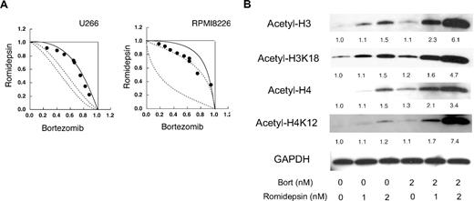
Romidepsin has proved to be one of the most effective HDAC inhibitors against hematologic malignancies both in vitro and in vivo.33,34 Initially, we examined the combination of romidepsin and bortezomib to develop an effective treatment strategy for MM, because bortezomib can enhance the effects of other anticancer drugs.6,30 As anticipated, an isobologram analysis of drug combination revealed that bortezomib and romidepsin showed synergistic cytotoxicity in U266 and RPMI8226 cells (Figure 1A). Next, we investigated the molecular basis of the synergistic effect of the 2 drugs. We speculated that bortezomib enhanced the HDAC inhibitory activities of romidepsin. To test this hypothesis, we determined cellular HDAC activity by monitoring the status of histone acetylation. As shown in Figure 1B, consistent with our hypothesis, bortezomib markedly enhanced romidepsin-induced hyperacetylation of histones H3 and H4. Moreover, we found here that bortezomib not only enhanced the effect of romidepsin but also induced histone hyperacetylation. These results suggest a novel mechanism of bortezomib action: it may induce cytotoxicity in MM cells by HDAC suppression. Although most cellular HDAC activities rely on class I HDACs, Hideshima et al35 showed that specific inhibitors of class II HDACs, such as tubacin, synergized with bortezomib via inhibition of HDAC6 activity. We therefore examined the effect of romidepsin on HDAC6 activity by monitoring the status of tubulin acetylation. As shown in supplemental Figure 1, tubulin acetylation was observed only at higher concentrations (> 2nM), indicating that romidepsin inhibits the activities of class I HDACs at lower concentrations and both class I and class II HDACs at higher concentrations. As additive cytotoxicity and histone hyperacetylation were observed with less than 1nM romidepsin, these effects are considered to be achieved mainly via the inhibition of class I HDACs. Thereafter, we focused on this novel property of bortezomib and investigated its molecular mechanisms and roles in cytotoxicity against MM.
Synergistic effects of romidepsin and bortezomib on cell viability and histone acetylation. (A) Isobolograms of simultaneous exposure of U266 and RPMI8226 cells to bortezomib and romidepsin are shown. The concentrations that produced 80% growth inhibition are expressed as 1.0 on the ordinate and abscissa of isobolograms. The envelope of additivity, surrounded by solid and broken lines, is constructed from dose-response curves of bortezomib and romidepsin. When the data points of the drug combination fall within the area surrounded by the envelope of additivity, the combination is regarded as additive. When the data points fall to the left of the envelope, the drug combination is regarded as supra-additive (synergism). When the data points fall to the right of the envelope, the combination is regarded as antagonistic. The isobolograms shown are representative of at least 3 independent experiments. Each point represents the mean value of at least 3 independent experiments; the SEMs were less than 25% and were omitted. (B) U266 cells were cultured in the absence or presence of either romidepsin (Romidepsin), bortezomib (Bort), or both agents for 48 hours at the indicated doses. Whole-cell lysates were subjected to immunoblotting. The membranes were reprobed with anti-GAPDH antibody to serve as a loading control. The signal intensities of each band were quantified, normalized to those of the corresponding GAPDH, and shown as relative values setting untreated controls to 1.0. Data shown are representative of multiple independent experiments.
Synergistic effects of romidepsin and bortezomib on cell viability and histone acetylation. (A) Isobolograms of simultaneous exposure of U266 and RPMI8226 cells to bortezomib and romidepsin are shown. The concentrations that produced 80% growth inhibition are expressed as 1.0 on the ordinate and abscissa of isobolograms. The envelope of additivity, surrounded by solid and broken lines, is constructed from dose-response curves of bortezomib and romidepsin. When the data points of the drug combination fall within the area surrounded by the envelope of additivity, the combination is regarded as additive. When the data points fall to the left of the envelope, the drug combination is regarded as supra-additive (synergism). When the data points fall to the right of the envelope, the combination is regarded as antagonistic. The isobolograms shown are representative of at least 3 independent experiments. Each point represents the mean value of at least 3 independent experiments; the SEMs were less than 25% and were omitted. (B) U266 cells were cultured in the absence or presence of either romidepsin (Romidepsin), bortezomib (Bort), or both agents for 48 hours at the indicated doses. Whole-cell lysates were subjected to immunoblotting. The membranes were reprobed with anti-GAPDH antibody to serve as a loading control. The signal intensities of each band were quantified, normalized to those of the corresponding GAPDH, and shown as relative values setting untreated controls to 1.0. Data shown are representative of multiple independent experiments.
Down-regulation of HDAC expression in MM cells during bortezomib treatment
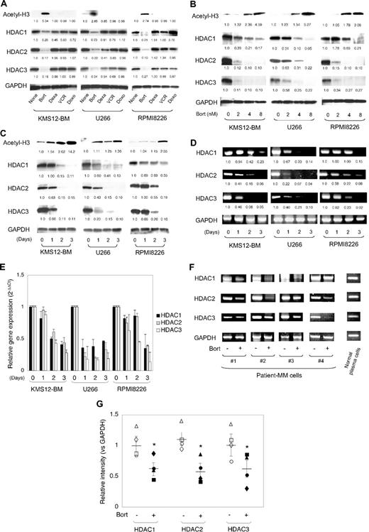
First, we investigated the molecular mechanisms of how bortezomib induced histone hyperacetylation in MM cells. We speculated that bortezomib did not inhibit HDAC activity like conventional HDAC inhibitors but down-regulated the expression of class I HDACs, because we and others have shown that bortezomib suppressed the expressions of various molecules, such as CD49d,30 HLA class I,36 and DNMT1,37 in MM cells. To verify this hypothesis, we determined the expression of class I HDACs (HDAC1, HDAC2, and HDAC3) in human MM cell lines (KMS12-BM, U266, and RPMI8226) during bortezomib treatment. We also treated MM cell lines with conventional antimyeloma drugs (vincristine, dexamethasone, and doxorubicin) to be used as control samples. As shown in Figure 2A, along with the induction of histone hyperacetylation, bortezomib readily down-regulated the expression of class I HDACs in all 3 MM cell lines. In contrast, there were no significant changes in histone acetylation and HDAC expression in cells treated with other drugs. The histone hyperacetylation and down-regulation of HDACs were reciprocally induced by bortezomib in a dose- and time-dependent fashion (Figure 2B-C). These results suggest that bortezomib induced histone hyperacetylation via the down-regulation of class I HDACs expression in MM cells.
Expression of class I HDACs in MM cells during bortezomib treatment. (A) MM cell lines (KMS12-BM, U266, and RPMI8226) were cultured in the absence or presence of either 4nM bortezomib (Bort), 50nM dexamethasone (Dexa), 1nM vincristine (VCR), or 100nM doxorubicin (Doxo) for 48 hours. Whole-cell lysates were subjected to immunoblotting. (B) MM cell lines were cultured in the absence or presence of bortezomib (Bort) at the indicated doses for 48 hours, or (C) cultured in the presence of 4nM bortezomib for up to 3 days. Whole-cell lysates were prepared at given time points and subjected to immunoblotting. (D) Total cellular RNA was isolated simultaneously in the experiments described in panel C and subjected to semiquantitative RT-PCR analysis for the expression of HDAC1, HDAC2, HDAC3, and GAPDH (internal control). The amplified products were visualized by ethidium bromide staining after 2% agarose gel electrophoresis. The results of suboptimal amplification cycles, 35 cycles, are shown. The signal intensities of each band were quantified, normalized to those of the corresponding GAPDH, and shown as relative values setting day 0 controls to 1.0. (E) Total cellular RNA was isolated simultaneously in the experiments described in panel C and subjected to real-time quantitative RT-PCR. The expression of HDAC1, HDAC2, and HDAC3 was normalized to that of GAPDH and quantified by the 2−ΔΔCt method. The means ± SD (bars) of 3 independent experiments are shown. (F) We cultured primary MM cells in the absence or presence of 2nM bortezomib for 48 hours, and determined the expression of HDAC1, HDAC2, HDAC3, and GAPDH (internal control) transcripts by semiquantitative RT-PCR. PCR amplification was carried out with 1 μL of cDNA solution (corresponding to 500 cells). PCR products were resolved on 2% agarose gels and visualized by staining with ethidium bromide. The results of suboptimal amplification cycles, 40 cycles, are shown. (G) The signal intensities of HDACs were quantified with a densitometer, and their means are shown as a ratio to those of GAPDH in corresponding samples. The values of individual samples isolated in the absence or presence of bortezomib are indicated as follows: patient no. 1, circles; patient no. 2, squares; patient no. 3, triangles; and patient no. 4, diamonds, respectively. Bars indicate the average values of each molecule. P values were calculated by 1-way analysis of variance (ANOVA) with the Student-Newman-Keuls multiple comparisons test. *P < .05.
Expression of class I HDACs in MM cells during bortezomib treatment. (A) MM cell lines (KMS12-BM, U266, and RPMI8226) were cultured in the absence or presence of either 4nM bortezomib (Bort), 50nM dexamethasone (Dexa), 1nM vincristine (VCR), or 100nM doxorubicin (Doxo) for 48 hours. Whole-cell lysates were subjected to immunoblotting. (B) MM cell lines were cultured in the absence or presence of bortezomib (Bort) at the indicated doses for 48 hours, or (C) cultured in the presence of 4nM bortezomib for up to 3 days. Whole-cell lysates were prepared at given time points and subjected to immunoblotting. (D) Total cellular RNA was isolated simultaneously in the experiments described in panel C and subjected to semiquantitative RT-PCR analysis for the expression of HDAC1, HDAC2, HDAC3, and GAPDH (internal control). The amplified products were visualized by ethidium bromide staining after 2% agarose gel electrophoresis. The results of suboptimal amplification cycles, 35 cycles, are shown. The signal intensities of each band were quantified, normalized to those of the corresponding GAPDH, and shown as relative values setting day 0 controls to 1.0. (E) Total cellular RNA was isolated simultaneously in the experiments described in panel C and subjected to real-time quantitative RT-PCR. The expression of HDAC1, HDAC2, and HDAC3 was normalized to that of GAPDH and quantified by the 2−ΔΔCt method. The means ± SD (bars) of 3 independent experiments are shown. (F) We cultured primary MM cells in the absence or presence of 2nM bortezomib for 48 hours, and determined the expression of HDAC1, HDAC2, HDAC3, and GAPDH (internal control) transcripts by semiquantitative RT-PCR. PCR amplification was carried out with 1 μL of cDNA solution (corresponding to 500 cells). PCR products were resolved on 2% agarose gels and visualized by staining with ethidium bromide. The results of suboptimal amplification cycles, 40 cycles, are shown. (G) The signal intensities of HDACs were quantified with a densitometer, and their means are shown as a ratio to those of GAPDH in corresponding samples. The values of individual samples isolated in the absence or presence of bortezomib are indicated as follows: patient no. 1, circles; patient no. 2, squares; patient no. 3, triangles; and patient no. 4, diamonds, respectively. Bars indicate the average values of each molecule. P values were calculated by 1-way analysis of variance (ANOVA) with the Student-Newman-Keuls multiple comparisons test. *P < .05.
Next, we determined whether the down-regulation of HDAC expression occurred at transcriptional or posttranscriptional levels. We performed semiquantitative RT-PCR and real-time quantitative RT-PCR in MM cells during bortezomib treatment. As shown in Figure 2D-E, bortezomib down-regulated mRNA levels of HDAC1, HDAC2, and HDAC3 in all 3 MM cell lines in a time-dependent fashion. The suppression pattern was quite similar to that of proteins. In addition, semiquantitative RT-PCR and Western blot analyses revealed that there were no significant changes in the expression of other classes of HDACs, such as HDAC4, HDAC5, HDAC6, and SIRT1 (supplemental Figure 2; data not shown). These results suggest that bortezomib transcriptionally repressed the expression of class I HDACs in MM cells.
In addition, we performed the same experiments using primary myeloma cells: CD138+ cells from BM mononuclear cells of patients with MM. First, primary MM cells (patient no. 3) were cultured in the absence or presence of 2, 4, and 8nM bortezomib for 2 days, followed by semiquantitative RT-PCR analysis. We could detect HDAC1, HDAC2, and HDAC3 transcripts in a semiquantitative manner between 30 and 40 PCR cycles (supplemental Figure 3A), and observed the down-regulation of HDAC expression at more than 2 nM bortezomib (supplemental Figure 3B-C). Based on these preliminary experiments, we cultured primary MM cells in the absence or presence of 2nM bortezomib for 2 days and detected the expression of HDAC genes at 40 PCR cycles. As shown in Figure 2F and G, bortezomib significantly down-regulated the expressions of HDAC1, HDAC2, and HDAC3 in primary myeloma cells, suggesting that bortezomib transcriptionally down-regulated the expressions of class I HDACs in primary MM cells as well as cell lines.
Caspase-8–dependent cleavage of Sp1 protein underlies transcriptional repression of HDAC genes by bortezomib
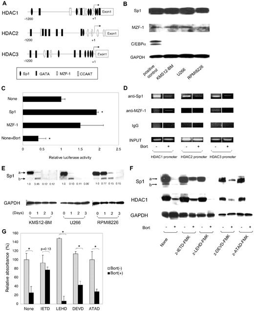
Next, we investigated the mechanisms of transcriptional repression of HDAC genes during bortezomib treatment in MM cells. As illustrated in Figure 3A, there are a number of putative transcription factor binding sites in the promoter regions of class I HDAC genes. We have previously shown that Sp1 and GATA1 are potent transcriptional activators, and MZF-1, GATA2, and C/EBPα are repressors for HDAC1 transcription in myeloid cells.15 We initially screened for the expression of these factors in MM cells, and identified the expression of Sp1 and MZF-1 but not GATA1, GATA2 and C/EBPα (Figure 3B; data not shown). Therefore, we focused on Sp1 and MZF-1 as transcriptional regulators of HDACs in MM cells. Because their binding sites are commonly observed in the promoter regions of 3 HDAC genes, we speculated that HDAC transcription was regulated in a similar manner in MM cells. First, we examined the transcriptional activity of the HDAC1 promoter in KMS12-BM cells by reporter assays using the segment between −1179 and +397 of the HDAC1 gene, which confers full promoter activity.15 As shown in Figure 3C, overexpression of Sp1 significantly increased HDAC1 promoter activity, whereas MZF-1 failed to do so, indicating that Sp1 acts as a transcriptional activator in KMS12-BM cells. In addition, bortezomib was shown to modify the transcriptional activity in this system (Figure 3C). We also examined the transcriptional activity of the HDAC3 promoter in KMS12-BM cells. Reporter assays revealed that promoter activity was enhanced by Sp1 but not MZF-1, suggesting that Sp1 transactivates HDAC3 as well as HDAC1 (data not shown). Furthermore, bortezomib also reduced HDAC3 promoter activity (data not shown). Taken together, Sp1 is a major transcriptional activator of class I HDAC genes in MM cells.
Regulation of HDAC1 promoter by Sp1 transcription factor. (A) Schematic representations of HDAC1, HDAC2, and HDAC3 promoter constructs are shown. Relative locations of the putative binding sites of hematopoietic transcription factors are approximated by the symbols shown in the box. (B) Whole-cell lysates were prepared from MM cell lines and subjected to immunoblotting. HEK293 cells were transduced with expression vectors encoding Sp1, MZF-1, and C/EBPα, and used as positive controls. (C) We transfected 10 μg of pGL4.10 plasmid containing HDAC1 promoter sequences between −1170 and +397 into KMS12-BM cells along with 10 μg of expression vectors encoding Sp1 and MZF-1, and measured luciferase activities after 48 hours. HDAC1 promoter activity was calculated as firefly luciferase activities of cells transfected with an empty expression vector set at 1.0 after normalization of transfection efficiencies using Renilla luciferase activities. Data shown are the means ± SD of 3 independent experiments. P values were calculated by 1-way ANOVA with Student-Newman-Keuls multiple comparisons test. *P < .05. (D) KMS12-BM cells were cultured in the absence or presence of bortezomib for 2 days and subjected to ChIP assays. Chromatin suspensions were immunoprecipitated with the indicated antibodies and corresponding control antibodies. The resulting precipitants were subjected to PCR to amplify the promoter regions of the HDAC genes. The amplified products were visualized by ethidium bromide staining after 2% agarose gel electrophoresis. Representative data of 50 cycles are shown. Input indicates that PCR was performed with genomic DNA. (E) Whole-cell lysates were isolated simultaneously in the experiments described in Figure 2C, and subjected to immunoblotting. Arrows “a” and “b” indicate the intact and cleaved bands of Sp1, respectively. The signal intensities of each band were quantified, normalized to those of the corresponding GAPDH, and shown as relative values setting day 0 controls to 1.0. (F) KMS12-BM cells were cultured with the indicated combinations of 8nM bortezomib (Bort), 100μM z-IETD-FMK (caspase-8 inhibitor), 100μM z-LEHD-FMK (caspase-9 inhibitor), 50μM z-DEVD-FMK (caspase-3 inhibitor), and 20μM z-ATAD-FMK (caspase-12 inhibitor) for 48 hours. Whole-cell lysates were subjected to immunoblotting. (G) Cell viability was determined with a Cell Counting Kit (WAKO) after culturing MM cells in the absence or presence of 8nM bortezomib (Bort) with or without either 100μM z-IETD-FMK (IETD) or 100μM z-LEHD-FMK (LEHD) for 48 hours. Absorbance at 450 nm was measured with a microplate reader, and expressed as a percentage of the value of the corresponding untreated cells. The means ± SD (bars) of 3 independent experiments are shown. P values were calculated by 1-way ANOVA with the Student-Newman-Keuls multiple comparisons test. *P < .05.
Regulation of HDAC1 promoter by Sp1 transcription factor. (A) Schematic representations of HDAC1, HDAC2, and HDAC3 promoter constructs are shown. Relative locations of the putative binding sites of hematopoietic transcription factors are approximated by the symbols shown in the box. (B) Whole-cell lysates were prepared from MM cell lines and subjected to immunoblotting. HEK293 cells were transduced with expression vectors encoding Sp1, MZF-1, and C/EBPα, and used as positive controls. (C) We transfected 10 μg of pGL4.10 plasmid containing HDAC1 promoter sequences between −1170 and +397 into KMS12-BM cells along with 10 μg of expression vectors encoding Sp1 and MZF-1, and measured luciferase activities after 48 hours. HDAC1 promoter activity was calculated as firefly luciferase activities of cells transfected with an empty expression vector set at 1.0 after normalization of transfection efficiencies using Renilla luciferase activities. Data shown are the means ± SD of 3 independent experiments. P values were calculated by 1-way ANOVA with Student-Newman-Keuls multiple comparisons test. *P < .05. (D) KMS12-BM cells were cultured in the absence or presence of bortezomib for 2 days and subjected to ChIP assays. Chromatin suspensions were immunoprecipitated with the indicated antibodies and corresponding control antibodies. The resulting precipitants were subjected to PCR to amplify the promoter regions of the HDAC genes. The amplified products were visualized by ethidium bromide staining after 2% agarose gel electrophoresis. Representative data of 50 cycles are shown. Input indicates that PCR was performed with genomic DNA. (E) Whole-cell lysates were isolated simultaneously in the experiments described in Figure 2C, and subjected to immunoblotting. Arrows “a” and “b” indicate the intact and cleaved bands of Sp1, respectively. The signal intensities of each band were quantified, normalized to those of the corresponding GAPDH, and shown as relative values setting day 0 controls to 1.0. (F) KMS12-BM cells were cultured with the indicated combinations of 8nM bortezomib (Bort), 100μM z-IETD-FMK (caspase-8 inhibitor), 100μM z-LEHD-FMK (caspase-9 inhibitor), 50μM z-DEVD-FMK (caspase-3 inhibitor), and 20μM z-ATAD-FMK (caspase-12 inhibitor) for 48 hours. Whole-cell lysates were subjected to immunoblotting. (G) Cell viability was determined with a Cell Counting Kit (WAKO) after culturing MM cells in the absence or presence of 8nM bortezomib (Bort) with or without either 100μM z-IETD-FMK (IETD) or 100μM z-LEHD-FMK (LEHD) for 48 hours. Absorbance at 450 nm was measured with a microplate reader, and expressed as a percentage of the value of the corresponding untreated cells. The means ± SD (bars) of 3 independent experiments are shown. P values were calculated by 1-way ANOVA with the Student-Newman-Keuls multiple comparisons test. *P < .05.
Next, we performed ChIP assays to investigate the binding of these transcriptional regulators to HDAC promoters in vivo and their changes during bortezomib treatment. We detected the binding of Sp1 to HDAC1, HDAC2, and HDAC3 promoter regions in untreated KMS12-BM cells, whereas Sp1 dissociated from all 3 promoters upon bortezomib treatment (Figure 3D). Although MM cells expressed MZF-1, we could not detect its binding to promoters. These results suggest that Sp1 confers the baseline expression of HDAC genes.
To clarify the mechanisms of changes in promoter binding, we detected the expression of Sp1 protein in MM cell lines during bortezomib treatment. As shown in Figure 3E, bortezomib markedly down-regulated the expression of Sp1 in all 3 MM cell lines in a time-dependent fashion. In addition, we found that an extra signal (indicated by arrow “b” in Figure 3E) appeared below the major signal of Sp1 protein (indicated by arrow “a” in Figure 3E) in bortezomib-treated cells. Previous studies showed that Sp1 protein was cleaved and degraded by caspases such as caspase-8, caspase-9, and caspase-3,38 and bortezomib activated caspase-8, caspase-9, caspase-3, and caspase-12.39-41 On this basis, we hypothesized that the extra signal was caspase-cleaved Sp1 protein. To confirm this hypothesis, we cultured KMS12-BM cells with peptide inhibitors of caspase-8 (z-IETD-FMK), caspase-9 (z-LETD-FMK), caspase-3 (z-DEVD-FMK), and caspase-12 (z-ATAD-FMK) in the absence or presence of bortezomib, and examined the expression of Sp1 protein. As shown in Figure 3F, IETD but not other inhibitors perturbed the bortezomib-induced down-regulation of Sp1. In KMS12-BM cells, caspase-8 activation occurred 12 hours after bortezomib exposure, which was accompanied by reciprocal down-regulation of Sp1 expression (supplemental Figure 4). These results suggest that caspase-8 is responsible for the cleavage and degradation of Sp1 protein in bortezomib-treated cells. In parallel with the restored Sp1 expression, the caspase-8 inhibitor blocked the down-regulation of HDAC expression in KMS12-BM cells. In addition, IETD but not other caspase inhibitors could abrogate bortezomib-induced cytotoxicity in KMS12-BM cells in a dose-dependent fashion (Figure 3G; supplemental Figure 5). These results suggest that bortezomib degraded Sp1 protein via the caspase-8–dependent pathways, leading in turn to the transcriptional repression of HDAC genes in MM cells. In line with our observation, Hideshima et al39 reported that caspase-8 is a primary initiating caspase in bortezomib-mediated cell death. Taken together, our results suggest that the caspase-8/Sp1/HDAC axis is a critical pathway for bortezomib-mediated cytotoxicity in MM cells.
Increased cytotoxicity of bortezomib by shRNA/siRNA-mediated knockdown of HDAC1 expression
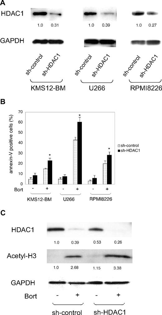
To confirm the roles of HDACs in bortezomib-induced cytotoxicity, we performed loss-of-function analysis using the shRNA/siRNA lentivirus system.30 We could achieve significant loss-of-function of HDACs by solely targeting HDAC1, because HDAC1 represents more than half of all cellular HDAC activities, and other HDACs cannot compensate for the loss of HDAC1.11 We constructed lentiviral shRNA/siRNA expression vectors (supplemental Figure 6A) and transfected them into 3 MM cell lines for further analyses. As shown in Figure 4A, specific reduction of HDAC1 expression was confirmed by Western blotting in GFP+ cells collected by a cell sorter. Upon transduction with shRNA, we determined apoptosis induction in the absence or presence of bortezomib. As shown in Figure 4B, HDAC1 knockdown slightly increased apoptosis in untreated MM cells, but the effect was not statistically significant compared with inactive sh-controls. With bortezomib treatment, however, HDAC1 knockdown significantly increased apoptosis in 3 MM cell lines; this effect was observed at various doses of bortezomib (supplemental Figure 7A). Cell proliferation assays and BrdU/7-AAD double staining revealed that HDAC1 knockdown only marginally affected growth rate and cell-cycle patterns (supplemental Figure 7B-C). These results indicate that the consequence of HDAC down-regulation is mainly apoptosis induction rather than growth inhibition. Histone hyperacetylation was enhanced by HDAC1 knockdown in bortezomib-treated RPMI8226 cells (Figure 4C). Therefore, it appears that shRNA-mediated knockdown of HDAC1 increases sensitivity to bortezomib via synergistic inhibition of HDAC activity in MM cells. These results suggest that bortezomib-induced cytotoxicity depends on cellular HDAC activities in MM cells.
Effects of shRNA-mediated knockdown of HDAC1 on bortezomib-induced apoptosis in MM cells. (A) MM cell lines were transfected with either pLL3.7-sh-HDAC1 (sh-HDAC1) or sh-control vector. Whole-cell lysates were prepared from GFP+ cells collected using a FACSAria flow cytometer and subjected to immunoblotting. The signal intensities of each band were quantified, normalized to those of the corresponding GAPDH, and shown as relative values setting the sh-control to 1.0. (B) MM cell lines transfected with shRNA vectors were cultured in the absence or presence of 2nM bortezomib. After 48 hours, MM cells were harvested, stained with annexin-V/APC, and subjected to flow cytometric analysis. The y-axis shows the proportion of annexin-V positivity in the GFP+ fraction. The means ± SD (bars) of 3 independent experiments are shown. P values were calculated by 1-way ANOVA with the Student-Newman-Keuls multiple comparisons test. *P < .05 against the sh-control. (C) shRNA-transduced RPMI8226 cells were cultured in the absence or presence of 2nM bortezomib. After 48 hours, whole-cell lysates were prepared from GFP+ cells collected by the FACSAria flow cytometer, and subjected to immunoblotting. The signal intensities of each band were quantified, normalized to those of the corresponding GAPDH, and shown as relative values setting the untreated sh-control to 1.0.
Effects of shRNA-mediated knockdown of HDAC1 on bortezomib-induced apoptosis in MM cells. (A) MM cell lines were transfected with either pLL3.7-sh-HDAC1 (sh-HDAC1) or sh-control vector. Whole-cell lysates were prepared from GFP+ cells collected using a FACSAria flow cytometer and subjected to immunoblotting. The signal intensities of each band were quantified, normalized to those of the corresponding GAPDH, and shown as relative values setting the sh-control to 1.0. (B) MM cell lines transfected with shRNA vectors were cultured in the absence or presence of 2nM bortezomib. After 48 hours, MM cells were harvested, stained with annexin-V/APC, and subjected to flow cytometric analysis. The y-axis shows the proportion of annexin-V positivity in the GFP+ fraction. The means ± SD (bars) of 3 independent experiments are shown. P values were calculated by 1-way ANOVA with the Student-Newman-Keuls multiple comparisons test. *P < .05 against the sh-control. (C) shRNA-transduced RPMI8226 cells were cultured in the absence or presence of 2nM bortezomib. After 48 hours, whole-cell lysates were prepared from GFP+ cells collected by the FACSAria flow cytometer, and subjected to immunoblotting. The signal intensities of each band were quantified, normalized to those of the corresponding GAPDH, and shown as relative values setting the untreated sh-control to 1.0.
HDAC1 overexpression rescues MM cells from bortezomib-induced apoptosis
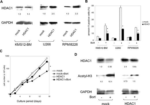
To confirm the dependency of bortezomib action on HDACs, we performed gain-of-function of HDAC1 in MM cells using the lentiviral transduction system (supplemental Figure 6B).27 CSII-DsRed (mock) and CSII-HDAC1-DsRed (HDAC1) vectors were lentivirally transduced into 3 MM cell lines. HDAC1 overexpression was confirmed by Western blotting in DsRed+ cells collected by a cell sorter (Figure 5A). Using this system, we examined the effects of HDAC1 overexpression on bortezomib-induced apoptosis. As shown in Figure 5B, there was no significant difference in the proportion of apoptosis between mock- and HDAC1-transduced cells in the absence of bortezomib. In contrast, bortezomib-induced apoptosis was significantly inhibited by HDAC1 overexpression. The inhibition of apoptosis was observed in increased doses of bortezomib (supplemental Figure 8). However, overexpression of HDAC2 and HDAC3 did not ameliorate the effect of the drug (supplemental Figure 9), suggesting that the HDAC1 levels determine the sensitivity to bortezomib. Next, we investigated the effect of HDAC1 overexpression on the growth of bortezomib-treated cells. As shown in Figure 5C, there was no significant difference in the growth rate between mock- and HDAC1-transduced RPMI8226 cells in the absence of bortezomib. In the presence of bortezomib, the growth of mock-transduced cells was significantly delayed due to the induction of apoptosis. In contrast, HDAC1-transduced cells grew as fast as untreated cells, suggesting that HDAC1 overexpression ameliorates the cytotoxic effects of bortezomib. In addition, we examined the status of histone acetylation in these cells. As anticipated, HDAC1 overexpression markedly diminished bortezomib-induced hyperacetylation of histones (Figure 5D) but not tubulin (supplemental Figure 11). Taken together, HDAC1 overexpression rescued MM cells from bortezomib-induced apoptosis by sustaining HDAC activity in MM cells.
Effects of HDAC1 overexpression on bortezomib-induced apoptosis in MM cells in vitro. (A) MM cell lines were lentivirally transfected with CSII-DsRed (mock) or CSII-DsRed-HDAC1 (HDAC1) vector. Whole-cell lysates were prepared from DsRed+ cells collected using a FACSAria flow cytometer and subjected to immunoblotting. The signal intensities of each band were quantified, normalized to those of the corresponding GAPDH, and shown as relative values with mock-transfected controls setting to 1.0. (B) MM cell lines transfected with mock or HDAC1 vector were cultured in the absence or presence of 2nM bortezomib. After 48 hours, MM cells were harvested, stained with annexin-V/APC, and subjected to flow cytometric analysis. The y-axis shows the proportion of annexin-V positivity in the DsRed+ fraction. The means ± SD (bars) of 3 independent experiments are shown. P values were calculated by 1-way ANOVA with the Student-Newman-Keuls multiple comparisons test. *P < .05 against the mock. (C) RPMI 8226 cells transfected with mock or HDAC1 vector were cultured in the absence or presence of 2nM bortezomib. Total numbers of DsRed+ cells were calculated by flow cytometer at the indicated time points. P values were calculated by 1-way ANOVA with the Student-Newman-Keuls multiple comparisons test. *P < .05 against the mock + Bort. (D) After 48 hours, whole-cell lysates were prepared from DsRed+ cells collected using a FACSAria flow cytometer and subjected to immunoblotting. The signal intensities of each band were quantified, normalized to those of the corresponding GAPDH, and shown as relative values with untreated mock-transfected controls setting to 1.0.
Effects of HDAC1 overexpression on bortezomib-induced apoptosis in MM cells in vitro. (A) MM cell lines were lentivirally transfected with CSII-DsRed (mock) or CSII-DsRed-HDAC1 (HDAC1) vector. Whole-cell lysates were prepared from DsRed+ cells collected using a FACSAria flow cytometer and subjected to immunoblotting. The signal intensities of each band were quantified, normalized to those of the corresponding GAPDH, and shown as relative values with mock-transfected controls setting to 1.0. (B) MM cell lines transfected with mock or HDAC1 vector were cultured in the absence or presence of 2nM bortezomib. After 48 hours, MM cells were harvested, stained with annexin-V/APC, and subjected to flow cytometric analysis. The y-axis shows the proportion of annexin-V positivity in the DsRed+ fraction. The means ± SD (bars) of 3 independent experiments are shown. P values were calculated by 1-way ANOVA with the Student-Newman-Keuls multiple comparisons test. *P < .05 against the mock. (C) RPMI 8226 cells transfected with mock or HDAC1 vector were cultured in the absence or presence of 2nM bortezomib. Total numbers of DsRed+ cells were calculated by flow cytometer at the indicated time points. P values were calculated by 1-way ANOVA with the Student-Newman-Keuls multiple comparisons test. *P < .05 against the mock + Bort. (D) After 48 hours, whole-cell lysates were prepared from DsRed+ cells collected using a FACSAria flow cytometer and subjected to immunoblotting. The signal intensities of each band were quantified, normalized to those of the corresponding GAPDH, and shown as relative values with untreated mock-transfected controls setting to 1.0.
Bortezomib resistance of HDAC1-transduced MM cells in NOD/SCID mice
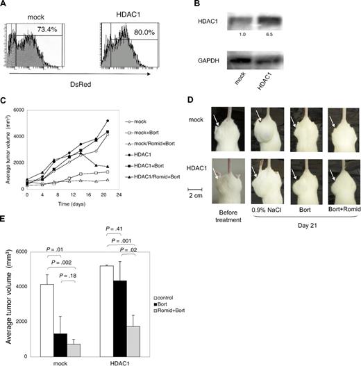
Finally, we confirmed the role of HDACs as target molecules of bortezomib in vivo. To this end, we established RPMI 8226 sublines lentivirally transduced with CSII-DsRed (mock) or CSII-HDAC1-DsRed (HDAC1). Whereas each subline expressed DsRed at equal levels (Figure 6A), HDAC1 was overexpressed solely in the HDAC1-transduced subline (Figure 6B). We inoculated them subcutaneously into nonobese diabetic/severe combined immunodeficiency (NOD/SCID) mice in the right thigh at 3 × 10⁷ cells.31 When measurable tumors developed after 10 to 14 days, mice were assigned to 3 groups: vehicle (0.9% NaCl) control, 0.5 mg/kg bortezomib–treated, and 0.25 mg/kg romidepsin and 0.5 mg/kg bortezomib–treated (n = 3-5 in each group). As shown in Figure 6C, tumor volume constantly increased in vehicle control mice given transplants of both mock and HDAC1 until 21 days. Bortezomib strikingly inhibited tumor growth in mice inoculated with mock-transduced cells, but failed to do so when HDAC1-transduced cells were inoculated. We compared the tumor volume between vehicle-control and bortezomib-treated groups on day 21. As shown in Figure 6D and E, the tumor volume of mock-transduced cells was significantly lower in the bortezomib-treated group than in the vehicle-control group. In contrast, there was no difference in the tumor volume of HDAC1-transduced cells between groups. These results suggest that HDAC1 overexpression confers bortezomib resistance to MM cells in vivo. To confirm the role of HDAC1 in bortezomib resistance, we examined the effects of an HDAC inhibitor on the sensitivity of HDAC1-transduced cells to bortezomib. The addition of romidepsin successfully regressed bortezomib-resistant tumor after 12 days (Figure 6C), and the tumor size was significantly smaller than that of the bortezomib-treated group at 21 days (Figure 6D-E). These results strongly suggest that HDACs are critical targets of bortezomib in vivo.
Effects of HDAC1 overexpression on bortezomib-induced apoptosis in RPMI 8226 cells in vivo. (A) RPMI 8226 cells were transfected with CSII-DsRed (mock) or CSII-DsRed-HDAC1 (HDAC1) vector. DsRed+ cells were collected using a FACSAria flow cytometer and seeded 1 cell/well in a 96-well plate; single-cell clones were then obtained. Each subline was analyzed by a flow cytometer, and representative histogram plots of whole cells are shown. (B) Whole-cell lysates were subjected to immunoblotting. The signal intensities of HDAC1 were quantified, normalized to those of the corresponding GAPDH, and shown as relative values. (C) NOD/SCID mice were inoculated subcutaneously with 3 × 10⁷ cells of RPMI 8226 sublines in the right thigh. The following treatments were started at day 0 when tumors were measurable: bortezomib intravenously twice a week, romidepsin intraperitoneally every other day, and vehicle (0.9% NaCl) alone at the same schedule. Caliper measurements of the longest perpendicular tumor diameters were performed on alternate days to estimate the tumor volume (mm3) using the following formula: 4/3π × (width/2)2 × (length/2). Mice inoculated with mock clones were treated with vehicle alone (mock; ○; n = 5), 0.5 mg/kg bortezomib (mock + Bort; □; n = 4), or 0.5 mg/kg bortezomib and 0.25 mg/kg romidepsin (HDAC1/Romid + Bort; ▵; n = 4). Mice inoculated with HDAC1 clones were treated with vehicle alone (HDAC1; ●; n = 4), 0.5 mg/kg bortezomib (HDAC1 + Bort; ■; n = 3), or 0.5 mg/kg bortezomib and 0.25 mg/kg romidepsin (HDAC1/Romid + Bort; ▲; n = 4). (D) Representative photographs of inoculated NOD/SCID mice at day 21 are shown (original magnification, ×2). Arrowheads indicate inoculated tumors. (E) The y-axis shows the tumor volume in inoculated mice at day 21. The means ± SD (bars) are shown. P values were calculated by 1-way ANOVA with the Student-Newman-Keuls multiple comparisons test.
Effects of HDAC1 overexpression on bortezomib-induced apoptosis in RPMI 8226 cells in vivo. (A) RPMI 8226 cells were transfected with CSII-DsRed (mock) or CSII-DsRed-HDAC1 (HDAC1) vector. DsRed+ cells were collected using a FACSAria flow cytometer and seeded 1 cell/well in a 96-well plate; single-cell clones were then obtained. Each subline was analyzed by a flow cytometer, and representative histogram plots of whole cells are shown. (B) Whole-cell lysates were subjected to immunoblotting. The signal intensities of HDAC1 were quantified, normalized to those of the corresponding GAPDH, and shown as relative values. (C) NOD/SCID mice were inoculated subcutaneously with 3 × 10⁷ cells of RPMI 8226 sublines in the right thigh. The following treatments were started at day 0 when tumors were measurable: bortezomib intravenously twice a week, romidepsin intraperitoneally every other day, and vehicle (0.9% NaCl) alone at the same schedule. Caliper measurements of the longest perpendicular tumor diameters were performed on alternate days to estimate the tumor volume (mm3) using the following formula: 4/3π × (width/2)2 × (length/2). Mice inoculated with mock clones were treated with vehicle alone (mock; ○; n = 5), 0.5 mg/kg bortezomib (mock + Bort; □; n = 4), or 0.5 mg/kg bortezomib and 0.25 mg/kg romidepsin (HDAC1/Romid + Bort; ▵; n = 4). Mice inoculated with HDAC1 clones were treated with vehicle alone (HDAC1; ●; n = 4), 0.5 mg/kg bortezomib (HDAC1 + Bort; ■; n = 3), or 0.5 mg/kg bortezomib and 0.25 mg/kg romidepsin (HDAC1/Romid + Bort; ▲; n = 4). (D) Representative photographs of inoculated NOD/SCID mice at day 21 are shown (original magnification, ×2). Arrowheads indicate inoculated tumors. (E) The y-axis shows the tumor volume in inoculated mice at day 21. The means ± SD (bars) are shown. P values were calculated by 1-way ANOVA with the Student-Newman-Keuls multiple comparisons test.
Discussion
In this study, we have clearly demonstrated that HDACs play a critical role in bortezomib-induced cytotoxicity against MM. The expression of class I HDACs was down-regulated by bortezomib at a transcriptional level via caspase-8–dependent degradation of Sp1 protein, the most potent transactivator of HDACs. As a result, HDAC activities were reduced in MM cells, leading to apoptotic cell death. Because Sp1 is a broadly acting transcription factor, many other target genes should be repressed by Sp1 down-regulation in bortezomib-treated MM cells. Using loss-of-function and gain-of-function analyses, however, we confirmed that sensitivity to bortezomib depends on the levels of HDAC1 expression in MM cells both in vitro and in vivo. This implies that HDACs are critical targets of Sp1 for bortezomib-induced cytotoxicity, although the involvement of other molecules cannot be ruled out. Taken together, our present findings may address the emerging question about the targets of bortezomib,24 add a novel and critical determinant in a list of effector molecules of bortezomib,42 and thus provide a novel rationale for the use of proteasome inhibitors in the treatment of patients with MM.
In support of our findings, recent evidence has suggested a close link between proteasome inhibitors and HDACs. Catley et al43 reported that a hydroxamic acid–derivative NVP-LAQ824, referred to an HDAC inhibitor, affects proteasome activities in MM cells. This compound has a unique ability in overcoming cell adhesion–mediated drug resistance (CAM-DR) of MM cells. Recently, we have shown that VLA-4 is a key molecule of CAM-DR in MM cells, and bortezomib can overcome CAM-DR via the down-regulation of VLA-4 expression.30 However, our preliminary experiments suggested that HDAC inhibitors, such as romidepsin and valproic acid, did not down-regulate VLA-4 expression but reversed CAM-DR in MM cells (data not shown). The characteristics of NVP-LAQ824 may not be like other HDAC inhibitors but rather resemble bortezomib; therefore, it is possible that NVP-LAQ824 inhibits HDAC activity via the down-regulation of HDAC expression. In addition, Miller et al44 reported that a novel proteasome inhibitor NPI-0052 induced caspase-8–dependent histone acetylation in leukemia cells. They demonstrated that ubiquitinated histones did not accumulate in response to NPI-0052, indicating that histone hyperacetylation is not solely due to the accumulation of acetylated histones. It is likely that NPI-0052 induces HDAC down-regulation and histone hyperacetylation via the same mechanism as bortezomib, because we have found that bortezomib down-regulated HDAC expression not only in MM cells but also in other hematologic malignant cell lines, such as HL-60 (acute myeloid leukemia), BJA-B (Burkitt lymphoma) and K562 (chronic myelogenous leukemia; data not shown). Therefore, proteasome inhibitors may generally reduce HDAC activity via the down-regulation of HDAC expression in hematologic malignancies. On the other hand, Fotheringham et al45 reported that HR23b, which shuttles ubiquitinated cargo proteins to the proteasome, is a mediator of HDAC inhibitor–induced cytotoxicity. HDAC inhibitors increased the activity of HR23b and provoked abnormal protein turnover, which resulted in the inhibition of proteasome activities by saturating the proteasome. Furthermore, Mitsiades et al5,46 screened for target molecules of the HDAC inhibitor SAHA and bortezomib using DNA microarrays. Several molecules (p21, CXCR4, syndecan-1, IGF-1, cyclin B2, cyclin F, and bcl-2) are commonly influenced by SAHA and bortezomib in MM cells. Therefore, HDAC inhibitors and proteasome inhibitors may exert cytotoxicity through overlapping or redundant pathways.
Previous studies showed that the unfolded protein response is a dominant mechanism of bortezomib-induced cytotoxicity.47 In this study, we have demonstrated that caspase-12 inhibitor, which could inhibit endoplasmic reticulum (ER) stress-induced apoptosis, did not affect bortezomib-induced apoptosis, but caspase-8 inhibitor did (Figure 3G). The caspase-8–mediated pathway is dominant more than the ER stress-mediated pathway in our system. This discrepancy may be attributable to the difference in doses of bortezomib; less than 8nM in our experiments versus more than 10nM in previous studies. Pharmacodynamic studies revealed that serum concentrations of bortezomib decrease to 2 ng/mL (approximately 5nM) or less 4 hours after administration of 1.3 mg/m2 bortezomib.48 Therefore, our data may reflect the cellular events in vivo more accurately, and support the notion that the caspase-8/Sp1/HDAC axis is more important than other pathways.
In recent clinical studies, bortezomib proved effective even in the context of heavily pretreated, relapsed and refractory MM; however, primary and secondary bortezomib resistance occurred in more than 50% of patients.4 The molecular bases of different individual responsiveness to bortezomib remain unclear. Several previous studies have suggested that adaptation or sensitivity to bortezomib depends on the activity of the ubiquitin-proteosome system in MM cells.49,50 We have demonstrated here that the overexpression of HDAC1 induced bortezomib resistance in vitro and in vivo. The current study is the second to demonstrate that adaptation to bortezomib can indeed be achieved in an MM cell line. Because of the variation of HDAC expression levels, the treatment outcome of bortezomib-treated patients may depend on HDAC activities of MM cells. Importantly, we could overcome the resistance to bortezomib by the combination of the HDAC inhibitor romidepsin in murine xenograft models. HDAC inhibitors, such as romidepsin, SAHA, and tubacin, are promising agents for patients with bortezomib-resistant MM for the following reasons: (1) bortezomib specifically down-regulated the expression of class I HDACs; (2) romidepsin inhibited both class I and II HDAC activities; and (3) HDAC6 knockdown enhanced bortezomib-induced apoptosis (supplemental Figure 10). Therefore, bortezomib is an indispensable agent for MM treatment and combination with other drugs, especially HDAC inhibitors, may be the best treatment strategy for MM treatment. Our findings reinforce the potential clinical utility of combining these 2 agents.
An Inside Blood analysis of this article appears at the front of this issue.
The online version of this article contains a data supplement.
The publication costs of this article were defrayed in part by page charge payment. Therefore, and solely to indicate this fact, this article is hereby marked “advertisement” in accordance with 18 USC section 1734.
Acknowledgments
We are grateful to Drs Ralph Mazitschek and Stuart L. Schreiber (Broad Institute of Harvard University and Massachusetts Institute of Technology) for providing tubacin and niltubacin. We thank Ms Akiko Yonekura for excellent technical assistance.
This work was supported in part by the High-Tech Research Center Project for Private Universities: Matching Fund Subsidy from MEXT 2002-2006. J.K., T.W., K.N.-H., and K.M. are winners of the Jichi Medical School Young Investigator Award.
Authorship
Contribution: J.K. designed and performed experiments, analyzed data, and drafted the manuscript; T.W., R.S., M.A., K.M., and M.N. performed experiments; T.I. and Y.K. provided clinical samples; K.N.-H. and K.O. provided materials and critically reviewed the manuscript; and Y.F. designed and supervised research and wrote the manuscript.
Conflict-of-interest disclosure: The authors declare no competing financial interests.
Correspondence: Yusuke Furukawa, Division of Stem Cell Regulation, Center for Molecular Medicine, Jichi Medical University, 3311-1 Yakushiji, Shimotsuke, Tochigi 329-0498, Japan; e-mail: furuyu@jichi.ac.jp.

This feature is available to Subscribers Only
Sign In or Create an Account Close Modal