In this issue of Blood, Winkler and colleagues use blood perfusion to define and characterize 2 distinct HSC populations in the BM, demonstrating that the most primitive HSCs reside in a BM niche with negligible perfusion.1
Over the past decade there has been great interest in in vivo regulatory mechanisms of hematopoietic stem cells (HSCs). While much has been learned regarding both the intrinsic and extrinsic determinants of stem cell function, pinpointing the location of the cells in the adult bone marrow (BM) has been elusive. Studies by different groups have shown that cells of the osteoblast lineage are key components of the HSC niche, correlating with previous studies demonstrating that HSCs are preferentially located at the endosteal surface of bone.2,3 However, the identification of the signaling lymphocyte activation molecule family of receptors as novel markers of HSCs suggested that the majority of these stem cells were actually positioned adjacent to cells of the endothelial lineage, thus implicating the vascular niche.4 The relevance of each niche, and its specific role in regulating HSC number and function has since been extensively discussed in the literature with no conclusive evidence for differing roles of these niches.5 Complicating matters further was a recent report suggesting that in some cases, the endosteal and vascular HSC niches may in fact be indistinct, and that HSCs are actually located adjacent to both osteoblastic and endothelial cells.6
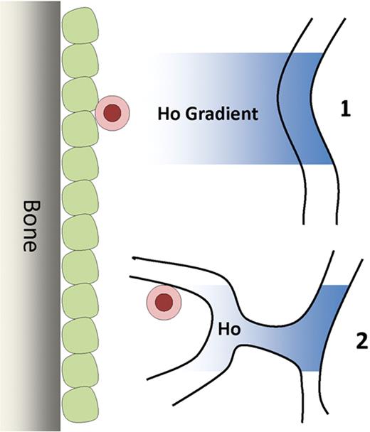
The most primitive hematopoietic stem cells are located in regions with the lowest blood perfusion, suggesting that these cells are either located in regions removed from vascular structures (1), or alternatively, the stem cells may be located next to specialized endothelial cells that do not allow the passive diffusion of various factors (2).
The most primitive hematopoietic stem cells are located in regions with the lowest blood perfusion, suggesting that these cells are either located in regions removed from vascular structures (1), or alternatively, the stem cells may be located next to specialized endothelial cells that do not allow the passive diffusion of various factors (2).
In this article, Winkler and colleagues1 used the properties of blood perfusion to define these potentially distinct niches. Specifically, the authors used the diffusion of the Hoechst 33342 (Ho) DNA dye after intravenous injection to identify the location of the HSCs relative to the perfusion of the dye in vivo. This technique had been previously used by Parmar and colleagues7 where they recognized that primitive HSCs reside in regions of the BM that are the least perfused by the Ho dye and have the lowest levels of oxygenation, suggesting that hypoxia may play a key role in the maintenance of stem cell function in the BM. Winkler and colleagues made an important advance using this technique to show that the HSCs could actually by subdivided into 2 distinct subpopulations dependent upon Ho uptake. The authors found that the cells resident in those areas least perfused by the Ho dye (Honeg) had the greatest degree of stem cell activity compared with those phenotypically defined HSCs resident in areas that were perfused to a slightly higher degree (Homed cells). The authors made 2 other interesting observations. First, during granulocyte colony-stimulating factor–induced mobilization, HSCs were found to be localized in regions with increased blood perfusion. These results suggested that the HSCs migrated from their hypoxic microenvironment to a location more accessible to vascular perfusion, possibly a perivascular location, where the cells then enter into the circulation. Second, as expected, immunophenotypically identified endothelial cells were located near the areas of highest blood perfusion, whereas osteoblastic cells were located in regions of negligible perfusion. However, interestingly, cells identified as mesenchymal stromal cells (CD45−Lin−CD31−Sca-1+CD51+) were actually located in areas of highest blood perfusion, again presumably perivascularly.
While not conclusively pinpointing the location of the most primitive HSCs in the adult BM, this article markedly narrows down the search. Their data confirm previous reports that used a more controversial method of identifying HSCs to suggest that the hematopoietic cells with the slowest turnover, potentially primitive HSCs, are located in a hypoxic zone, removed from capillary structures.8 The question of the relative role of the endosteal niche versus the vascular niche remains open. The most primitive stem cells may in fact not be present next to vascular structures. However, endothelial cells may control perfusion (particularly oxygen) to regulate HSC function, or the HSCs may be located next to vessels where the blood flow is so low that diffusion is very poor (see figure). This would suggest that there may be specialized vascular structures that comprise the vascular niche.
Conflict-of-interest disclosure: The author declares no competing financial interests. ■
REFERENCES
National Institutes of Health


This feature is available to Subscribers Only
Sign In or Create an Account Close Modal