Abstract
DNA-damaging chemotherapy is the backbone of cancer treatment, although it is not clear how such treatments kill tumor cells. In nontransformed lymphoid cells, the combined loss of 2 proapoptotic p53 target genes, Puma and Noxa, induces as much resistance to DNA damage as loss of p53 itself. In Eμ-Myc lymphomas, however, lack of both Puma and Noxa resulted in no greater drug resistance than lack of Puma alone. A third B-cell lymphoma-2 homology domain (BH)3-only gene, Bim, although not a direct p53 target, was up-regulated in Eμ-Myc lymphomas incurring DNA damage, and knockdown of Bim levels markedly increased the drug resistance of Eμ-Myc/Puma−/−Noxa−/− lymphomas both in vitro and in vivo. Remarkably, c-MYC–driven lymphoma cell lines from Noxa−/−Puma−/−Bim−/− mice were as resistant as those lacking p53. Thus, the combinatorial action of Puma, Noxa, and Bim is critical for optimal apoptotic responses of lymphoma cells to 2 commonly used DNA-damaging chemotherapeutic agents, identifying Bim as an additional biomarker for treatment outcome in the clinic.
Introduction
Defects in apoptosis are major contributors to the development of cancer and the impaired response of tumor cells to cancer therapy.1 The apoptotic response to a diverse range of damage signals requires the intrinsic (also called stress, mitochondrial, or B-cell lymphoma [BCL]-2–regulated) pathway, which is controlled by the interaction of pro- and antiapoptotic members of the BCL-2 family. The prosurvival members (BCL-2, BCL-XL, BCL-W, myeloid cell leukemia [MCL]-1, and A1) inhibit apoptosis by keeping the multi-B-cell lymphoma-2 homology (BH) domain proapoptotic BCL-2 family members, BCL-2–associated X protein (BAX) and BCL-2 homologous antagonist/killer (BAK), in check, thereby preventing mitochondrial outer membrane permeabilization (MOMP) and activation of the destructive caspase cascade.2 The proapoptotic BH3-only BCL-2 family members (BIM, PUMA/BBC3, NOXA, BID, BAD, BMF, BlK/BLK/NBK, HRK/DP5) are essential for apoptosis initiation and are transcriptionally and/or posttranslationally activated in a death stimulus– and cell type–specific manner.3 Some BH3-only proteins, including PUMA and BIM, bind avidly to all prosurvival BCL-2–like proteins, whereas others display selective binding activity. For example, NOXA binds only MCL-1 and A1, whereas BAD interacts selectively with BCL-2, BCL-XL, and BCL-W.4 The role of BH3-only proteins for activation of BAX and BAK remains controversial. One view is that BAX/BAK activation is caused by direct binding of BIM, truncated BID (tBID), and, perhaps, PUMA, in a “hit–and–run” mechanism,5 while another holds that they have no direct activating role, but trigger apoptosis by binding to and neutralizing prosurvival BCL-2 family members, thereby liberating BAX and BAK.6
Commonly used chemotherapy drugs, such as cyclophosphamide (CTX) and etoposide, unleash the intrinsic apoptotic pathway by causing DNA damage, thereby activating the tumor suppressor, p53. The BH3-only genes, Puma and Noxa, are direct transcriptional targets of p53, with Puma playing a major, and Noxa a more restricted, role in DNA damage–induced p53-mediated apoptosis of normal (ie, nontransformed) cells.7-12
How DNA-damaging drugs kill malignant cells is currently poorly defined. We have taken a genetic approach to define which BH3-only proteins are required, by crossing gene-targeted mice with Eμ-Myc transgenic mice, in which all animals spontaneously develop clonal pre-B (sIg−) or B-cell (sIg+) lymphomas within 1 year of age.13,14 Unexpectedly, not only Puma and Noxa, but also Bim proved critical for lymphoma cell killing, revealing the importance of understanding the status of all 3 BH3-only proteins for the prediction of drug response. This study has revealed the genetic basis for optimal apoptotic responses to 2 commonly used DNA-damaging chemotherapeutic agents, and suggests that levels of these proteins may be used as biomarkers for therapeutic outcome.
Methods
Materials and expression constructs
Drugs used are described in supplemental Methods (available on the Blood Web site; see the Supplemental Materials link at the top of the online article). Expression constructs for human FLAG-tagged BCL-2,15 the Bim short hairpin RNA (shRNA),16 and a control scrambled shRNA construct17 were described previously.
Experimental animals
All experiments with animals were performed according to the guidelines of the institutional Walter and Eliza Hall Institute of Medical Research Animal Ethics Committee. Generation and genotyping of mice have been described previously: Noxa−/−,9 Puma−/−,9 Bim−/−,18 p53−/−,19 and Eμ-Myc.13,14 For the present study, Eμ-Myc transgenic BH3-deficient strains were generated as described in supplemental Methods. Eμ-Myc lymphomas were generated to lack Bad (CJV), Bim, Bmf, or Bid (kindly provided by Drs A. Egle, A. Villunger, and R. Johnstone, respectively). Mice were generated to lack Noxa, Puma, and Bim (triple knockout) by crossing Noxa−/−Bim−/− double-knockout males with Noxa−/−Puma−/− double-knockout females and then intercrossing Noxa−/−Puma+/−Bim+/− mice. Primer sequences for allele-specific polymerase chain reaction (PCR) genotyping are contained in the supplemental Methods.
Eμ-Myc lymphomas and cell lines
Eμ-Myc lymphomas were characterized by immunophenotype by flow cytometry and defined as either pre-B lymphomas (B220+sIgM−) or B-cell lymphomas (B220+sIgM+). Frozen lymphoma stocks were thawed for either direct transplantation into C57BL/6 recipient mice for expansion and in vivo drug sensitivity analysis or for in vitro culture in order to obtain stable cell lines. Cell lines were generated as described in supplemental Methods.
Flow cytometric analysis
Flow cytometry was performed as detailed previously.9 For cytoplasmic immunofluorescent staining, cells were fixed and permeabilized as described in supplemental Methods, then stained with a monoclonal antibody (mAb), 3C5 (Enzo Life Sciences), to detect BIM. FLAG-tagged proteins were detected by cytoplasmic immunofluorescence staining with anti-FLAG antibody (M2; Sigma-Aldrich) as detailed previously.20
shRNA-mediated knockdown of Bim in lymphomas and cell lines
For retroviral infection of primary lymphoma cells or cell lines, a spin-infection protocol was used,21 with details in supplemental Methods. Retroviral constructs, based on murine stem cell virus (MSCV) Puro internal ribosome entry site/green fluorescent protein (PURO-IRES-GFP) (pM-PIG)22 and MSCV/LTRmiR30-PIG (LMP),22 were used, including 2 independent Bim shRNA hairpin constructs and control constructs, as described in supplemental Methods.
Lymphomas derived by c-Myc retroviral transduction
Day E13.5 fetal liver cells from the relevant gene-targeted mice were infected with a pMIG retroviral construct,23 containing human c-MYC cDNA generated in our laboratory, as described in supplemental Methods. Following infection, cells were transferred onto OP9 stromal cells24 for 48 hours to reduce myeloid differentiation, transplanted, lymphomas harvested, cell lines established, and their genotype confirmed as described in supplemental Methods. Analysis of the Cdkn2a locus and p53 mutation status of lymphoma cell lines was performed as described in supplemental Methods.
Western blotting
Western blotting methods and mAbs used are described in supplemental Methods. Where indicated, for example, for experiments including 18-hour etoposide treatment time points, quinolone-Val-Asp-CH2-difluorophenoxy (QVD-OPH; 25μM) was added to prevent a caspase-mediated degradation of proteins. To date, we have not identified an antibody suitable for the detection of endogenous mouse NOXA protein.
Quantitative reverse transcription (qRT)-PCR
qRT-PCR was performed as described in supplemental Methods. Data analyses were performed by the comparative threshold cycle method.25 qRT-PCR analysis primer sequences are contained in the supplemental Methods.
Cell death assays
Cell death was assessed by flow cytometric analysis as described in supplemental Methods. The extent of apoptosis induced specifically by DNA damage treatment (ie, percent specific apoptosis) was calculated using the following equation: [% apoptosis − % spontaneous apoptosis]/[100 − % spontaneous apoptosis]. Each independent cell line was analyzed in at least 3 independent experiments.
BAX and BAK activation assays
Activation of BAX and BAK was assessed as detailed previously either by following the movement of BAX from the cytosolic to the membrane fraction using subcellular fractionation or by examining the formation of BAX or BAK oligomers using cross-linking assays.26
In vivo drug response following knockdown of Bim in lymphomas
Following retroviral transduction with control pM-PIG or shBim-pM-PIG constructs, primary lymphoma samples were transplanted into Rag-1−/− animals (as the puromycin expression from the pM-PIG vector results in selection against the vector in immunocompetent C57BL/6 mice). Lymphoma cells were harvested from the spleen, transplanted a second time into Rag-1−/− mice in order to further select for lymphoma cells expressing the Bim knockdown construct, and utilized in in vivo treatment studies as described in supplemental Methods. BIM protein levels were analyzed by Western blotting and by cytoplasmic immunofluorescence staining. Lymphomas were utilized in in vivo treatment studies as described in supplemental Methods.
In vivo survival analysis post DNA damaging drug
For drug sensitivity studies, 6-8-week-old C57BL/6 female mice were injected (intravenously) on day 0 with 1-3 × 10⁶ lymphoma cells. When spleens were palpable (usually days 10-16 post-transplantation), mice were randomly assigned to treatment with CTX (200-300 mg/kg; Sigma-Aldrich) or vehicle alone. The mice were culled when deemed unwell (lethargy, tremor, hind-leg paralysis, > 5% weight loss, and palpable tumor) in accordance with ethical guidelines or in the absence of symptoms of lymphoma at 180 days post-treatment. Mice treated with vehicle died within 1-21 days (supplemental Table 1).
Statistical analysis
Prism (Version 5; GraphPad Software) and R (Version 2.9.0; www.r-project.org; accessed September 4, 2009) software were used for statistical analysis. Two-group comparisons were made using 2-tailed t tests assuming equal variances. Trend tests were performed by linear regression. Animal survival data were plotted using Kaplan-Meier curves. Differences in survival time between cohorts of mice bearing shBim-GFP or GFP-control lymphoma were tested using log-rank tests (each independent lymphoma analyzed separately). P values less than .05 were considered to indicate statistical significance.
The in vivo survival experiments (other than those involving shBim knockdown lymphomas) produced data on multiple independent lymphomas per genotype and multiple recipient mice per lymphoma. To ensure that the survival values could be treated as statistically independent and to ensure statistical significance was assessed relative to biological variation between the lymphomas for each genotype, the data were summarized at the lymphoma level. Two approaches were used. In the first, each lymphoma was awarded an ordinal score (0, 1, or 2), depending on whether none, some, or all of the mice injected with that lymphoma survived to 180 days. The genotypes were then compared using proportional odds logistic regression models and likelihood ratio tests.27 This approach was used to assess statistical significance between the survival curves (see Figures 2–3 and supplemental Figure 3).
In the second approach, a Weibull survival model28 was used to predict a median survival time for each lymphoma. These median survival times were then converted to ranks and presented in box plots (supplemental Figure 4). This approach was used to test whether the survival times tend to be ranked by genotype in the hypothesized order. Pairwise comparisons between groups of genotypes were made using Wilcoxon tests. An overall test for trend was conducted by correlating the ranked survival times with the hypothesized ordering and computing a P value by permuting the genotype labels 100 000 times.
Results
To assess the contribution of individual proapoptotic BH3-only proteins in the response of lymphoid tumors to therapeutic agents that induce DNA damage, we have collected a panel of primary pre-B and B-cell lymphomas from Eμ-Myc mice or Eμ-Myc mice generated to lack one or more BH3-only genes. Primary lymphomas were cultured in vitro to generate stable cell lines (hereafter termed Eμ-Myc cell lines) and/or expanded by transplantation into multiple syngeneic recipients and cryopreserved for subsequent in vivo studies of sensitivity to therapeutic regimes.
In vitro response of Eμ-Myc cell lines to DNA damaging drugs
Eμ-Myc cell lines were assessed for sensitivity in vitro to the DNA damaging drugs, etoposide (Figure 1A) or 4-hydroperoxy-cyclophosphamide (4-OOH-CTX) (supplemental Figure 1). The majority (13/16) were sensitive, with apoptosis being evident within 4 hours of treatment and associated with cleavage and activation of caspases-9, -3, and -7 (eg, Figure 1C). Apoptosis could be blocked by retrovirus-mediated overexpression of BCL-2 (Eμ-Myc/rvBCL-2 cells; Figure 1A) or by the caspase inhibitor, QVD-OPH (eg, Figure 1B). The resistance of the other 3 cell lines correlated with impairment of the p19Arf/p53 pathway, as evidenced by the failure to up-regulate the p53 targets, Noxa and Puma (data not shown29 ). As expected,30 cell lines generated from Eμ-Myc lymphomas lacking p53 (Eμ-Myc/p53−/−; see the Methods) proved highly resistant to etoposide- and 4-OOH-CTX–induced apoptosis (Figure 1A, supplemental Figure 1). In the sensitive cell lines, caspase processing was preceded by activation of BAX/BAK, as evidenced by changes in conformation and subcellular localization and by the cytosolic release of cytochrome c (eg, Figure 1D). Collectively, these data demonstrate that DNA damage triggered by etoposide (Figure 1), 4-OOH-CTX (supplemental Figure 1), or γ-irradiation (data not shown) kills Eμ-Myc cell lines via the intrinsic apoptotic pathway, with p53 playing a major role.
Response of Eμ-Myc cell lines to DNA-damaging drugs in vitro. (A) Eμ-Myc cell lines with functional p53 (Eμ-Myc, n = 6 independently derived lymphomas; black circles), harboring either mutant p53 (Eμ-Myc/p53 mutant, n = 3; red diamonds) or a targeted p53-deleted allele (with subsequent loss of heterozygosity of wild-type allele) (Eμ-Myc/p53−/−, n = 4; red triangles), or retrovirally overexpressing BCL-2 (Eμ-Myc/rvBCL-2, n = 3; blue squares) were left untreated or treated for 24 hours with etoposide (0-1 μg/mL). Cell death expressed as percent specific apoptosis. Mean ± SEM from 3-6 independent cell lines for each genotype, from 3 independent experiments per cell line. (B) Eμ-Myc/p53+/+ cell line AF47-T2 treated with etoposide (1 μg/mL) in the presence or absence of QVD-OPH (25μM) with cell death assessed at 2, 4, and 6 hours. Mean ± SEM from 3 independent experiments. Similar results for 2 further Eμ-Myc/p53+/+ cell lines (AH29 and AH54). (C) The Eμ-Myc/p53+/+ cell line, AF47-T2, was treated with etoposide (E; 1 μg/mL) in the presence or absence of QVD-OPH (QVD, 25μM) for 6 hours, prior to the assessment for caspase processing by Western blot analysis. Comparable results for the Eμ-Myc cell line AH29. NT = not treated. (D) Subcellular redistribution assay demonstrating, for Eμ-Myc cell line AF47-T2, the movement of BAX to the mitochondrial fraction (“M”) and concomitant translocation of cytochrome c from mitochondria into the cytosol (“C”) after treatment with etoposide (1 μg/mL) for 6 hours. BAK and HSP70 provide loading controls. Comparable results were obtained with the Eμ-Myc cell line, AH29. (E) Eμ-Myc/p53+/+ and Eμ-Myc/p53−/− cell lines (n = 3 for each genotype) were treated with etoposide (1 μg/mL) for 3 hours and assessed by RT-PCR for BH3-only gene mRNA expression. Mean fold change ± SEM from 3 independent experiments. (F) Eμ-Myc/p53+/+ and Eμ-Myc/p53−/− cell lines (n = 2 for each genotype) were treated with etoposide (1 μg/mL) for 6 hours and assessed by Western-blot analysis for levels of PUMA and p53 proteins.
Response of Eμ-Myc cell lines to DNA-damaging drugs in vitro. (A) Eμ-Myc cell lines with functional p53 (Eμ-Myc, n = 6 independently derived lymphomas; black circles), harboring either mutant p53 (Eμ-Myc/p53 mutant, n = 3; red diamonds) or a targeted p53-deleted allele (with subsequent loss of heterozygosity of wild-type allele) (Eμ-Myc/p53−/−, n = 4; red triangles), or retrovirally overexpressing BCL-2 (Eμ-Myc/rvBCL-2, n = 3; blue squares) were left untreated or treated for 24 hours with etoposide (0-1 μg/mL). Cell death expressed as percent specific apoptosis. Mean ± SEM from 3-6 independent cell lines for each genotype, from 3 independent experiments per cell line. (B) Eμ-Myc/p53+/+ cell line AF47-T2 treated with etoposide (1 μg/mL) in the presence or absence of QVD-OPH (25μM) with cell death assessed at 2, 4, and 6 hours. Mean ± SEM from 3 independent experiments. Similar results for 2 further Eμ-Myc/p53+/+ cell lines (AH29 and AH54). (C) The Eμ-Myc/p53+/+ cell line, AF47-T2, was treated with etoposide (E; 1 μg/mL) in the presence or absence of QVD-OPH (QVD, 25μM) for 6 hours, prior to the assessment for caspase processing by Western blot analysis. Comparable results for the Eμ-Myc cell line AH29. NT = not treated. (D) Subcellular redistribution assay demonstrating, for Eμ-Myc cell line AF47-T2, the movement of BAX to the mitochondrial fraction (“M”) and concomitant translocation of cytochrome c from mitochondria into the cytosol (“C”) after treatment with etoposide (1 μg/mL) for 6 hours. BAK and HSP70 provide loading controls. Comparable results were obtained with the Eμ-Myc cell line, AH29. (E) Eμ-Myc/p53+/+ and Eμ-Myc/p53−/− cell lines (n = 3 for each genotype) were treated with etoposide (1 μg/mL) for 3 hours and assessed by RT-PCR for BH3-only gene mRNA expression. Mean fold change ± SEM from 3 independent experiments. (F) Eμ-Myc/p53+/+ and Eμ-Myc/p53−/− cell lines (n = 2 for each genotype) were treated with etoposide (1 μg/mL) for 6 hours and assessed by Western-blot analysis for levels of PUMA and p53 proteins.
Transcriptional activation of BH3-only genes was assessed by qRT-PCR analysis in Eμ-Myc cell lines treated for 3 hours with etoposide. Puma and Noxa showed robust induction, except in the p53-mutant lines (Figure 1E). An increase in PUMA protein was also observed, coincident with the increase in p53 protein (Figure 1F; no suitable antibodies to mouse NOXA are available). Thus, Puma and Noxa are activated by DNA damage in Eμ-Myc cell lines, and their induction is p53 dependent.
Eμ-Myc cell lines require Puma for optimal response to DNA damage–inducing drugs
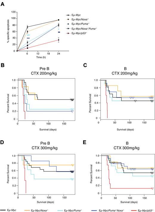
To directly test the role of Puma and Noxa in the DNA damage response of Eμ-Myc lymphomas in vitro, we analyzed cell lines derived from multiple independent lymphomas arising in Eμ-Myc/Puma−/− or Eμ-Myc/Noxa−/− mice. Puma deficiency resulted in a significant resistance of Eμ-Myc cell lines to etoposide (Figure 2A), 4-OOH-CTX (supplemental Figure 1), and γ-irradiation (data not shown), although the resistance was not as marked as that seen with p53 loss. In contrast, Eμ-Myc/Noxa−/− cell lines were almost as sensitive as conventional Eμ-Myc cell lines (Figure 2A). Furthermore, the concomitant lack of both Puma and Noxa (Eμ-Myc/Puma−/−Noxa−/−) did not afford greater protection than did the loss of Puma alone (Figure 2A; Table 1). Similar results were obtained for cell lines from both pre-B and B-cell lymphomas for all these genotypes (supplemental Figure 2B).
Loss of Puma and combined loss of Puma and Noxa promotes resistance to etoposide-induced apoptosis, but to a lesser extent than p53 deficiency. (A) Eμ-Myc cell lines of the indicated genotypes were treated with etoposide (0.2 μg/mL) for the indicated times in vitro, and cell death was assessed by flow cytometry. Apoptosis of Eμ-Myc/Puma−/− cells was significantly lower than for control Eμ-Myc cells at 6 and 24 hours (P < .0001 and P < .05, respectively). Similar protection was observed for Eμ-Myc/Noxa−/−Puma−/− cells (P < .0001 and P < .001). ***P < .0001; **P < .001; *P < .05. Data represent mean ± SEM from 3-7 independent Eμ-Myc cell lines for each genotype, with each cell line analyzed in at least 3 independent experiments. (B-E) Response of primary Eμ-Myc lymphomas to CTX treatment in vivo: Kaplan-Meier survival curves of mice transplanted (day 0) with Eμ-Myc lymphomas and treated, once lymphomas became palpable, from around d12. Pre-B-cell (B) or B-cell (C) Eμ-Myc lymphomas of the relevant genotypes, including those lacking Noxa and/or Puma or p53, were treated with 200 mg/kg CTX. (D-E) Same as for (B-C), but treated with 300 mg/kg CTX. Data were pooled from 3-13 independent lymphomas (supplemental Table 1) of each of 7 genotypes and 2 immunophenotypes (total 276 independently derived lymphomas); 2-14 recipient mice per treatment per independent lymphoma.
Loss of Puma and combined loss of Puma and Noxa promotes resistance to etoposide-induced apoptosis, but to a lesser extent than p53 deficiency. (A) Eμ-Myc cell lines of the indicated genotypes were treated with etoposide (0.2 μg/mL) for the indicated times in vitro, and cell death was assessed by flow cytometry. Apoptosis of Eμ-Myc/Puma−/− cells was significantly lower than for control Eμ-Myc cells at 6 and 24 hours (P < .0001 and P < .05, respectively). Similar protection was observed for Eμ-Myc/Noxa−/−Puma−/− cells (P < .0001 and P < .001). ***P < .0001; **P < .001; *P < .05. Data represent mean ± SEM from 3-7 independent Eμ-Myc cell lines for each genotype, with each cell line analyzed in at least 3 independent experiments. (B-E) Response of primary Eμ-Myc lymphomas to CTX treatment in vivo: Kaplan-Meier survival curves of mice transplanted (day 0) with Eμ-Myc lymphomas and treated, once lymphomas became palpable, from around d12. Pre-B-cell (B) or B-cell (C) Eμ-Myc lymphomas of the relevant genotypes, including those lacking Noxa and/or Puma or p53, were treated with 200 mg/kg CTX. (D-E) Same as for (B-C), but treated with 300 mg/kg CTX. Data were pooled from 3-13 independent lymphomas (supplemental Table 1) of each of 7 genotypes and 2 immunophenotypes (total 276 independently derived lymphomas); 2-14 recipient mice per treatment per independent lymphoma.
Response of Eμ-Myc lymphomas of different genotypes to DNA-damaging drugs in vitro and in vivo
| . | Etoposide response in vitro* . | Cyclophosphamide response in vivo . | |||
|---|---|---|---|---|---|
| Pre-B lymphomas . | B-cell lymphomas . | ||||
| 200 mg/kg . | 300 mg/kg . | 200 mg/kg . | 300 mg/kg . | ||
| Noxa−/− | S | S | S | S | S |
| Puma−/− | M | M | S† | S | S |
| Noxa−/−Puma+/− | S | S | S | S | S |
| Noxa−/−Puma−/− | M | M | S† | S | S |
| Bim−/− | S | S | S | (M)‡ | S |
| p53−/− | R | R | R | R | R |
S indicates similar sensitivity to that seen for control (wild-type) Eμ-Myc lymphomas; M, midresistant, compared with control Eμ-Myc lymphomas; R, resistant, as seen for p53 loss. See Figures 2A-E, 3C-E, and supplemental Figure 3A-D.
Similar responses were observed in vitro for lymphoma cell lines of Pre-B and B-cell phenotype.
Significant resistance (P < .05 relative to control Eμ-Myc lymphomas) was observed with the loss of Puma; however, even with a concomitant loss of Noxa, this was not as severe as the resistance seen with the loss of p53. Pre-B lymphomas were more dependent on Puma, whereas B-cell lymphomas showed a trend toward dependence on Bim for optimal drug response (P < .08).
Relative to Eμ-Myc lymphomas lacking Puma, those lacking both Puma and Noxa were relatively more sensitive to CTX in vivo when treated with 300 mg/kg (see also Figure 2D; ordinal regression, P = .04).
Lack of Puma confers resistance in Eμ-Myc pre-B lymphomas to DNA damaging drugs in vivo
In order to analyze the contribution of BH3-only proteins to the response of Eμ-Myc lymphomas to DNA damaging drugs in vivo, primary tumors were transplanted into multiple immunocompetent (ie, nonirradiated) C57BL/6-recipient mice and, once tumors became palpable (generally after 12 days), the mice were treated with CTX at either 200 mg/kg body weight or 300 mg/kg (the maximum tolerated dose; MTD). Kaplan-Meier survival curves are shown in Figure 2B-E (see also supplemental Table 1). Pre-B (Figure 2B,D) and B-cell (Figure 2C,E) lymphomas were analyzed separately, because loss of BH3-only proteins can affect the proportion of pre-B versus B-cell lymphomas that develop in Eμ-Myc mice,29,31 and human lymphomas of different subtypes are known to differ in response to treatment.32 CTX treatment significantly extended the survival of mice transplanted with Eμ-Myc lymphomas that were wild type for Noxa and Puma (black curves) (median survival of 176 days post CTX 300 mg/kg, compared with 4 days for mice treated with vehicle alone; data not shown). All Eμ-Myc lymphomas lacking p53 (red curves) were highly resistant to CTX therapy, however, and died within 25 days of the tumors becoming palpable. For the pre-B lymphomas (Figure 2B,D), lack of Noxa (orange curves) had no impact, but the lack of Puma (light blue curves) conferred significant resistance—the median survival of mice bearing Eμ-Myc/Puma−/− pre-B lymphomas treated with 200 mg/kg CTX was only 23.5 days, as compared with 62 days for those bearing control Eμ-Myc lymphomas (Figure 2B; supplemental Table 1). Ordinal regression analysis confirmed the significance of this result (P = .04; supplemental Table 2). In contrast, for B-cell lymphomas (Figure 2C,E), neither loss of Noxa nor loss of Puma provided resistance (Table 1).
As anticipated from the in vitro analysis, pre-B lymphomas lacking both Puma and Noxa were no more resistant than those lacking Puma alone (median survival 22 and 23.5 days, respectively, following treatment with 200 mg/kg of CTX; Figure 2B; supplemental Table 1). By regression analysis, the relative resistance of these Puma-deficient Eμ-Myc tumors, with or without Noxa, was intermediate between p53−/− and all other genotypes (P = 1e−5; supplemental Figure 4; supplemental Table 1). Most unexpectedly, however, at the higher dose of CTX (300 mg/kg), pre-B lymphomas lacking both Puma and Noxa were markedly more sensitive than lymphomas lacking only Puma (median survival 177 vs. 42 days, Figure 2D; ordinal regression, P = .04; permutation test, P = .004). Thus, at the MTD of CTX, DNA damage–induced apoptosis can still proceed efficiently in vivo, despite the absence of both Puma and Noxa. This outcome stands in marked contrast to that obtained in vitro for Eμ-Myc pre-B lymphomas (see above) and in vivo for nontransformed thymocytes, where the combined loss of both Puma and Noxa afforded more profound resistance to whole-body γ-irradiation than loss of Puma alone.29
Bim contributes to cyclophosphamide-induced killing of B lymphomas in vivo
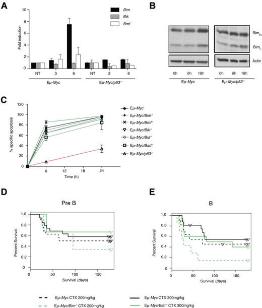
In view of these unexpected results, we investigated whether other BH3-only proteins contribute to DNA damage–induced apoptosis of Eμ-Myc cell lines. In control Eμ-Myc cell lines, the level of Bim mRNA increased ∼7-fold after 6 hours of exposure to etoposide, after a negligible change at 3 hours (Figure 3A; supplemental Figure 5A). Thus, the kinetics of Bim induction were slower than for Puma and Noxa, which were clearly elevated at 3 hours (Figure 1E). A modest increase in BIM protein was also observed at 18 hours (Figure 3B). In order to determine whether the induction of Bim mRNA was p53 dependent, 3 independent Eμ-Myc cell lines were retrovirally infected with shp53 or control expression vector, resulting in the impaired induction of p53 protein and p53 target genes (p21 and Mdm2) following etoposide treatment. In 2 of the 3 cell lines, we also observed diminished Bim induction following exposure to etoposide (supplementary Figure 5). Presumably, residual p53 expression was still capable of inducing Bim. Indeed, the increase in Bim mRNA was not detected in Eμ-Myc/p53−/− cell lines (Figure 3A), suggestive of a potential involvement for p53 in DNA damage–elicited Bim induction. Presumably, the role of p53 must be indirect because Bim has not been identified as a direct target of p53 in a whole genomewide screen.33
Bim contributes to DNA-damage response of Eμ-Myc lymphomas in vivo. (A) Eμ-Myc cell lines or Eμ-Myc cell lines lacking p53 were treated with etoposide (1 μg/mL) for 3 or 6 hours and assessed for the levels of BH3-only gene mRNA. Mean ± SEM from 3 independent experiments. (B) Eμ-Myc cell lines or Eμ-Myc cell lines lacking p53 were left untreated or treated with etoposide (1 μg/mL) in the presence of QVD-OPH (25μM) for 6 or 18 hours and assessed for the levels of BIM protein by Western blotting, with β-actin serving as a loading control. (C) Cell lines derived from independent Eμ-Myc lymphomas of the indicated genotypes (lacking Bik, Bad, Bim, Bid, Bmf, or p53) were treated with etoposide (0.2 μg/mL) for the indicated times in vitro and cell death was assessed by flow cytometry. Mean ± SEM from 3-7 independent cell lines per genotype, with each cell line analyzed in at least 3 independent experiments. No significant differences were noted, except for the Eμ-Myc/p53−/− cell lines. (D-E) Response of Eμ-Myc lymphomas to CTX in vivo, shown as Kaplan-Meier survival curves. Mice were transplanted with Eμ-Myc lymphoma cells (d0) and treated, once spleens were palpable, from around d12. Pre-B (D) or B-cell (E) Eμ-Myc and Eμ-Myc/Bim−/− lymphomas were treated with 200 or 300 mg/kg CTX. Data were pooled from 2-13 independent lymphomas of each genotype; 2-10 recipient mice per treatment per independent lymphoma (supplemental Table 1).
Bim contributes to DNA-damage response of Eμ-Myc lymphomas in vivo. (A) Eμ-Myc cell lines or Eμ-Myc cell lines lacking p53 were treated with etoposide (1 μg/mL) for 3 or 6 hours and assessed for the levels of BH3-only gene mRNA. Mean ± SEM from 3 independent experiments. (B) Eμ-Myc cell lines or Eμ-Myc cell lines lacking p53 were left untreated or treated with etoposide (1 μg/mL) in the presence of QVD-OPH (25μM) for 6 or 18 hours and assessed for the levels of BIM protein by Western blotting, with β-actin serving as a loading control. (C) Cell lines derived from independent Eμ-Myc lymphomas of the indicated genotypes (lacking Bik, Bad, Bim, Bid, Bmf, or p53) were treated with etoposide (0.2 μg/mL) for the indicated times in vitro and cell death was assessed by flow cytometry. Mean ± SEM from 3-7 independent cell lines per genotype, with each cell line analyzed in at least 3 independent experiments. No significant differences were noted, except for the Eμ-Myc/p53−/− cell lines. (D-E) Response of Eμ-Myc lymphomas to CTX in vivo, shown as Kaplan-Meier survival curves. Mice were transplanted with Eμ-Myc lymphoma cells (d0) and treated, once spleens were palpable, from around d12. Pre-B (D) or B-cell (E) Eμ-Myc and Eμ-Myc/Bim−/− lymphomas were treated with 200 or 300 mg/kg CTX. Data were pooled from 2-13 independent lymphomas of each genotype; 2-10 recipient mice per treatment per independent lymphoma (supplemental Table 1).
In vitro, lack of Bim alone had little impact on sensitivity to DNA damage, as cell lines derived from Eμ-Myc/Bim−/− lymphomas were as sensitive as control Eμ-Myc cell lines to etoposide in vitro, as were Eμ-Myc cell lines lacking either Bmf, Bid, Bad, or Bik (Figure 3C). However, mice bearing Eμ-Myc/Bim−/− B-cell lymphomas treated in vivo with 200 mg/kg CTX (dotted green line) showed a trend toward poorer survival (median survival, 37 days), compared with mice bearing control Eμ-Myc B-cell lymphomas (Figure 3E dotted black line; median survival, 49 days; P < .08; supplemental Table 1) and were intermediate between all other genotypes and those lacking p53 (permutation test: P = .004, CTX 200 mg/kg; P = 2e−5, CTX 300 mg/kg). In contrast, a trend for loss of Bim to cause drug resistance was not observed in Eμ-Myc pre-B lymphomas in vivo (Figure 3D). Thus, in vivo, the loss of Bim alone may confer a modest resistance to CTX in Eμ-Myc B-cell lymphomas, but not pre-B lymphomas (Table 1).
Reduction in Bim confers profound resistance to DNA damaging chemotherapy in lymphomas lacking both Puma and Noxa in vitro and in vivo
We next examined the combinatorial roles of Bim, Puma, and Noxa in the response of Eμ-Myc cell lines to DNA damage–inducing cancer therapy. First, mRNA and protein levels of Bim were measured following treatment with etoposide in vitro. A similar induction of Bim mRNA and BIM protein was observed for both control Eμ-Myc and Eμ-Myc/Noxa−/−Puma−/− cell lines (supplemental Figure 6A-B). Thus, loss of Puma and Noxa does not induce obvious up-regulation of Bim.
To explore the impact of the combined loss of Puma, Noxa, and Bim, independent Eμ-Myc/Noxa−/−Puma−/− and control Eμ-Myc cell lines were retrovirally transduced with Bim shRNA or control expression vectors (supplemental Figure 6C). An appropriate reduction in BIM protein levels was confirmed by Western blotting (supplemental Figure 7A). Bim knockdown caused a minor reduction in etoposide-induced killing in control Eμ-Myc cell lines and modest protection in Eμ-Myc cell lines lacking Puma (Figure 4B; P = .009 shBim vs control at 6 hours), but had a greater impact in cell lines lacking both Puma and Noxa, where significantly more protection was observed than in cell lines lacking Puma alone (shBim relative to control vector for Eμ-Myc/Noxa−/−Puma−/− vs. Eμ-Myc/Puma−/− cell lines P = .008 at 6 hours) (Figure 4A-C). These results imply an important combinatorial role for Bim, Puma, and Noxa in the response to etoposide in vitro.
Loss of Bim increases resistance of Eμ-Myc lymphomas lacking both Puma and Noxa to DNA-damaging drugs both in vitro and in vivo. (A) Eμ-Myc/Noxa−/−Puma−/− cell lines, transduced with either shBim-GFP, GFP, or shCaspase-12-GFP retroviral vectors, were treated with etoposide (0.04, 0.2, or 1 μg/mL). Percent of drug-specific apoptosis in cells transduced with the shBim-GFP vector, compared with control vectors, at 6 (left-hand panel) and 24 hours (right-hand panel). Mean ± SEM from 3 independent experiments; representative of 5 Eμ-Myc/Noxa−/−Puma−/− cell lines analyzed. (B) Percent specific apoptosis for Eμ-myc (shown for 2 independently generated lymphomas), Eμ-myc/Puma−/− (derived from n = 4 independently generated lymphomas) and Eμ-myc/Noxa−/−Puma−/− (derived from n = 5 independently generated lymphomas) cell lines retrovirally transduced with a shBim-GFP vector (black bars) or a control-GFP vector (white bars) and treated with etoposide (0.2 μg/mL) for 6 (left-hand panel) or 24 hours (right-hand panel). Mean ± SEM from 4-5 independent cell lines per genotype (for Eμ-myc/Puma−/−, Eμ-myc/Noxa−/−Puma−/−), with each cell line analyzed in at least 3 independent experiments. (C) Data from (B) represented as the percent reduction in apoptosis after 6 hours (left panel) or 24 hours (right panel) of etoposide treatment of lymphoma cell lines transduced with a shBim-GFP vector, compared with that observed for control-GFP vector. Mean ± SEM from 4-5 independent cell lines per genotype (for Eμ-myc/Puma−/−, Eμ-myc/Noxa−/−Puma−/−), with each cell line analyzed in at least 3 independent experiments. (D-F) Primary Eμ-Myc/Noxa−/−Puma−/− lymphomas transduced with shBim-GFP or GFP retroviral vectors were injected into recipient Rag-1−/− mice (see supplemental Figure 6C). When spleens were palpable, mice were treated with CTX (250 mg/kg), monitored, and culled if unwell. Kaplan-Meier curves for 2 independent lymphomas: (D) shBim-GFP vs. GFP-control vector: median survival = 25 days, 20 days respectively, n = 13 mice; P = .0007; (E) shBim-GFP vs. GFP-control vector: median survival 15 and 24.5 days, respectively, n = 10 mice; P < .0001); and (F) shBim-GFP vs. GFP-control vector: median survival = 18 days and 42.5 days, respectively, n = 10 mice, P = .0042.
Loss of Bim increases resistance of Eμ-Myc lymphomas lacking both Puma and Noxa to DNA-damaging drugs both in vitro and in vivo. (A) Eμ-Myc/Noxa−/−Puma−/− cell lines, transduced with either shBim-GFP, GFP, or shCaspase-12-GFP retroviral vectors, were treated with etoposide (0.04, 0.2, or 1 μg/mL). Percent of drug-specific apoptosis in cells transduced with the shBim-GFP vector, compared with control vectors, at 6 (left-hand panel) and 24 hours (right-hand panel). Mean ± SEM from 3 independent experiments; representative of 5 Eμ-Myc/Noxa−/−Puma−/− cell lines analyzed. (B) Percent specific apoptosis for Eμ-myc (shown for 2 independently generated lymphomas), Eμ-myc/Puma−/− (derived from n = 4 independently generated lymphomas) and Eμ-myc/Noxa−/−Puma−/− (derived from n = 5 independently generated lymphomas) cell lines retrovirally transduced with a shBim-GFP vector (black bars) or a control-GFP vector (white bars) and treated with etoposide (0.2 μg/mL) for 6 (left-hand panel) or 24 hours (right-hand panel). Mean ± SEM from 4-5 independent cell lines per genotype (for Eμ-myc/Puma−/−, Eμ-myc/Noxa−/−Puma−/−), with each cell line analyzed in at least 3 independent experiments. (C) Data from (B) represented as the percent reduction in apoptosis after 6 hours (left panel) or 24 hours (right panel) of etoposide treatment of lymphoma cell lines transduced with a shBim-GFP vector, compared with that observed for control-GFP vector. Mean ± SEM from 4-5 independent cell lines per genotype (for Eμ-myc/Puma−/−, Eμ-myc/Noxa−/−Puma−/−), with each cell line analyzed in at least 3 independent experiments. (D-F) Primary Eμ-Myc/Noxa−/−Puma−/− lymphomas transduced with shBim-GFP or GFP retroviral vectors were injected into recipient Rag-1−/− mice (see supplemental Figure 6C). When spleens were palpable, mice were treated with CTX (250 mg/kg), monitored, and culled if unwell. Kaplan-Meier curves for 2 independent lymphomas: (D) shBim-GFP vs. GFP-control vector: median survival = 25 days, 20 days respectively, n = 13 mice; P = .0007; (E) shBim-GFP vs. GFP-control vector: median survival 15 and 24.5 days, respectively, n = 10 mice; P < .0001); and (F) shBim-GFP vs. GFP-control vector: median survival = 18 days and 42.5 days, respectively, n = 10 mice, P = .0042.
To investigate whether Bim reduction could increase drug resistance in vivo, primary Eμ-Myc/Noxa−/−Puma−/− lymphoma cells were stably transduced with a Bim shRNA or control expression vector coexpressing GFP (supplemental Figure 5C). Lymphoma cells infected with a retrovirus expressing shBim, expressed lower levels of BIM protein, as documented by Western blotting and fluorescence-activated cell sorting than did paired GFP-control–infected lymphoma cells (supplemental Figure 7B-C). Three independent Eμ-Myc/Noxa−/−Puma−/− shBim-GFP or paired control GFP-infected lymphomas were transplanted into recipient mice (> 50% GFP-positive cells injected; supplemental Figure 6C), and, once lymphomas were palpable in spleens (around d12), mice were treated with CTX (250 mg/kg) or carrier. All mice bearing Eμ-Myc/Noxa−/−Puma−/− lymphomas transduced with the shBim vector died rapidly due to severe drug resistance (Figure 4D-F). When 100% of shBimGFP-expressing lymphoma-bearing mice had succumbed, the proportion of control GFP-expressing lymphoma-bearing mice remaining alive for all 3 independently derived pairs of lymphomas was 58% (7/12 mice; Figure 4D), 100% (8/8 mice; Figure 4E), and 50% (5/10 mice; Figure 4F). For all 3 independently derived pairs of lymphomas, median survival was impaired by the retroviral induction of shBim, compared with GFP-control (P = .0007, P < .0001, and P = .0042, respectively; see Figure 4D-F). Thus, in vivo studies support a critical combinatorial role for Bim, Puma, and Noxa in promoting apoptosis of lymphoma cells in response to DNA damaging therapy.
Myc-driven lymphomas lacking Puma, Noxa, and Bim are as drug resistant as those lacking p53
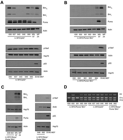
To generate c-Myc–driven B-lymphoid tumors entirely deficient of Noxa, Puma, and Bim, we infected fetal liver cells (FLCs) from Noxa−/−Puma−/−Bim−/− triple-knockout mice with a c-MYC retrovirus and transplanted them into lethally irradiated C57BL/6-Ly5.1 mice. (It is not feasible to generate Eμ-Myc lymphomas lacking Noxa, Puma, and Bim by conventional breeding due to fertility issues and early lethality of intermediate genotypes.) Once palpable, lymphomas were harvested, cultured to establish cell lines, and phenotyped by flow cytometry (eg, supplemental Figure 7A). Genotypes of primary lymphomas and cell lines were confirmed by Western blot analysis (Figure 5A-C). PUMA and BIM proteins were both absent in relevant genotypes. As expected, no p53 protein was detectable in p53−/− cell lines, which uniformly expressed p19Arf due to loss of negative feedback by p53.34 Stabilized p53 protein was not detected in cell lines from any of the other genotypes, which, together with lack of induction of p19Arf, is suggestive of the presence of wild-type p53 (further addressed in Figure 6).
c-MYC-retrovirus driven lymphomas lacking Puma, Noxa, and Bim. (A) Western blot analysis of PUMA, BIM, p19Arf, p53, and β-actin or heat shock protein 70 (loading controls) of cell lines derived from rv c-MYC–driven lymphomas lacking p53 (A), or lacking both PUMA and BIM (B), or lacking NOXA, PUMA and BIM (C), confirmed allele-specific genotyping data (data not shown). BIM protein was variably expressed in cell lines lacking p53. Immortalized mouse embryo fibroblasts (MEFs) were included as positive controls for p19Arf overexpression. MN97 = Eμ-Myc/Noxa−/− lymphoma was included as a positive control for PUMA expression, and MP24 = Eμ-Myc/Puma−/− lymphoma was included as a negative control for PUMA expression. (D) Genomic PCR for the Cdkn2a locus of rv c-MYC–driven cell lines of the different genotypes: exons 1α, 1β, 2. For 2 of the 5 rv c-MYC–driven Puma−/−Bim−/− cell lines tested, deletion of both exons 1α and 2 was observed, consistent with the loss of p16Ink4a and p19Arf.
c-MYC-retrovirus driven lymphomas lacking Puma, Noxa, and Bim. (A) Western blot analysis of PUMA, BIM, p19Arf, p53, and β-actin or heat shock protein 70 (loading controls) of cell lines derived from rv c-MYC–driven lymphomas lacking p53 (A), or lacking both PUMA and BIM (B), or lacking NOXA, PUMA and BIM (C), confirmed allele-specific genotyping data (data not shown). BIM protein was variably expressed in cell lines lacking p53. Immortalized mouse embryo fibroblasts (MEFs) were included as positive controls for p19Arf overexpression. MN97 = Eμ-Myc/Noxa−/− lymphoma was included as a positive control for PUMA expression, and MP24 = Eμ-Myc/Puma−/− lymphoma was included as a negative control for PUMA expression. (D) Genomic PCR for the Cdkn2a locus of rv c-MYC–driven cell lines of the different genotypes: exons 1α, 1β, 2. For 2 of the 5 rv c-MYC–driven Puma−/−Bim−/− cell lines tested, deletion of both exons 1α and 2 was observed, consistent with the loss of p16Ink4a and p19Arf.
c-MYC retrovirus-driven lymphomas lacking Puma, Noxa, and Bim are as drug resistant as those lacking p53. (A-B) Cell lines derived from rv c-MYC–driven lymphomas of the different genotypes were treated with etoposide (0.04, 0.2, or 1.0 μg/mL for 6 (A) or 24 hours (B). Data represent mean ± SEM from at least 3 independent experiments (each independent cell line represented by an individual line). (C) Induction of p53 protein following DNA damage with etoposide. More p53 protein is evident at 6 hours postexposure to etoposide in rv c-MYC-Noxa−/−Puma−/−Bim−/−, consistent with the fact that cell death is not as great at this time point in this genotype, relative to the rv c-MYC-Puma−/−Bim−/− cell line. (D) Induction of the p53 target genes, p21 and Mdm2, following exposure to etoposide relative to untreated controls. Data for individual lymphoma cell lines represent mean ± SEM 3 independent experiments).
c-MYC retrovirus-driven lymphomas lacking Puma, Noxa, and Bim are as drug resistant as those lacking p53. (A-B) Cell lines derived from rv c-MYC–driven lymphomas of the different genotypes were treated with etoposide (0.04, 0.2, or 1.0 μg/mL for 6 (A) or 24 hours (B). Data represent mean ± SEM from at least 3 independent experiments (each independent cell line represented by an individual line). (C) Induction of p53 protein following DNA damage with etoposide. More p53 protein is evident at 6 hours postexposure to etoposide in rv c-MYC-Noxa−/−Puma−/−Bim−/−, consistent with the fact that cell death is not as great at this time point in this genotype, relative to the rv c-MYC-Puma−/−Bim−/− cell line. (D) Induction of the p53 target genes, p21 and Mdm2, following exposure to etoposide relative to untreated controls. Data for individual lymphoma cell lines represent mean ± SEM 3 independent experiments).
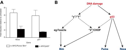
Retrovirally derived c-MYC cell lines (here denoted as retrovirus [rv] c-MYC) lacking both Puma and Bim displayed midresistance to etoposide, similar to that seen for Eμ-Myc cell lines lacking Puma alone (Figure 6A-B and supplemental Figure 8C, compared with Figure 2A). Strikingly, however, both rv c-MYC cell lines lacking Noxa, Puma, and Bim displayed resistance to etoposide comparable to that seen for rv c-MYC cell lines lacking p53 at all concentrations of etoposide and time points tested (Figure 6A-B; supplemental Figure 8A). No increase in the expression of Bcl-2 family prosurvival proteins, BCL-2, BCL-XL, or MCL-1, was detected in rv c-MYC cell lines lacking Noxa, Puma, and Bim (supplemental Figure 8B). Rather, the apparent low expression levels of BCL-2 and MCL-1 protein in rv c-MYC cell lines lacking NOXA, PUMA, and BIM protein may reflect the reduced availability of proapoptotic family members. Importantly, the p53-p19Arf pathway was intact in both cell lines lacking Noxa, Puma, and Bim, as evidenced by an intact Cdkn2a locus (genomic PCR; Figure 5D), the absence of mutation in p53 (determined by sequencing of the p53 gene), and normal induction of both p53 protein and the p53 target genes, p21 and Mdm2, following DNA damage with etoposide (Figure 6C-D). As expected, Noxa mRNA was induced following exposure to etoposide in rv c-MYC cell lines lacking Puma and Bim, but not in those lacking p53 (Figure 7A), indicating that Noxa is available for killing in this less resistant genotype. Taken together, these results show that Puma, Noxa, and, unexpectedly, Bim are all required for optimal apoptotic responses of c-Myc-driven lymphomas to 2 DNA-damaging chemotherapeutic agents commonly used in the clinic.
Regulation of Noxa, Puma, and Bim following DNA damage. (A) rv c-MYC-driven Puma−/−Bim−/− and p53−/− cell lines (derived from n = 3 independent lymphomas per genotype) were treated with etoposide (0.2 μg/mL) for 6 hours in the presence of QVD-OPH (25μM) and assessed for levels of mRNA of the genes indicated. Data represent mean ± SEM from 3 independent experiments. (B) Regulation of Noxa, Puma, and Bim following DNA damage: Regulation of Bim may be p53 dependent or independent. p53 activation directly induces the BH3-only genes, Noxa and Puma. p53 activation (possibly indirect) may also trigger the induction of C/EBP-homologous protein, which, in turn, may elevate Bim. X, Y, and Z represent unidentified potential regulators of Bim transcriptional induction.
Regulation of Noxa, Puma, and Bim following DNA damage. (A) rv c-MYC-driven Puma−/−Bim−/− and p53−/− cell lines (derived from n = 3 independent lymphomas per genotype) were treated with etoposide (0.2 μg/mL) for 6 hours in the presence of QVD-OPH (25μM) and assessed for levels of mRNA of the genes indicated. Data represent mean ± SEM from 3 independent experiments. (B) Regulation of Noxa, Puma, and Bim following DNA damage: Regulation of Bim may be p53 dependent or independent. p53 activation directly induces the BH3-only genes, Noxa and Puma. p53 activation (possibly indirect) may also trigger the induction of C/EBP-homologous protein, which, in turn, may elevate Bim. X, Y, and Z represent unidentified potential regulators of Bim transcriptional induction.
Discussion
Successful anticancer therapy with DNA-damaging chemotherapeutic drugs requires efficient killing of tumor cells.35 This is thought to occur largely through the activation of the intrinsic apoptotic pathway by the tumor suppressor, p53. The precise mechanisms underlying the induction of apoptosis of tumor cells by commonly used DNA-damaging chemotherapeutic drugs and, in particular, the identification of the critical BH3-only proteins have not been resolved. Puma and Noxa are direct transcriptional targets of p53, and their combined loss renders primary thymocytes as resistant to DNA damage as p53 deficiency, both in vitro and in vivo.12 We have shown here that, unexpectedly, however, DNA damage–induced death of lymphoma cells involves not only Puma and Noxa, but also Bim, which is not known to be a direct p53 target.33
Our preclinical mouse model recapitulates certain aspects of human Burkitt lymphoma,36 but is likely more broadly relevant, as many human tumors (∼ 70%) are characterized by abnormally high c-MYC expression levels,37 and c-MYC up-regulation is a feature of a proportion of follicular lymphomas undergoing aggressive transformation to diffuse large B-cell lymphoma (DLBCL).38 Furthermore, this model provides a syngeneic, genetically pliable, and spontaneous tumor model for drug-response analysis and has been extremely useful in establishing other key paradigms in lymphoma biology21,39-43
We compared the responsiveness of multiple independent c-Myc–driven murine lymphomas lacking 1, 2, or all 3 of the genes encoding Puma, Noxa, and Bim, to DNA damage–inducing agents. In vitro, using Eμ-Myc lymphoma-derived cell lines, we found that Puma was the most critical of these 3 BH3-only proteins, since lymphoma cells lacking Puma alone displayed midlevel drug resistance (supplemental Figure 2A), but those lacking only Noxa (Figure 2A) or Bim (Figure 3C) were normally sensitive (Table 1). Interestingly, although pre-B and B-cell lymphomas were comparable in their in vitro responses, pre-B-cell lymphomas were more dependent on Puma in vivo than B-cell (sIgM+) lymphomas for DNA damage–induced apoptosis (compare Figure 2B with 2C). The combined lack of Puma and Noxa did not further increase the resistance of Eμ-Myc pre-B lymphomas to DNA-damaging drugs in vivo, compared with the loss of Puma alone. Indeed, surprisingly, with the loss of both Puma and Noxa, lymphomas became more sensitive to DNA damage at the MTD of CTX (300 mg/kg) (compare Figure 2B with 2D). Thus, most mice transplanted with the Eμ-Myc/Puma−/−Noxa−/− tumors achieved a complete remission (disease free at 180 days post-treatment) at 300 mg/kg CTX, in contrast to the profound drug resistance observed for Eμ-Myc/p53−/− lymphomas and the relatively impaired response of Eμ-Myc/Puma−/− lymphomas treated with this dose (Figure 2D; Table 1). We hypothesized that this could be due to an increase in the availability of other BH3-only protein(s) in lymphoma cells lacking both Puma and Noxa in vivo, perhaps because of a rebalancing of the ratio of prosurvival Bcl-2 family members to BH3-only proteins.
Indeed, surprisingly, the induction of a third BH3-only gene, Bim, was also observed in lymphomas following DNA damage, including those lacking both Puma and Noxa. By introducing Bim shRNA into Eμ-Myc lymphomas lacking both Puma and Noxa and by generating c-MYC retrovirus-induced lymphomas from Noxa−/−Puma−/−Bim−/− triple-knockout mice, we demonstrated that these 3 BH3-only proteins cooperate in killing lymphoma cells both in vitro and in vivo. Our analysis of the combinatorial effects of BH3-only proteins involved more than 300 independently derived primary lymphomas of different genotypes and immunophenotypes, as well as more than 80 independently derived lymphoma cell lines. Remarkably, the extent of drug resistance observed in these triple-knockout lymphomas was as severe as that seen with the loss of p53. These data are striking as Bim has not previously been implicated in the DNA damage response of tumor cells using gene deletion models, although the loss of Bim was observed to afford nontransformed CD4+8+ thymocytes protection, albeit only modest, against γ-irradiation in vitro18 and in vivo.44 p53-mediated effects on Bim are, presumably, indirect, as Bim is not a known direct target of p53,33 and recent studies suggest that potential consensus binding sites for p53 identified in the Bim promoter are actually likely to be specific for p73, rather than p53.45 Indeed, malignant cells may differ from primary nontransformed cells in their response to DNA-damaging drugs as the former, through oncogenic activation of prosurvival signaling pathways, may express a broader range of prosurvival Bcl-2 family members than nontransformed cells (eg, thymocytes). Therefore, the induction of a broader range of BH3-only proteins may be needed for efficient killing of tumor cells.
The outcome of CTX treatment in vivo was dose dependent, with only the higher dose (300 mg/kg, the MTD30,43 ) being sufficient to trigger efficient therapeutic responses in pre-B lymphomas lacking Puma and Noxa (Figure 2D; Table 1). Thus, there may be a threshold level of DNA damage required for the sufficient induction of Bim. The induction of Bim may not occur in vitro to the same extent due to cell line adaptation mechanisms. In vivo, the mechanism may be p53 dependent, at least in part, since Bim mRNA induction post-DNA damage was less apparent in Eμ-Myc cell lines lacking p53 (Figure 3A). The transcription factor, C/EBP-homologous protein/growth-arrest– and DNA-damage–inducible gene 153 (CHOP/GADD153), one of the first defined transcriptional targets of p53,46 has recently been shown to bind to the alternate Bim promoter, leading to its activation in response to endoplasmic reticulum stress.47 Therefore, our data are consistent with the notion that p53 activation (possibly indirectly) triggers the induction of CHOP, which, in turn, results in the elevation of Bim. Other mechanisms may also be involved. For example, c-Myc itself was shown to induce Bim,31,48 although the mechanism remains undefined. Other potential regulators of Bim transcription following DNA damage include the forkhead box O (FoxO) transcription factors, such as FoxO3a, although whether these are p53 dependent or independent reportedly varies according to cell type and status49,50 (reviewed in Fu and Tindall51 ). Thus, p53-dependent and -independent mechanisms of Bim induction in response to DNA damage may exist (Figure 7B). However, the relative contributions of these pathways are likely to vary from tumor to tumor. Investigation of the response of lymphomas lacking both p53 and Bim to DNA damage would be of great interest.
We have shown that combinatorial action of Puma, Noxa, and Bim is required for optimal apoptotic responses of c-Myc–driven lymphomas to DNA damaging chemotherapeutic drugs. In human tumors, mutations of PUMA or NOXA have rarely been documented, but loss of heterozygosity has been described for NOXA and multiple modes of abnormal reduction, including epigenetic silencing, overexpression of inhibitory microRNA, and deletion have been described for PUMA and BIM (reviewed in Happo et al52 ). Our data indicate that inactivation of all 3 genes may result in drug resistance as severe as that seen for the loss of p53 or overexpression of Bcl-218 and thereby demonstrate the importance of understanding the status of these 3 BH3-only proteins for the prediction of drug response.
In conclusion, this study has demonstrated the genetic basis for the efficient induction of apoptosis of lymphoma cells by 2 chemotherapeutic drugs commonly used in the clinic. Our data have revealed an unexpected role for Bim in the response of malignant lymphoma following p53-inducing, DNA-damaging chemotherapy and provide important insights into the relative importance of Puma and Noxa. Similar to the implications of loss of p53 or overexpression of Bcl-2 in a tumor, concomitant loss or inactivation of Noxa, Puma, and Bim in a lymphoma should be considered to be potential biomarkers of drug resistance.
The online version of this article contains a data supplement.
The publication costs of this article were defrayed in part by page charge payment. Therefore, and solely to indicate this fact, this article is hereby marked “advertisement” in accordance with 18 USC section 1734.
Acknowledgments
We are grateful to Drs R. Lindeman, R. Johnstone, A. Egle, A. Villunger, and R. Dickins for provision of lymphomas, constructs, and reagents. We thank Ms Lin Tai for expert technical assistance and all of our colleagues in the Molecular Genetics of Cancer division for their gifts of reagents, scientific advice, and technical support.
This work was supported by fellowships and grants from the National Health and Medical Research Council (Australia; program #257502; R.D. Wright Biomedical CDA 406675), the Leukemia and Lymphoma Society (New York; SCOR grant #7015; CLS Special Fellow Grant 3209-04), the National Cancer Institute (National Institutes of Health; CA 80188 and CA 43540), the Leukemia and Lymphoma Research UK (fellowship to M.S.C.), the DFG (German Science Foundation) (fellowship to M.J.H.), the Leukemia Foundation Australia (PhD scholarship to L.H.), and the University of Melbourne (PhD scholarship for B.P.).
National Institutes of Health
Authorship
Contribution: C.L.S., M.S.C., A.S., and S.C. initiated the studies; L.H., M.S.C., J.H., E.J., and G.D. performed experiments; E.M., C.V., and M.J.H. provided critical reagents and advice; L.H., M.S.C., A.S., B.P., G.S., S.C., and C.L.S. analyzed data; and M.S.C. and C.L.S. wrote the paper, with contributions from S.C. and A.S.
Conflict-of-interest disclosure: The authors declare no competing financial interests.
The current affiliation for M.S.C. is Cancer Sciences Division, University of Southampton School of Medicine, Southampton, United Kingdom. The current affiliation for J.M.H. is Institute of Immunology, Rikshospitalet University Hospital, Oslo, Norway and The University of Oslo, Oslo, Norway. The current affiliation for E.M.M. is The Netherlands Cancer Institute, Amsterdam, The Netherlands.
Correspondence: Clare L. Scott, The Walter and Eliza Hall Institute of Medical Research, 1G Royal Parade, Parkville, 3050 Victoria, Australia; e-mail: scottc@wehi.edu.au.
References
Author notes
L.H. and M.S.C. contributed equally.


