Abstract
Bortezomib therapy has proven successful for the treatment of relapsed, relapsed/refractory, and newly diagnosed multiple myeloma (MM). At present, bortezomib is available as an intravenous injection, and its prolonged treatment is associated with toxicity and development of drug resistance. Here we show that the novel proteasome inhibitor ONX 0912, a tripeptide epoxyketone, inhibits growth and induces apoptosis in MM cells resistant to conventional and bortezomib therapies. The anti-MM activity of ONX-0912 is associated with activation of caspase-8, caspase-9, caspase-3, and poly(ADP) ribose polymerase, as well as inhibition of migration of MM cells and angiogenesis. ONX 0912, like bortezomib, predominantly inhibits chymotrypsin-like activity of the proteasome and is distinct from bortezomib in its chemical structure. Importantly, ONX 0912 is orally bioactive. In animal tumor model studies, ONX 0912 significantly reduced tumor progression and prolonged survival. Immununostaining of MM tumors from ONX 0912–treated mice showed growth inhibition, apoptosis, and a decrease in associated angiogenesis. Finally, ONX 0912 enhances anti-MM activity of bortezomib, lenalidomide dexamethasone, or pan-histone deacetylase inhibitor. Taken together, our study provides the rationale for clinical protocols evaluating ONX 0912, either alone or in combination, to improve patient outcome in MM.
Introduction
The Ubiquitin-Proteasome Signaling (UPS) pathway regulates normal cellular processes including cell cycle, DNA replication, transcription, and cell death via proteolysis of regulatory proteins. Alterations in UPS are linked to the pathogenesis of various human diseases,1 and therefore, targeting UPS components such as proteasomes offers great promise as a novel therapeutic strategy. Bortezomib is the first-in-class proteasome inhibitor, approved by the Food and Drug Administration for the treatment of relapsed, relapsed/refractory, and newly diagnosed multiple myeloma (MM).1-5 Although bortezomib therapy is a major advance,3,4 it has been associated with possible off-target toxicities and the development of drug resistance.6,7
On the heels of clinical success of bortezomib, many recent studies have focused on developing other proteasome inhibitors as therapeutics in cancer. In this context, recent reports demonstrated that carfilzomib (PR-171), a structural analog of the microbial natural product epoxomicin, triggers potent antitumor activity8,9 associated with inhibition of chymotrypsin-like (CT-L) proteasome activity.9-12 A Phase II clinical trial of carfilzomib in relapsed-refractory MM has shown promising single agent activity, including responses in patients relapsed from or refractory to bortezomib therapy.13 However, like bortezomib, carfilzomib is also administered intravenously in patients. More recently, an orally bioavailable analog of carfilzomib, ONX 0912, was discovered during a medicinal chemistry effort using tripeptide epoxyketones.14 Importantly, the clinical applicability of ONX 0912 will allow for enhanced dosing flexibility and patient convenience.
In the present study, we examined the antitumor activity of ONX 0912 using MM cell lines and primary patient cells, as well as animal models. ONX 0912, like carfilzomib and bortezomib, triggers marked anti-MM activity associated with inhibition of proteasome CT-L activity. In vivo studies, using 2 distinct human MM xenograft mouse models, showed that ONX 0912 is well tolerated, inhibits tumor growth, and prolongs survival in mice. The combination of ONX 0912 with bortezomib, lenalidomide, pan-histone deacetylase (HDAC) inhibitor MS-275 or dexamethasone induces synergistic/additive anti-MM activity. Current bortezomib or carfilzomib therapy requires intravenous administration, whereas ONX 0912 is orally bioactive. Overall, our preclinical data provide the framework for clinical trials of ONX 0912, either alone or in combination with other anti-MM agents, to inhibit tumor cell growth, overcome drug resistance, limit toxicity profiles, and improve patient outcome in MM.
Methods
Cell culture
MM.1S (Dexamethasone [Dex]-sensitive), MM.1R (Dex-resistant), RPMI-8226, Doxorubicin (Dox)–resistant (Dox-40), KMS12, and INA-6 (interleukin-6 [IL-6]-dependent) human MM cell lines were cultured in complete medium (RPMI-1640 media supplemented with 10% fetal bovine serum [FBS], 100 U/mL penicillin, 100 μg/mL streptomycin, and 2mM l-glutamine). Tumor cells from MM patients were purified (greater than 95% purity) by CD138 positive selection using the Auto MACS magnetic cell sorter (Miltenyi Biotec). Informed consent was obtained from all patients in accordance with the Helsinki protocol. Peripheral blood mononuclear cells (PBMCs) from normal healthy donors were maintained in culture medium, as above. Drug sources were as follows: ONX 0912 (Onyx Pharmaceuticals), lenalidomide (discarded patient drug), Bortezomib (discarded patient drug), MS-275 (Sigma-Aldrich), and Dex (Calbiochem).
Cell viability, proliferation, and apoptosis assays
Cell viability was assessed by 3-(4,5-dimethylthiozol-2-yl)-2,5-diphenyltetrazolium (MTT) bromide (Chemicon International), as previously described.15 Percent cell death in control versus treated cells was obtained using trypan blue exclusion assay. Apoptosis was quantified using annexin V/propidium iodide (PI) staining assay kit, as per manufacturer's instructions (R&D Systems), and analyzed on a FACSCalibur (BD Biosciences).
In vitro migration and capillary-like tube structure formation assays
Transwell Insert Assays (Chemicon International) were used to measure migration as previously described.16 In vitro angiogenesis was assessed by Matrigel capillary-like tube structure formation assay.17 For endothelial tube formation assay, human umbilical vein endothelial cells (HUVECs) were obtained from Clonetics and maintained in endothelial cell growth medium-2 (EGM2 MV SingleQuots, Clonetics) containing 5% fetal bovine serum. After 3 passages, HUVEC viability was measured using trypan blue exclusion assay, and < 5% of cell death was observed with ONX 0912 treatment.
Immunoblotting and in vitro proteasome activity assays
Western blot analysis was performed as previously described.16 Briefly, equal amounts of proteins were resolved by 10% sodium dodecyl sulfate–polyacrylamide gel electrophoresis (SDS-PAGE) and transferred onto nitrocellulose membranes. Membranes were blocked by incubation in 5% nonfat dry milk in PBST (0.05% Tween-20 in phosphate-buffered saline [PBS]), and probed with specific antibodies against poly(ADP) ribose polymerase (PARP; BD Biosciences Pharmingen), glyceraldehyde-3-phosphate dehydrogenase (GAPDH; BD Biosciences Pharmingen), or caspase-8, caspase-9, or caspase-3 (Cell Signaling Technology). Blots were then developed by enhanced chemiluminescence (ECL; Amersham). Proteasome activity assays were performed using fluorogenic peptide substrates, as previously described.17
Human plasmacytoma xenograft and SCID-hu model
All animal experiments were approved by and conform to the relevant regulatory standards of the Institutional Animal Care and Use Committee at the Dana-Farber Cancer Institute and Onyx Pharmaceuticals. The severe combined immunodeficiency (SCID)–hu model has been described previously.18 For SCID-hu model studies, INA-6 cells (IL-6–dependent MM cell line; 2.5 × 10⁶) were injected directly into human bone chips implanted subcutaneously in SCID mice. Tumor growth was assessed every tenth day by measuring circulating levels of soluble interleukin-6 receptor (shIL-6R) in mouse blood using enzyme-linked immunosorbent assay (ELISA; R&D Systems). The human plasmacytoma (MM1.S) xenograft tumor model was performed as previously described with slight modifications.17,19,20 Female beige nude xid (BNX) mice (Charles River Laboratories) were implanted with 3 × 10⁷ MM1.S cells in matrigel (1:1) and randomized to treatment groups when tumors reached 250-300 mm3.
In situ detection of apoptosis and assessment of MVD
Implanted human bone chips were excised from mice and subjected to immunohistochemistry (IHC) analysis. Apoptotic cells in resected mice tumors were identified by IHC staining for caspase-3 activation.17 Ubiquitination was assessed by staining with Ubiquitin antibodies. Microvessel density (MVD), reflective of tumor angiogenesis, was quantified by IHC-staining of factor VIII. Vascular endothelial growth factor receptor 1 (VEGFR1)/(fms-like tyrosine kinase 1 (FLT-1) expression was examined by IHC staining with specific VEGFR1 Abs (Abcam), as previously described.17,21
Statistical analysis
Statistical significance of differences observed in drug-treated versus control cultures was determined using the Student t test. The minimal level of significance was P < .05. Statistical significance in animal studies was determined using a Student t test. Tumor volume and survival of mice were measured using the GraphPad Prism 4 (GraphPad Software). Isobologram analysis22 was performed using the CalcuSyn software program Version 2.0 (Biosoft). Combination index (CI) values of < 1.0 indicate synergism, and values of > 1.0 indicate antagonism.
Results
ONX 0912 is structurally distinct from both carfilzomib and bortezomib and inhibits proteasome CT-L activity in vitro
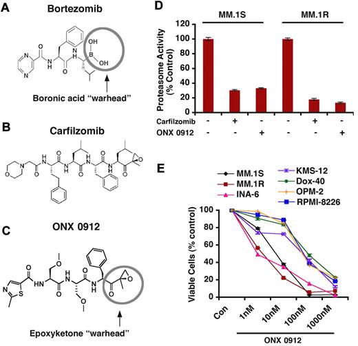
The synthetic and naturally occurring inhibitors of the UPS pathway include peptide aldehydes, peptide boronates, nonpeptide inhibitors, peptide vinyl sulfones, and peptide epoxyketones.1,12,23 Bortezomib is a boronic acid dipeptide derivative and inhibits proteasome function reversibly through interaction of boronic acid at the C-terminal of bortezomib with an active threonine site in the proteasome12 (Figure 1A). In contrast to bortezomib, carfilzomib is a structural analog of the microbial natural product epoxomicin and inhibits proteasome activity irreversibly via formation of a dual covalent morpholino adduct9,10,14,24,25 (Figure 1B). Although both agents are equipotent, carfilzomib shows a greater selectivity than bortezomib against the CT-L, β5 proteasome activity and immunoproteasome-subunit low molecular mass peptide 7 (LMP7) (β5i).8 Nonetheless, both carfilzomib and bortezomib are currently administered intravenously. Carfilzomib (Figure 1B) has limited oral bioavailability, likely due to its tetrapeptide structure; in this context, a systematic structure–activity relationship (SAR) study led to the development of an orally bioavailable tripeptide epoxyketone proteasome inhibitor, ONX 0912 (Figure 1C). The effects of ONX 0912 in both in vitro and in human MM xenograft mouse models remain undefined.
Proteasome inhibitor ONX 0912 is structurally distinct from bortezomib and carfilzomib, inhibits proteasome activity in vitro, and triggers MM cell death. (A-C) Chemical structures of proteasome inhibitors bortezomib, carfilzomib, and ONX 0912. (D) ONX 0912 and carfilzomib inhibit CT-L proteasome activities in human MM cell lines. MM.1S and MM.1R cells were treated with carfilzomib (5nM) and ONX 0912 (3nM) for 48 hours and harvested; cytosolic extracts were then analyzed for CT-L proteasome activities. Results are represented as percent inhibition in proteasome activities in drug-treated versus vehicle control. (E) MM cell lines were treated with or without ONX 0912 at the indicated doses for 48 hours, followed by assessment for cell viability using MTT assays. Data presented are means ± SD (n = 3; P < .05 for all cell lines).
Proteasome inhibitor ONX 0912 is structurally distinct from bortezomib and carfilzomib, inhibits proteasome activity in vitro, and triggers MM cell death. (A-C) Chemical structures of proteasome inhibitors bortezomib, carfilzomib, and ONX 0912. (D) ONX 0912 and carfilzomib inhibit CT-L proteasome activities in human MM cell lines. MM.1S and MM.1R cells were treated with carfilzomib (5nM) and ONX 0912 (3nM) for 48 hours and harvested; cytosolic extracts were then analyzed for CT-L proteasome activities. Results are represented as percent inhibition in proteasome activities in drug-treated versus vehicle control. (E) MM cell lines were treated with or without ONX 0912 at the indicated doses for 48 hours, followed by assessment for cell viability using MTT assays. Data presented are means ± SD (n = 3; P < .05 for all cell lines).
We first examined the ability of ONX 0912 versus carfilzomib to inhibit CT-L proteasome activity using 2 different MM cell lines. MM.1S and MM.1R were treated with ONX 0912 (3nM) and carfilzomib (5nM), and protein lysates were subjected to proteasome activity assays using CT-L–specific fluorogenic peptide substrates. Results showed that ONX 0912 triggers a significant and similar degree of proteasome activity inhibition as carfilzomib (Figure 1D; P < .05 for both cell lines). These in vitro findings suggest that ONX 0912, like carfilzomib, targets proteasomes and, importantly, retains the potency and selectivity of carfilzomib against CT-L proteasome activity.14
Anti-MM activity of ONX 0912 in vitro
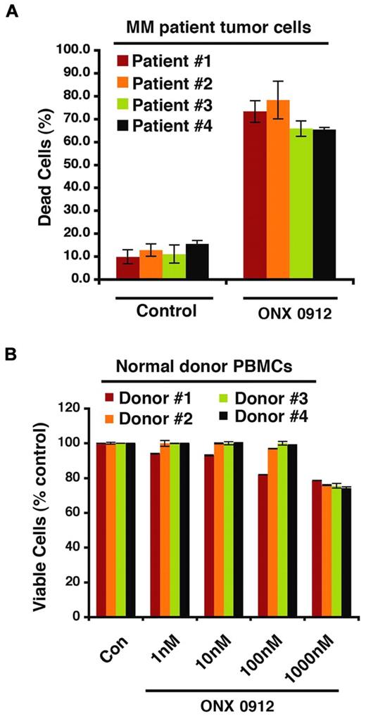
Human MM cell lines (MM.1S, INA-6, RPMI-8226, MM.1R, Dox-40, KMS12, and OPM2) were treated with ONX 0912 for 48 hours, followed by assessment for cell viability using MTT assays. A significant decrease in viability of all cell lines was observed in response to treatment with ONX 0912 (P < .05; n = 3; Figure 1E). To determine whether ONX 0912 similarly affects purified patient MM cells, tumor cells from 4 MM patients relapsing after multiple prior therapies, including lenalidomide and Dex (patients 1 and 3) and bortezomib (patients 2 and 4), were treated with ONX 0912. Patients were considered refractory to specific therapy when disease progressed on therapy or relapsed within 2 months of stopping therapy. Of note, 2 of 4 patients studied were refractory to bortezomib therapy, and 2 were refractory to lenalidomide and Dex therapies. A significant increase in cell death of all patient MM cells was noted after ONX 0912 treatment (P < .05 for all patients; Figure 2A). ONX 0912 did not trigger a significant decrease in the viability of normal PBMCs, even at higher concentration (1μM; P < .05; n = 3; Figure 2B).
ONX 0912 triggers antitumor activity in MM patient cells. (A) Purified patient MM cells (CD138-positive) were treated with ONX 0912 (10nM), and cell death was measured using trypan blue exclusion assay. Data presented are mean ± SD of triplicate samples (P < .05 for all patient samples). (B) PBMCs from healthy donors were treated with indicated concentrations of ONX 0912 and then analyzed for viability using MTT assay. Data presented are mean ± SD (n = 3; P < .05).
ONX 0912 triggers antitumor activity in MM patient cells. (A) Purified patient MM cells (CD138-positive) were treated with ONX 0912 (10nM), and cell death was measured using trypan blue exclusion assay. Data presented are mean ± SD of triplicate samples (P < .05 for all patient samples). (B) PBMCs from healthy donors were treated with indicated concentrations of ONX 0912 and then analyzed for viability using MTT assay. Data presented are mean ± SD (n = 3; P < .05).
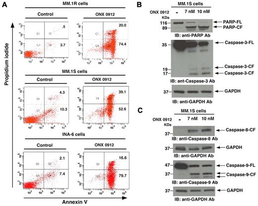
We next examined whether the ONX 0912–triggered decrease in viability is due to apoptosis. ONX 0912 (3nM) induced a significant increase in the annexin V+/PI− apoptotic cell population in MM.1R, MM.1S, and INA-6 cells (Figure 3A; P < .04, n = 3). In addition, treatment of MM.1S cells with ONX 0912 (3nM) triggered a marked increase in proteolytic cleavage of PARP,26 a signature event during apoptosis (Figure 3B). Similarly, ONX 0912 induced cleavage of caspase-3 (Figure 3B), an upstream activator of PARP.27 Previous studies have established that mitochondria participate in mediating apoptotic signaling via activation of the cell death initiator caspase, pro-caspase 9.28 Similarly, the Fas-associated death-domain (FADD) protein is an essential part of the death-inducing signaling complexes (DISCs) that assemble upon engagement of tumor necrosis factor (TNF) receptor family members, such as Fas, resulting in proteolytic processing and autoactivation of pro-caspase-8.29 Our data show that ONX 0912 induces activation of both casapse-8 (extrinsic) and caspase-9 (intrinsic) apoptotic pathways (Figure 3C). These findings suggest that ONX 0912–induced apoptosis in MM cells occurs via both extrinsic (mitochondria-independent) and intrinsic (mitochondria-dependent) apoptotic-signaling pathways.
ONX 0912 triggers apoptosis in MM cells, associated with PARP cleavage and caspase activation. (A) MM cell lines were treated with ONX 0912 for 48 hours and analyzed for apoptosis using annexin V/PI staining assay. (B-C) MM.1S cells were treated with ONX 0912 at the indicated doses for 48 hours and harvested; whole-cell lysates were subjected to immunoblot analysis with anti-PARP, anti–caspase-3, anti–caspase-8, anti–caspase-9, or anti-GAPDH Abs. FL indicates full-length; CF denotes cleaved fragment. Blots shown are representative of 3 independent experiments.
ONX 0912 triggers apoptosis in MM cells, associated with PARP cleavage and caspase activation. (A) MM cell lines were treated with ONX 0912 for 48 hours and analyzed for apoptosis using annexin V/PI staining assay. (B-C) MM.1S cells were treated with ONX 0912 at the indicated doses for 48 hours and harvested; whole-cell lysates were subjected to immunoblot analysis with anti-PARP, anti–caspase-3, anti–caspase-8, anti–caspase-9, or anti-GAPDH Abs. FL indicates full-length; CF denotes cleaved fragment. Blots shown are representative of 3 independent experiments.
ONX 0912 blocks migration of MM cells and angiogenesis
Previous reports have shown that migration and angiogenesis play an important role in the progression of MM.16,30-34 The effect of ONX 0912 on these events was therefore examined using Transwell insert systems and in vitro tubule formation assays. Serum alone markedly increased MM.1S cell migration; importantly, ONX 0912 (10nM) significantly inhibited serum-dependent MM.1S cell migration, as reflected by a decrease in the number of crystal violet–stained cells (Figure 4A). Vascular endothelial growth factor (VEGF) is elevated in the MM bone marrow (BM) microenvironment, and prior studies showed that VEGF triggers growth, migration, and angiogenesis in MM.16,30-34 We examined whether ONX 0912 affects VEGF-induced MM cell migration. VEGF alone markedly increases MM.1S cell migration; conversely, ONX 0912 significantly (P = .03) inhibits VEGF-dependent MM cell migration (Figure 4B). These cells were > 95% viable before and after performing the migration assay, excluding the possibility that drug-induced inhibition of migration was due to cell death. These results suggest that ONX 0912 may negatively regulate homing of MM cells to the BM, as well as their egress into the peripheral blood.
ONX 0912 blocks migration and tube formation. (A) MM.1S cells were pretreated with 10nM ONX 0912 for 24 hours; the cells were > 90% viable at this time point. The cells were washed and cultured in serum-free medium. After 2 hours of incubation, cells (viability > 90%) were plated on a fibronectin-coated polycarbonate membrane in the upper chamber of Transwell inserts and exposed for 4 hours to serum-containing medium in the lower chamber. Cells migrating to the bottom face of the membrane were fixed with 90% ethanol and stained with crystal violet (×10 original magnification /0.25 numeric aperture [NA] oil). A total of 3 randomly selected fields were examined for cells that had migrated from top to bottom chambers. Image is representative of 3 experiments with similar results. (B) Cells were plated as in panel A, and ONX 0912 effect on migration was assessed in the presence or absence of rVEGF. The bar graph represents quantification of migrated cells. Data presented are means ± SD (n = 3; P = .03 for control versus ONX 0912–treated cells). Error bars represent SD. (C) HUVECs were cultured in the presence or absence of ONX 0912 for 24 hours and then assessed for in vitro angiogenesis using Matrigel capillary-like tube structure formation assays (×4 original magnification/0.10 NA oil, media: endothelium basal medium-2 [EBM-2]). Image is representative from 3 experiments with similar results. The in vitro angiogenesis is reflected by capillary tube branch formation (dark brown). Data represent means ± SD (n = 2; P < .05). (D) In vitro angiogenesis using Matrigel capillary tube formation assay was performed using HUVECs as in panel C. The bar graph represents quantification of capillary-like tube structure formation in response to vehicle alone and ONX 0912–treated cells: Branch points in several random view fields/wells were counted; values were averaged, and statistically significant differences were measured using Student t test. Data presented are means ± SD (n = 3; P = .01 for control versus ONX 0912–treated cells). Error bars represent SD.
ONX 0912 blocks migration and tube formation. (A) MM.1S cells were pretreated with 10nM ONX 0912 for 24 hours; the cells were > 90% viable at this time point. The cells were washed and cultured in serum-free medium. After 2 hours of incubation, cells (viability > 90%) were plated on a fibronectin-coated polycarbonate membrane in the upper chamber of Transwell inserts and exposed for 4 hours to serum-containing medium in the lower chamber. Cells migrating to the bottom face of the membrane were fixed with 90% ethanol and stained with crystal violet (×10 original magnification /0.25 numeric aperture [NA] oil). A total of 3 randomly selected fields were examined for cells that had migrated from top to bottom chambers. Image is representative of 3 experiments with similar results. (B) Cells were plated as in panel A, and ONX 0912 effect on migration was assessed in the presence or absence of rVEGF. The bar graph represents quantification of migrated cells. Data presented are means ± SD (n = 3; P = .03 for control versus ONX 0912–treated cells). Error bars represent SD. (C) HUVECs were cultured in the presence or absence of ONX 0912 for 24 hours and then assessed for in vitro angiogenesis using Matrigel capillary-like tube structure formation assays (×4 original magnification/0.10 NA oil, media: endothelium basal medium-2 [EBM-2]). Image is representative from 3 experiments with similar results. The in vitro angiogenesis is reflected by capillary tube branch formation (dark brown). Data represent means ± SD (n = 2; P < .05). (D) In vitro angiogenesis using Matrigel capillary tube formation assay was performed using HUVECs as in panel C. The bar graph represents quantification of capillary-like tube structure formation in response to vehicle alone and ONX 0912–treated cells: Branch points in several random view fields/wells were counted; values were averaged, and statistically significant differences were measured using Student t test. Data presented are means ± SD (n = 3; P = .01 for control versus ONX 0912–treated cells). Error bars represent SD.
We next used in vitro capillary-like tube structure formation assays to examine the antiangiogenic activity of ONX 0912. Angiogenesis was measured in vitro using Matrigel capillary-like tube structure formation assays: HUVECs plated onto Matrigel differentiate and form capillary-like tube structures similar to in vivo neovascularization—a process dependent on cell-matrix interaction, cellular communication, and cellular motility. This assay therefore provides evidence for antiangiogenic effects of drugs. HUVECs were seeded in 96-well culture plates precoated with Matrigel, treated with vehicle (dimethyl sulfoxide [DMSO]) and ONX 0912 for 24 hours, and then examined for tube formation using an inverted microscope. As seen in Figure 4C and D, tubule formation was markedly decreased in the ONX 0912–treated cells versus vehicle control. HUVEC viability was assessed using trypan blue exclusion assay, and < 5% cell death was observed with ONX 0912 treatment. Taken together, our findings suggest that ONX 0912 blocks migration and angiogenesis.
ONX 0912 inhibits human MM cell growth in vivo and prolongs survival in mouse models
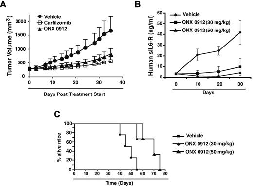
Having shown that ONX 0912 induced apoptosis in MM cells in vitro, we next examined the in vivo efficacy of ONX 0912 using 2 distinct mouse models.17,19 In the first study, MM.1S tumor–bearing mice were treated intravenously with carfilzomib (5 mg/kg) or orally with vehicle or ONX 0912 (30 mg/kg). Animals were treated for 2 consecutive days, and treatment was repeated weekly for 7 weeks. As seen in Figure 5A, a marked reduction (P = .002) in tumor growth was noted in ONX 0912–treated mice versus mice receiving vehicle alone. The extent of tumor growth inhibition was similar in mice treated orally with ONX 0912 or intravenously with carfilzomib.
ONX 0912 inhibits growth of xenografted human MM cells in mice. (A) MM.1S cells alone (3 × 10⁷ cells/mouse) were implanted in the rear flank of female beige nude xid mice (5-7 weeks of age at the time of tumor challenge). On Day 30-33, mice were randomized to treatment groups and treated intravenously with carfilzomib (5 mg/kg) or orally with vehicle or ONX 0912 (30 mg/kg). Mice were treated for 2 consecutive days and the treatment repeated weekly for 7 weeks. Data are presented as mean tumor volume ± SEM (n = 9-10/group). One of the 2 representative experiments is shown. Bars indicate means ± SD. (B) Human fetal bone grafts (size range: 0.5-1.5 cm) were subcutaneously implanted into CB-17 SCID mice. Four weeks after bone implantation, INA-6 cells (2.5 × 10⁶) were injected directly into the human fetal bone implant in SCID mice (5 mice each group), and mouse sera samples were serially analyzed for levels of secreted human soluble IL-6R (shIL-6R) by enzyme-linked immunosorbent assay as a measure of tumor burden. Upon detection of measurable shIL-6R in mice (3-4 wks after injection of cells), mice were treated with either vehicle alone or indicated doses of ONX 0912 (orally, 2 consecutive days every week for 4 weeks); mouse serum samples were subjected to analysis for shIL-6R (mean ± SD; P = .03 for both doses; n = 2). Bars indicate means ± SD (C) Kaplan-Meier survival plot shows a significant increase (P = .03) in survival of mice receiving ONX 0912 (30 or 50 mg/kg) compared with vehicle-treated control. The mean overall survival (OS) was 47.5 days (95% confidence interval; 40-55) in the untreated versus 70 days (95% confidence interval; 65-70) in ONX 0912–treated cohorts.
ONX 0912 inhibits growth of xenografted human MM cells in mice. (A) MM.1S cells alone (3 × 10⁷ cells/mouse) were implanted in the rear flank of female beige nude xid mice (5-7 weeks of age at the time of tumor challenge). On Day 30-33, mice were randomized to treatment groups and treated intravenously with carfilzomib (5 mg/kg) or orally with vehicle or ONX 0912 (30 mg/kg). Mice were treated for 2 consecutive days and the treatment repeated weekly for 7 weeks. Data are presented as mean tumor volume ± SEM (n = 9-10/group). One of the 2 representative experiments is shown. Bars indicate means ± SD. (B) Human fetal bone grafts (size range: 0.5-1.5 cm) were subcutaneously implanted into CB-17 SCID mice. Four weeks after bone implantation, INA-6 cells (2.5 × 10⁶) were injected directly into the human fetal bone implant in SCID mice (5 mice each group), and mouse sera samples were serially analyzed for levels of secreted human soluble IL-6R (shIL-6R) by enzyme-linked immunosorbent assay as a measure of tumor burden. Upon detection of measurable shIL-6R in mice (3-4 wks after injection of cells), mice were treated with either vehicle alone or indicated doses of ONX 0912 (orally, 2 consecutive days every week for 4 weeks); mouse serum samples were subjected to analysis for shIL-6R (mean ± SD; P = .03 for both doses; n = 2). Bars indicate means ± SD (C) Kaplan-Meier survival plot shows a significant increase (P = .03) in survival of mice receiving ONX 0912 (30 or 50 mg/kg) compared with vehicle-treated control. The mean overall survival (OS) was 47.5 days (95% confidence interval; 40-55) in the untreated versus 70 days (95% confidence interval; 65-70) in ONX 0912–treated cohorts.
Our previous studies have shown that the MM-host BM microenvironment confers growth, survival, and drug resistance in MM cells.35,36 We therefore next examined whether the anti-MM activity of ONX 0912 is retained in the presence of the human BM microenvironment. For these studies, we used the SCID-hu model,18 which recapitulates the human BM milieu in vivo. In this model, INA-6 MM cells are injected directly into human bone chips that are implanted subcutaneously in SCID mice, and MM cell growth is assessed by serial measurements of circulating levels of soluble human IL-6R in mouse serum. A more robust growth inhibition of tumor occurred in mice receiving oral doses (30 mg/kg or 50 mg/kg) of ONX 0912 than in mice injected with vehicle alone (Figure 5B). Importantly, treatment of tumor-bearing mice with ONX 0912, but not vehicle alone, significantly prolonged survival (P = .03; Figure 5C).
We next examined the effect of ONX 0912 on in vivo apoptosis using immunostaining of implanted human bone for caspase-3 activation. ONX 0912 dramatically increased the number of caspase-3 cleavage-positive cells compared with vehicle treatment alone (Figure 6A). Similarly, we noted a marked decrease in Factor VIII and VEGFR1 expression in bone sections from mice injected with ONX 0912 versus vehicle alone (Figure 6B-C). These in vivo IHC data confirm the apoptotic and antiangiogenic activity of ONX 0912 in MM cells. Given that ONX 0912 inhibits proteasome activity (Figure 1D) and proteasome inhibition results in accumulation of ubiquitinated proteins, we also examined human bone sections from mice for alterations in the ubiquitination pattern. Bone chips were excised 2 hours after the last dose, and IHC was performed using antiubiquitin Abs. ONX 0912 treatment markedly increased ubiquitin staining versus control (Figure 6D). These in vivo data, together with our in vitro results, confirm that the anti-MM activity of ONX 0912 is associated with inhibition of proteasome activity.
Effect of ONX 0912 on apoptosis, neovascularization, and ubiquitination in vivo in xenografted MM tumors. (A-D) Human bone chips were removed from mice after the last treatment and immunostained with Abs against Caspase-3, Factor VIII, VEGFR1, and Ubiquitin. Scale bar, 10μm. Dark brown represents marker-positive cells in all cases. Micrographs are representative of bone sections from 2 different mice in each group. (E) Mice in vehicle-treated control and ONX 0912–treated groups were weighed every week. The average changes in body weight are shown.
Effect of ONX 0912 on apoptosis, neovascularization, and ubiquitination in vivo in xenografted MM tumors. (A-D) Human bone chips were removed from mice after the last treatment and immunostained with Abs against Caspase-3, Factor VIII, VEGFR1, and Ubiquitin. Scale bar, 10μm. Dark brown represents marker-positive cells in all cases. Micrographs are representative of bone sections from 2 different mice in each group. (E) Mice in vehicle-treated control and ONX 0912–treated groups were weighed every week. The average changes in body weight are shown.
The doses of ONX 0912 administered were well tolerated by mice because no significant weight loss was noted in these studies (Figure 6E). Moreover, no neurological behavioral changes were observed after ONX 0912 treatment (data not shown). Together, our findings from 2 distinct human MM xenograft models demonstrate the potent in vivo antitumor activity of ONX 0912 at doses that are well tolerated. Results from the SCID-hu model provide in vivo evidence for the ability of ONX 0912 to trigger apoptosis of tumor cells even in the presence of the BM microenvironment.
Combined treatment with ONX 0912 and bortezomib, Dex, lenalidomide or MS-275 induces synergistic/additive anti-MM activity
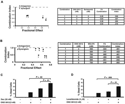
Having shown the single-agent anti-MM activity of ONX 0912, we next examined whether it can be combined with other drugs to enhance cytotoxicity. Proteasome inhibitors bortezomib and ONX 0912 are distinct in their structure and mechanism of action: ONX 0912 is a more selective inhibitor of CT-L activity than bortezomib, providing the rationale for combining these agents to enhance anti-MM activity. MM.1S cells were treated with both ONX 0912 and bortezomib simultaneously across a range of concentrations for 24 hours and then analyzed for viability using MTT assay. An analysis of synergistic anti-MM activity of combined agents was determined using the Chou and Talalay22 analysis of data from cell viability assays. Isobologram analysis showed that the combination of low concentrations of ONX 0912 and bortezomib triggered synergistic anti-MM activity, with a CI < 1.0 (Figure 7A graph and table). Shown in Figure 7A is cell viability expressed as the fraction of cells killed by each drug alone or in combination in drug-treated versus untreated cells. Data are expressed as Fraction Affected (FA) values (eg, FA = 0.5 corresponds to 50% decrease in viability or IC50 for agent or combination).
Combination of low doses of ONX 0912 and bortezomib, MS-275, Dex, or lenalidomide enhances MM cell death. (A) Low doses of ONX 0912 and bortezomib trigger synergistic anti-MM activity in MM cells. MM.1S cells were treated for 24 hours with indicated concentrations of ONX 0912, bortezomib, or ONX 0912 plus bortezomib and then assessed for viability using MTT assays. Isobologram analysis shows the synergistic cytotoxic effect of ONX 0912 and bortezomib in MM.1S cell line. The graph (left) is derived from the values given in the table (right). The numbers 1-4 in the graph represent combinations shown in the table. CI < 1 indicates synergy. Numbers represent FA (Fraction Affected) values (eg, FA = 0.5 corresponds to 50% decrease in viability or IC50 for agent). All experiments were performed in triplicate and mean value is shown. FACom = fraction of cells showing decrease in viability with ONX 0912 plus bortezomib treatment. (B) Low doses of ONX 0912 and MS-275 trigger synergistic anti-MM activity in MM cells. MM.1S cells were treated for 48 hours with indicated concentrations of ONX 0912, MS-275, or ONX 0912 plus MS-275 and then assessed for viability using MTT assays. Isobologram analysis shows the synergistic cytotoxic effect of ONX 0912 and MS-275 in MM.1S cell line. The graph (left) is derived from the values given in the table (right). The numbers 1-6 in the graph represent combinations shown in the table. CI < 1 indicates synergy. (C) MM.1S cells were treated for 48 hours with indicated concentrations of ONX 0912, Dex, or ONX 0912 plus Dex and then assessed for viability using MTT assays. Shown is mean ± SD of 3 independent experiments. (D) MM.1S cells were treated for 48 hours with indicated concentrations of ONX 0912, lenalidomide, or ONX 0912 plus lenalidomide and then assessed for viability using MTT assays. Shown is mean ± SD of 3 independent experiments.
Combination of low doses of ONX 0912 and bortezomib, MS-275, Dex, or lenalidomide enhances MM cell death. (A) Low doses of ONX 0912 and bortezomib trigger synergistic anti-MM activity in MM cells. MM.1S cells were treated for 24 hours with indicated concentrations of ONX 0912, bortezomib, or ONX 0912 plus bortezomib and then assessed for viability using MTT assays. Isobologram analysis shows the synergistic cytotoxic effect of ONX 0912 and bortezomib in MM.1S cell line. The graph (left) is derived from the values given in the table (right). The numbers 1-4 in the graph represent combinations shown in the table. CI < 1 indicates synergy. Numbers represent FA (Fraction Affected) values (eg, FA = 0.5 corresponds to 50% decrease in viability or IC50 for agent). All experiments were performed in triplicate and mean value is shown. FACom = fraction of cells showing decrease in viability with ONX 0912 plus bortezomib treatment. (B) Low doses of ONX 0912 and MS-275 trigger synergistic anti-MM activity in MM cells. MM.1S cells were treated for 48 hours with indicated concentrations of ONX 0912, MS-275, or ONX 0912 plus MS-275 and then assessed for viability using MTT assays. Isobologram analysis shows the synergistic cytotoxic effect of ONX 0912 and MS-275 in MM.1S cell line. The graph (left) is derived from the values given in the table (right). The numbers 1-6 in the graph represent combinations shown in the table. CI < 1 indicates synergy. (C) MM.1S cells were treated for 48 hours with indicated concentrations of ONX 0912, Dex, or ONX 0912 plus Dex and then assessed for viability using MTT assays. Shown is mean ± SD of 3 independent experiments. (D) MM.1S cells were treated for 48 hours with indicated concentrations of ONX 0912, lenalidomide, or ONX 0912 plus lenalidomide and then assessed for viability using MTT assays. Shown is mean ± SD of 3 independent experiments.
Recent preclinical studies provide evidence for potential clinical utility of targeting HDAC enzymes, also known as lysine deacetylases (KDAC), in MM37,38 and other hematologic malignancies.39-42 Recent clinical trials combining bortezomib and the HDAC inhibitor vorinostat showed a promising outcome in relapsed and refractory MM, including activity among some patients with prior exposure to bortezomib.43 Another study showed that vorinostat adds to the antitumor activity of the proteasome inhibitor carfilzomib in human diffuse large B-cell lymphoma (DLBCL) cells.42 In the light of these studies, we examined whether a combination of ONX 0912 with an HDAC inhibitor triggered synergistic or additive anti-MM activity. MM.1S cells were simultaneously treated with both ONX 0912 and MS-275, a pan-HDAC inhibitor, across a range of concentrations for 48 hours and then analyzed for viability. Isobologram analysis showed that the combination of low concentrations of ONX 0912 and MS-275 triggered synergistic anti-MM activity, with a CI < 1.0 (Figure 7B graph and table).
We next examined whether ONX 0912 enhances the cytotoxicity triggered by the conventional anti-MM agent, Dex. ONX 0912 plus Dex triggered additive anti-MM activity, as evidenced by a significant decrease in viability of MM.1S cells (Figure 7C; n = 3). Similarly, ONX 0912 adds to the anti-MM activity of the immunomodulatory agent lenalidomide (Figure 7D; n = 3). Combining low doses of ONX 0912 with bortezomib, MS-275, Dex, or lenalidomide does not significantly affect the viability of normal lymphocytes (data not shown). While the definitive evidence of decreased toxicity of combination therapy awaits results of careful clinical trials, the synergy observed in vitro may allow for use of lower doses and decreased toxicity.
Discussion
The improved MM patient outcome with proteasome inhibitor therapy necessitates frequent dosing and prolonged treatment. Both bortezomib and carfilzomib are administered intravenously on biweekly dosing schedules with treatment periods extending for 6 months or more. This notion led to the development of equipotent orally bioavailable proteasome inhibitors, allowing for flexibility in both the dosing schedule and patient convenience. In the present study, we evaluated the efficacy of one such novel orally bioactive proteasome inhibitor, ONX 0912,14 in MM using well-established in vitro and in vivo models.
We first showed that ONX 0912 decreases viability of MM cell lines and primary patient tumor cells without affecting normal PBMCs viability. Importantly, our data demonstrate anti-MM activity of ONX 0912 in MM cell lines, including those sensitive and resistant to therapies, as well as distinct cytogenetic profiles. For example, we examined isogenic cell lines Dex-sensitive MM.1S and Dex-resistant MM.1R with t(14;16) translocation and c-maf overexpression, KMS12PE t(11:14) with cyclin D1 deregulation, RPMI-8266 with TP53, K-Ras and EGFR mutations, and INA-6, an IL-6-dependent cell line, with N-Ras activating mutation.44-50 Although,ONX 0912 treatment decreased viability in all MM cell lines, the IC50 of ONX 0912 for each cell line varied; this may be due to the genetic heterogeneity and drug-resistant characteristics of MM.44-47 As with MM cell lines, our data showed similar responses even in patient-derived MM cells resistant to therapies such as Dex, lenalidomide, or bortezomib.
A previous report showed a selective inhibitory ability of ONX 0912 against CT-L activity in cell-free systems using purified human 20S proteasomes.14 Here we examined whether ONX 0912 similarly affected the CT-L proteasome activity in human MM cells. Our data show that ONX 0912 inhibits CT-L activity in MM cells; an in vitro comparative analysis of intravenously administered drug carfilzomib and its oral analog, ONX 0912, showed a similar extent of inhibition in CT-L (β5) activity in MM cells.
In addition to our in vitro studies, we also examined anti-MM activity of ONX 0912 in vivo. A marked growth inhibitory effect of ONX 0912 was observed in 2 distinct human MM xenograft mouse models. A head-to-head analysis of carfilzomib and ONX 0912 showed equipotent antitumor activity in a human plasmacytoma xenograft mouse model. Our findings are consistent with reported antitumor activity of ONX 0912 in nonHodgkin lymphoma (NHL) tumor xenograft and mouse syngeneic models.14 In our study, we were further able to examine the anti-MM activity of ONX 0912 in the human BM microenvironment using our SCID-hu mouse model. In this model, MM cells are injected directly into human bone chips that are implanted subcutaneously in SCID mice, and MM cell growth is measured by the circulating levels of soluble human IL-6R in mouse serum. Results provided evidence for a significant anti-MM activity of ONX 0912 in the presence of the BM milieu, suggesting that ONX 0912 not only directly targets MM cells but also overcomes the cytoprotective effects of the MM-host BM microenvironment.
Our data show that ONX 0912 treatment markedly prolongs survival in mice. ONX 0912 treatment was not associated with any toxicity, because no differences in body weight and overall appearance were noted. The remarkable anti-MM activity of ONX 0912 in vivo was confirmed by IHC analysis of implanted human bone harvested from control and ONX 0912–treated mice using molecular markers of apoptosis (caspase-3 cleavage) and associated angiogenesis (Factor VIII and VEGFR1). The apoptotic and antiangiogenic effect of ONX 0912 observed in our in vivo study further confirmed our in vitro findings shown in Figures 3 and 4. Together, these data demonstrated a dual effect of ONX 0912: increased MM cell apoptosis and decreased angiogenesis. Furthermore, analysis of the bone sections showed accumulation of ubiquitinated proteins in ONX 0912–treated mice but not in mice treated with vehicle alone, reflecting a blockade of proteasome function in tumor cells. These in vivo findings, coupled with our in vitro data showing minimal toxicity of ONX 0912 against normal cells, confirms that MM cells are more sensitive to proteasome inhibition than normal cells.
Finally, we also examined whether ONX 0912 can be combined with other conventional (Dex) and novel (bortezomib, MS-275, and lenalidomide) anti-MM agents. Prior studies have shown that bortezomib predominantly inhibits CT-L and, more recently defined, caspase-like (C-L) activities within the proteasome.20 Carfilzomib is a more selective inhibitor of CT-L activity than bortezomib.8 It is therefore likely that ONX 0912 (an oral analog of carfilzomib), by virtue of its ability to more selectively block CT-L activity, can be combined with bortezomib to confer proteasome inhibition at lower, and potentially safer, doses. Our data demonstrate synergistic anti-MM activity of ONX 0912 plus bortezomib. The mechanisms mediating the combined anti-MM activity of ONX 0912 and bortezomib remain to be defined. One possibility is that using low doses of bortezomib may limit its off-target activity and associated adverse effects,6,7,51-53 while allowing for a more efficient, synergistic, and specific blockade of CT-L proteasome activity in combination with ONX 0912. Alternatively, the overall effective cell killing by ONX 0912 plus bortezomib may involve activation of other apoptotic signaling pathways, due to their distinct chemical structures. Importantly, our data provide the rationale for combining ONX 0912 with bortezomib to achieve optimal proteasome inhibition and potent antitumor activity, thereby allowing for use of lower doses of bortezomib and reduced side effects.
Besides proteasomes, intracellular protein catabolism is also mediated via an HDAC-dependent aggressome-autophagic signaling pathway.37 Our study showed that inhibition of both mechanisms of protein catabolism by combining bortezomib and HDAC inhibitor induced significant cytotoxicity in MM cells.37,38 In agreement with these findings, our data also show synergistic anti-MM activity of ONX 0912 with the pan-HDAC inhibitor MS-275. A similar observation was also reported in the context of carfilzomib in combination with another pan-HDAC inhibitor, vorinostat.42 Together, these data confirm the potential clinical benefit of combining proteasome inhibitors and HDAC inhibitors. Nonetheless, therapeutic strategies combining a specific inhibitor of proteasome activities (CT-L, trypsin-like [T-L], C-L, or their corresponding immunoproteasome subunits), together with a class-specific HDAC inhibitor, may yield a more potent and less toxic drug regimen. Finally, we also found that ONX 0912 adds to the anti-MM activity of Dex and lenalidomide, which are frequently used in the treatment of MM.
Collectively, our preclinical studies demonstrate potent in vitro and in vivo anti-MM activity of ONX 0912 at doses that are well tolerated in human MM xenograft mouse models. These findings provide the framework for clinical trials of ONX 0912 to increase response, overcome drug resistance, reduce side effects, and improve patient outcome in MM.
The publication costs of this article were defrayed in part by page charge payment. Therefore, and solely to indicate this fact, this article is hereby marked “advertisement” in accordance with 18 USC section 1734.
Acknowledgments
We thank Jing Jiag (Onyx Pharmaceuticals) for her assistance with xenograft studies.
This work was supported by National Institutes of Health grants SPORE-P50100707, PO1-CA078378, and RO1CA050947. K.C.A. is an American Cancer Society Clinical Research Professor.
National Institutes of Health
Authorship
Contribution: D.C. designed research, analyzed/interpreted data, and wrote the manuscript; A.V.S. performed the experiments and interpreted data; M.B. processed PBMCs; B.C., N.R., and P.R. provided clinical samples; M.A. and C.J.K. provided ONX 0912; and K.C.A. analyzed data and critiqued the manuscript.
Conflict-of-interest disclosure: D.C. and K.C.A. are consultants to Onyx Pharmaceuticals; M.A. and C.K. are employees of Onyx Pharmaceuticals. The remaining authors declare no competing financial interests.
Correspondence: Dharminder Chauhan, Dana-Farber Cancer Institute, Mayer Building Rm 561, 44 Binney St, Boston, MA 02115; e-mail: Dharminder_Chauhan@dfci.harvard.edu.
References
Author notes
D.C. and A.V.S. contributed equally to the work.

![Figure 4. ONX 0912 blocks migration and tube formation. (A) MM.1S cells were pretreated with 10nM ONX 0912 for 24 hours; the cells were > 90% viable at this time point. The cells were washed and cultured in serum-free medium. After 2 hours of incubation, cells (viability > 90%) were plated on a fibronectin-coated polycarbonate membrane in the upper chamber of Transwell inserts and exposed for 4 hours to serum-containing medium in the lower chamber. Cells migrating to the bottom face of the membrane were fixed with 90% ethanol and stained with crystal violet (×10 original magnification /0.25 numeric aperture [NA] oil). A total of 3 randomly selected fields were examined for cells that had migrated from top to bottom chambers. Image is representative of 3 experiments with similar results. (B) Cells were plated as in panel A, and ONX 0912 effect on migration was assessed in the presence or absence of rVEGF. The bar graph represents quantification of migrated cells. Data presented are means ± SD (n = 3; P = .03 for control versus ONX 0912–treated cells). Error bars represent SD. (C) HUVECs were cultured in the presence or absence of ONX 0912 for 24 hours and then assessed for in vitro angiogenesis using Matrigel capillary-like tube structure formation assays (×4 original magnification/0.10 NA oil, media: endothelium basal medium-2 [EBM-2]). Image is representative from 3 experiments with similar results. The in vitro angiogenesis is reflected by capillary tube branch formation (dark brown). Data represent means ± SD (n = 2; P < .05). (D) In vitro angiogenesis using Matrigel capillary tube formation assay was performed using HUVECs as in panel C. The bar graph represents quantification of capillary-like tube structure formation in response to vehicle alone and ONX 0912–treated cells: Branch points in several random view fields/wells were counted; values were averaged, and statistically significant differences were measured using Student t test. Data presented are means ± SD (n = 3; P = .01 for control versus ONX 0912–treated cells). Error bars represent SD.](https://ash.silverchair-cdn.com/ash/content_public/journal/blood/116/23/10.1182_blood-2010-04-276626/4/m_zh89991061390004.jpeg?Expires=1763503152&Signature=Pe7jt5vZWnDJqif7XUAdtflazEALiq-hEYjKOg9asM8g8D3bGag6prs01sKL1uQxFbdd2xn3DDelKuTbT81NIdnC~ww-q7W5SfElXTrJpxLTFFTFc4bmtLDUFNFdTG3F2YKEuRN23wNQWF1gjK04YFxfvByF5eFtJ~NN2gqKeukMlI4hcOzp6kH9WmsulobWUz8LL0iYJb3JgejrPdQUPvBxeBzGCGD4ufZH9hLTYx3WWIrmCIe6Mp5vAde3WEzRH1CXPhjqgoDG91lE-xnB0XNydk9XyoBewx5FMNexVTUkoNcHS4D2K6jV0OQTdL72u~1x6sZQacBE7kWDpKn5Bg__&Key-Pair-Id=APKAIE5G5CRDK6RD3PGA)
