Abstract
Abstract 4264
β-thalassemia major is a genetic disorder characterized by the absence (β°) or reduced output (β+) of the β chains of haemoglobin. This disorder displays a great deal of phenotypic heterogeneity, not fully investigated in terms of cause-effect (Refaldi C et al, Blood Transf 2005; Thein SL, Haematologica 2005). The aim of our study was to detect if different phenotypes could be related to different levels of cardiac impairments, evaluated by cardiovascular magnetic resonance (CMR).
We performed a retrospective review of the CMR results and of clinical data about 328 TM patients (age 29 ± 9 years, 52% females) enrolled in the Myocardial Iron Overload in Thalassemia (MIOT) project (Meloni A et al, Int J Med Inform). In the MIOT network all CMR and thalassemia centers are linked by a web-based network, configured to collect patients' anamnestic, clinical, and diagnostic data. Myocardial iron overload was assessed by using a multislice multiecho T2* approach (Pepe A et al, Eur J Haem 2006). Three short axis views (basal, medium, and apical) of the left ventricle (LV) were acquired and analyzed using a custom-written, previous validated software (HIPPO MIOT®). The myocardium was automatically segmented into a 16-segments standardized LV model and the T2* value on each segment was calculated as well as the global T2* value. Cine sequences were obtained to quantify biventricular morphological and functional parameters.
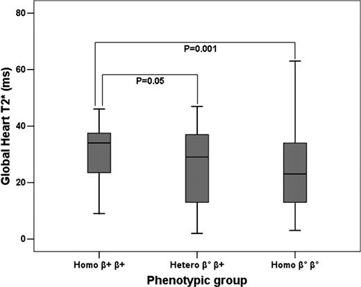
Three groups of patients were identified: heterozygous (N=172), homozygous β+ (N=58), homozygous β° (N=98). No significant differences for sex and age were found among the groups. The homozygous β+ group showed higher global heart T2* values than the heterozygous and homozygous β° group (32 ± 11 ms vs 26 ± 13 ms vs 23 ± 13 ms, P=0.001) (see figure). The homozygous β+ group showed lower left LV mass index than the heterozygous group (54 ± 10 g/m2 vs 60 ± 14 g/m2, P=0.045). The homozygous β+ group showed higher LV ejection fraction (EF) values than the heterozygous group (64 ± 5 % vs 60 ± 7 %, P<0.0001) and higher right ventricular EF values than the heterozygous and homozygous β° group (64 ± 5 % vs 59 ± 8 % vs 60 ± 7 %, P<0.0001).
The homozygous β+ TM patients showed less myocardial iron overload and a concordant better global systolic heart function and cardiac remodelling. These data support the knowledge of the different phenotypes in the clinical and instrumental management of the TM patients suggesting stricter monitoring by CMR in homozygous β° TM patients.
No relevant conflicts of interest to declare.
Author notes
Asterisk with author names denotes non-ASH members.


This feature is available to Subscribers Only
Sign In or Create an Account Close Modal