Abstract
The function of the mechanosensitive, multimeric blood protein von Willebrand factor (VWF) is dependent on its size. We tested the hypothesis that VWF may self-associate on the platelet glycoprotein Ibα (GpIbα) receptor under hydrodynamic shear. Consistent with this proposition, whereas Alexa-488–conjugated VWF (VWF-488) bound platelets at modest levels, addition of unlabeled VWF enhanced the extent of VWF-488 binding. Recombinant VWF lacking the A1-domain was conjugated with Alexa-488 to produce ΔA1-488. Although ΔA1-488 alone did not bind platelets under shear, this protein bound GpIbα on addition of either purified plasma VWF or recombinant full-length VWF. The extent of self-association increased with applied shear stress more than ∼ 60 to 70 dyne/cm2. ΔA1-488 bound platelets in the milieu of plasma. On application of fluid shear to whole blood, half of the activated platelets had ΔA1-488 bound, suggesting that VWF self-association may be necessary for cell activation. Shearing platelets with 6-μm beads bearing either immobilized VWF or anti-GpIbα mAb resulted in cell activation at shear stress down to 2 to 5 dyne/cm2. Taken together, the data suggest that fluid shear in circulation can increase the effective size of VWF bound to platelet GpIbα via protein self-association. This can trigger mechanotransduction and cell activation by enhancing the drag force applied on the cell-surface receptor.
Introduction
von Willebrand factor (VWF) is a large, multidomain glycoprotein found in normal blood at concentrations of approximately 10 μg/mL.1 The protein plays an important role in hemostasis by both carrying the coagulation protein factor VIII (FVIII) in circulation and by regulating the adhesion of platelets to sites of vascular injury. Whereas the D′D3 domain of VWF binds FVIII, the A1 and C1 domains engage platelet receptors glycoprotein Ibα (GPIbα) and αIIbβ3 (GPIIb-IIIa), respectively. Monomeric VWF has a molecular mass of approximately 250 kDa. This unit further polymerizes, via disulfide linkage formation in the endoplasmic reticulum and Golgi of endothelial cells and megakaryocytes. Multimeric VWF size ranges from 0.5 to 20 MDa.2 Ultra/unusually-large VWF is secreted from the Weibel-Palade bodies of endothelial cells on stimulation with a variety of agonists associated with inflammation and thrombosis, including thrombin, histamine, and tumor necrosis factor-α.
The hemostatic potential of VWF increases with protein size and the magnitude of the applied hydrodynamic shear.3,4 Ultra/unusually-large VWF secreted from endothelial cells under shear is extended in the form of strings or bundles on the vessel wall.5,6 Shear-mediated extension enhances cleavage of the cryptic Y1605-M1606 bond within the VWF-A2 domain by the constitutively active blood metalloprotease, ADAMTS13. In addition to cleavage when immobilized on the endothelium, VWF subjected to fluid shear in flowing blood7 and on platelets8 is also susceptible to proteolysis by ADAMTS13. Together, these mechanisms reduce and regulate the VWF multimer distribution in circulation. In support of a role for ADAMTS13 in regulating VWF activity in blood, ADAMTS13−/− mice exhibit prothrombotic phenotype.9,10 ADAMTS13 deficiency in humans is also associated with thrombotic thrombocytopenic purpura, a condition characterized by the existence of ultra/unusually-large VWF in circulation, thrombocytopenia, and thrombosis in the microvasculature.3,11
Although a number of studies have focused on the role of ADAMTS13 in regulating VWF size under hydrodynamic shear, fewer investigations examine the contributions of VWF self-association to this process. The self-association of VWF was first reported by Savage et al12 who demonstrated that the homotypic interaction between VWF in solution and VWF immobilized on substrates contributes to platelet adhesion in a parallel plate flow chamber-based model of vascular injury. Studies performed in a cone-plate viscometer where purified VWF was subjected to shear and then analyzed using light scattering demonstrated that VWF self-association in solution proceeds in a fluid shear-dependent manner and can be detected at shear rates more than 2000/s.13 Later studies demonstrate the formation of bundles or networks of VWF both on collagen14 and on αvβ3 integrin on the endothelial cell surface.6 Such interactions may also involve the self-association of VWF. These data suggest that the self-association of VWF may be an additional mechanism regulating protein size in circulation.
In the current manuscript, we tested the hypothesis that VWF self-association can take place on the surface of the platelet receptor GpIbα. Using experimental conditions where the applied shear rate and media viscosity were varied independently, we demonstrate that the extent of VWF self-association depends on the magnitude of applied shear stress. It proceeds optimally above 60 to 70 dyne/cm2 in both reconstituted systems and whole blood, conditions observed in some arterioles, stenotic vessels, and prosthetic cardiovascular devices.15,16 Thus, in addition to regulating VWF-GpIbα binding affinity, fluid shear driven VWF self-association may also alter the avidity of this molecular interaction. Using a model of shear-induced platelet activation (SIPAct),13 we show that the self-associated VWF formed on the mechanosensitive receptor GpIbα is functional. This self-associated protein complex effectively increases the apparent size of the globular domain of GpIbα, and it facilitates platelet activation under shear.
Methods
Reagents
Unless stated otherwise, all monoclonal antibodies (mAbs) were IgGs from mouse origin directed against human antigens. Anti-VWF Abs include rabbit polyclonal Ab (Dako North America), nonfunction blocking mAb AVW-1 against the C-terminus (GTI Diagnostics), mAb AVW-3 against VWF-A1 domain (GTI Diagnostics), mAb SZ-12317 against VWF-A3 domain (gift from Dr C. Ruan, Suzhou University, Suzhou, China) and 242.618 against VWF propeptide (gift from Dr R. Montgomery, BloodCenter of Wisconsin, Milwaukee, WI). Anti-GpIbα/CD42b function blocking mAbs are AP-1 (GTI Diagnostics), AK2 (Millipore), and HIP1 (BD Biosciences). Anti-GpIbα VM16d (Cell Sciences) does not block GpIbα binding to VWF. Anti-FVIII mAb ESH-4 blocks VWF binding to FVIII (American Diagnostica). Function blocking anti–GpIIb-IIIa (CD41b/CD61; αIIbβ3) mAbs include HIP8 (eBioscience), 7E3 (ATCC), and humanized mAb c7E3 Fab (ReoPro, Eli Lilly). Fluorescent-conjugated mAbs used to label platelets are CD61–peridinin chlorophyll protein (PerCP; BD Biosciences) and anti-CD31 (platelet endothelial cell adhesion molecule-1) mAb WM59 that was either conjugated with phycoerythrin (PE; BD Biosciences) or PerCP-eFluor-710 (eBioscience). Annexin V conjugated with PE (BD Biosciences) and PE/Cy5 (BioVision) were used in this study.
Human plasma and recombinant VWF
Multimeric human plasma VWF (pVWF) was purified from plasma cryoprecipitate obtained from the Community Blood Bank.19
Several molecular biology steps described in supplemental Methods (available on the Blood Web site; see the Supplemental Materials link at the top of the online article) were undertaken to express full-length VWF (rVWF) and VWF lacking the A1-domain (ΔA1-VWF) in Chinese Hamster Ovary (CHO) cells. To this end, a CHO cell line overexpressing furin called “CHO-furin” was established. The 8.5-kb VWF cDNA available in pCDNA 3.1 (neomycin) from our earlier study19 was transferred into pCDNA 3.1 Hygro(+) (Invitrogen) to generate the vector pCDNAΔ3-VWF. In addition, the A1 domain of VWF was deleted in pCDNAΔ3-VWF, and this resulted in pCDNAΔ3-ΔA1VWF. rVWF and ΔA1-VWF were expressed in CHO-furin cells by transient transfection of pCDNAΔ3-VWF and pCDNAΔ3-VWFΔA1 using Fugene 6 (Roche Diagnostics). Twelve to 18 hours after transfection, media was changed to PROCHO-AT serum-free media (Lonza Walkersville). A total of 200 mL of culture supernatant collected after 48 hours was centrifuged to remove cells and cell debris, and VWF was purified using Fractogel EMD-TMAE (EMD Biosciences) ion-exchange chromatography (supplemental Methods). This procedure yielded 5 to 6 mL of each protein at approximately 12 μg/mL.
When necessary, primary amine residues in pVWF, rVWF, and ΔA1-VWF were fluorescently labeled using either an Alexa-488 conjugation kit or fluorescein isothiocyanate (FITC; Invitrogen) as described earlier.20 VWF multimer distribution was similar before and after labeling.
Characterizing VWF
Agarose-Western blot analysis and silver staining of sodium dodecyl sulfate–polyacrylamide gel electrophoresis gels were performed as described previously21 to determine VWF multimer distribution and to assay for protein purity. Protein/VWF concentration was measured using both the Coomassie/Bradford protein assay kit (Thermo-Pierce), and the cytometry-bead assay described earlier.19
Function assays described in supplemental Methods were performed to characterize the recombinant proteins. Here, enzyme-linked immunosorbent assay measured: (1) the efficiency with which VWFpp (VWF propeptide) was removed from recombinant VWF when expressed in CHO-furin cells, (2) the efficiency of VWF-D′D3 domain binding to FVIII, (3) the binding of VWF to human collagen type III, and (4) the absence of A1 domain in purified ΔA1-VWF. Other assays confirmed that beads coupled to ΔA1-VWF do not bind platelets under shear. Finally, experiments were performed to confirm cleavage of VWF by recombinant ADAMTS13. For the last study, recombinant ADAMTS13 was produced using the vector pCAGG-hADAMTS13,22 a kind gift from Dr Kenji Soejima (Chemo-Sero-Therapeutic Research Institute, Kaketsuken, Japan).
Beads bearing antibodies
Carbodiimide chemistry was applied to covalently couple a variety of antibodies directed against human antigens onto 6-μm carboxylate polystyrene beads (Polysciences)23 (supplemental Methods). The immobilized reagents include polyclonal anti-VWF Ab (Dako North America) and mAbs AVW-1 and AP-1. “VWF-beads” were made by incubating 25 μg/mL pVWF with 3 × 10⁷ AVW-1 bearing beads/mL for 20 minutes at room temperature, followed by blocking with 2% bovine serum albumin for more than 2 hours. Beads with polyclonal Ab were used to quantify VWF concentrations in complex mixtures using the cytometry-bead assay.19 The “anti-GpIbα-beads” with immobilized mAb AP-1 were applied in the SIPAct assay described next.
SIPAct
Functional studies were performed with blood drawn in sodium citrate from healthy nonsmoking human volunteers. The protocol used was approved by the University at Buffalo Institutional Review Board. All studies were completed within 2 to 2.5 hours of blood draw.
Platelet-rich plasma (PRP) was obtained by centrifugation of blood at 150g for 12 minutes.13 In typical runs, PRP was diluted to 7.5 to 10 × 106 platelets/mL in HEPES buffer (30mM N-2-hydroxyethylpiperazine-N′-2-ethanesulfonic acid, 110mM NaCl, 10mM KCl, 1mM MgCl2, 0.1% human serum albumin, pH 7.3) containing 1.5mM CaCl2 along with 5 μg/mL Alexa-488/FITC–conjugated pVWF (or rVWF) and 5 to 15 μg/mL unconjugated pVWF. A VT550 cone-plate viscometer (Thermo-Haake) with a 0.5° cone was used to mix this solution at shear rates up to 9600/s. A total of 5 μL of sample withdrawn at indicated time points were either diluted in 100 to 200 μL of HEPES buffer and read immediately (within 1 minute) using a FACSCalibur flow cytometer (BD Biosciences), or these were incubated with 1 μL of PE/PE-Cy5–labeled annexin V and 5 μL of HEPES buffer containing 10mM CaCl2 for additional 5 minutes at 37°C. After incubation, samples containing annexin V–labeled platelets were diluted using 200 μL of HEPES buffer with 5mM CaCl2, and analyzed using a flow cytometer. Control experiments confirmed that, once bound, pVWF conjugated with Alexa-488 or FITC (pVWF-488/FITC) did not dissociate from platelets in 5 minutes. Platelets were labeled with CD61-PerCP or fluorescent anti-CD31 mAbs either before shear application or during the 5-minute incubation step. All analyses were performed on a population of singlet platelets. VWF-488/FITC binding and platelet activation quantify the percentage of platelets binding greater than baseline (time = 0) levels of VWF-488/FITC and annexin V, respectively.
Many variations to the aforementioned protocol were applied. In blocking studies, platelets were incubated with 25 μg/mL anti-GpIbα or GpIIb-IIIa mAbs for more than 10 minutes at room temperature before shear application. In some runs, 1.5% (wt/vol) dextran (molecular mass 2 × 10⁶) was added to the mixture to double media viscosity. In other runs, VWF-deficient plasma (Aniara Diagnostics) replaced HEPES buffer. VWF concentration in this deficient plasma was less than 1% normal levels. In whole blood studies, 100 μL of blood was mixed with 4 to 5 μg/mL ΔA1-488 at various shear rates in the viscometer. Typically, blood was diluted to 66% of its original concentration in this assay. In studies where VWF- or anti-GpIbα–bearing polystyrene beads were shear mixed with platelets, 107 platelets/mL were mixed with 2 × 10⁶ beads/mL in the viscometer over a range of shear rates.
Confocal microscopy
A total of 150 μL of PRP was sheared with 4 μg/mL ΔA1-488 at 9600/s for 2 minutes. The sheared sample was fixed in 0.5% paraformaldehyde overnight at 4°C. Cells were then pelleted at 800g for 10 minutes and washed thrice in HEPES modified Tyrode buffer (12mM NaHCO3, 138mM NaCl, 5.5mM glucose, 2.9mM KCl, 10mM N-2-hydroxyethylpiperazine-N′-2-ethanesulfonic acid, pH 7.4). Then, 10 μg/mL anti-GpIbα mAb VM16d was added for 20 minutes at 4°C. Cells were again washed with HEPES-modified Tyrode buffer before incubation with 1:1000 diluted Alexa-647 conjugated anti–mouse Ab (Invitrogen) for 20 minutes. Samples were then washed once more and prepared for microscopy examination. ProLong Gold antifade reagent (Invitrogen) was used to cure the samples during the mounting step. Images were acquired using a Zeiss LSM 510 Meta NLO confocal microscope with plan-apochromat 63×/1.4 oil objective.
Statistics
Data are mean plus or minus SEM for more than or equal to 3 independent experiments. Analysis of variance was applied for comparison between multiple treatments. P less than .05 was considered significant.
Results
Exogenous unlabeled pVWF augments pVWF-488 binding to platelets under fluid shear
Flow cytometry was applied to measure the binding of purified pVWF to human blood platelets at a shear rate of 9600/s in a cone-plate viscometer (Figure 1). Studies were performed using Alexa-488–conjugated pVWF (pVWF-488) either in the presence or absence of additional exogenous unlabeled pVWF. In control runs, 5μg/mL pVWF-488 activated platelets to a similar extent as 5 μg/mL unlabeled pVWF (supplemental Figure 1). Thus, Alexa-488 labeling of pVWF does not alter protein function. Annexin V was applied as a marker of SIPAct because this is superior to other measures, including microparticle formation,13 platelet P-selectin expression13 (supplemental Figure 2), and GpIIb-IIIa conformation-sensitive mAb PAC-1 (supplemental Figure 2). Platelet activation is quantified as the percentage of cells binding greater than a threshold level of annexin V because only cells with high cytosolic calcium expose phosphatidylserine on their surface.24 These activated cells bind annexin V at 2 to 3 orders of magnitude greater levels compared with unactivated cells (Figure 1A). In contrast to annexin V, similar to previous reports,25 shear-induced pVWF-488 binding only causes a modest increase in fluorescence signal (Figure 1A). In this case also, we quantify pVWF-488 binding to platelets based on the cell fraction that binds greater than baseline (time = 0) levels of pVWF-488. Presentation of the results based on the measured mean fluorescence intensity yields similar conclusions (supplemental Figure 3).
Cytometry detection of VWF binding and platelet activation. A total of 7.5 × 10⁶ human platelets/mL was sheared with Alexa-488–conjugated plasma VWF (pVWF-488) in the absence or presence of unlabeled pVWF in a cone-plate viscometer at 9600/s. While pVWF-488 and pVWF were fixed at 5 μg/mL and 15 μg/mL, respectively, in panels A to C, this was varied over a wider range in panels D and E. In all cases, samples withdrawn at indicated times were incubated with CD31 PerCP-eFluor 710 and annexin-PE for an additional 5 minutes before cytometry analysis. (A) VWF (pVWF-488 or pVWF) presence and time of shear application are indicated in individual panels. All panels have 5000 events/dots. pVWF-488 and annexin V binding increases the number of events in top quadrants (top left plus top right) and right quadrants (top right and bottom right), respectively. (B) Platelets with pVWF-488 quantifies percentage of platelets having more than basal (time = 0) green fluorescence in top quadrants. (C) Platelet activation is a measure of percentage of platelets binding annexin-PE in right quadrants. (D-E) Identical to panels B and C, respectively, except for the use of a wider concentration range of pVWF-488/pVWF. As seen, pVWF-488 binding and platelet activation are augmented on addition of unlabeled pVWF. This is blocked by 25 μg/mL anti-GpIbα but not anti–GpIIb-IIIa mAb. *P < .05 with respect to all other treatments except blocking with GpIIb-IIIa mAb alone.
Cytometry detection of VWF binding and platelet activation. A total of 7.5 × 10⁶ human platelets/mL was sheared with Alexa-488–conjugated plasma VWF (pVWF-488) in the absence or presence of unlabeled pVWF in a cone-plate viscometer at 9600/s. While pVWF-488 and pVWF were fixed at 5 μg/mL and 15 μg/mL, respectively, in panels A to C, this was varied over a wider range in panels D and E. In all cases, samples withdrawn at indicated times were incubated with CD31 PerCP-eFluor 710 and annexin-PE for an additional 5 minutes before cytometry analysis. (A) VWF (pVWF-488 or pVWF) presence and time of shear application are indicated in individual panels. All panels have 5000 events/dots. pVWF-488 and annexin V binding increases the number of events in top quadrants (top left plus top right) and right quadrants (top right and bottom right), respectively. (B) Platelets with pVWF-488 quantifies percentage of platelets having more than basal (time = 0) green fluorescence in top quadrants. (C) Platelet activation is a measure of percentage of platelets binding annexin-PE in right quadrants. (D-E) Identical to panels B and C, respectively, except for the use of a wider concentration range of pVWF-488/pVWF. As seen, pVWF-488 binding and platelet activation are augmented on addition of unlabeled pVWF. This is blocked by 25 μg/mL anti-GpIbα but not anti–GpIIb-IIIa mAb. *P < .05 with respect to all other treatments except blocking with GpIIb-IIIa mAb alone.
When 5 μg/mL pVWF-488 was applied alone under shear, approximately 15% of the single platelets bound the protein at 4 minutes (Figure 1B) and 7% of these cells were activated (Figure 1C). Although the addition of unlabeled 15 μg/mL pVWF may be expected to compete away the binding of pVWF-488, we instead observed a significant increase in the binding of the fluorescent molecule. Accompanying this increased pVWF-488 binding was an approximately 30% increase in annexin-PE binding to cells. A fraction of platelets with bound pVWF-488 remained inactive (top left quadrant), suggesting that VWF binding alone is insufficient to trigger platelet activation. Similar augmentation of pVWF-488 binding and platelet activation was observed on addition of unlabeled pVWF over a range (Figure 1D-E). Thus, our observations are not limited to a narrow range of pVWF-488/pVWF concentrations. Binding of both pVWF-488 and annexin to platelets was blocked by anti-GpIbα mAb (clone AK2) but not blocking mAb against GpIIb-IIIa (HIP8). In control studies, neither the addition of unlabeled bovine serum albumin in place of unlabeled pVWF nor the addition of bovine serum albumin-488 in place of pVWF-488 resulted in such an increase in fluorescence signal (supplemental Figure 4).
ΔA1-488 binds platelets only in the presence of full-length VWF and fluid shear
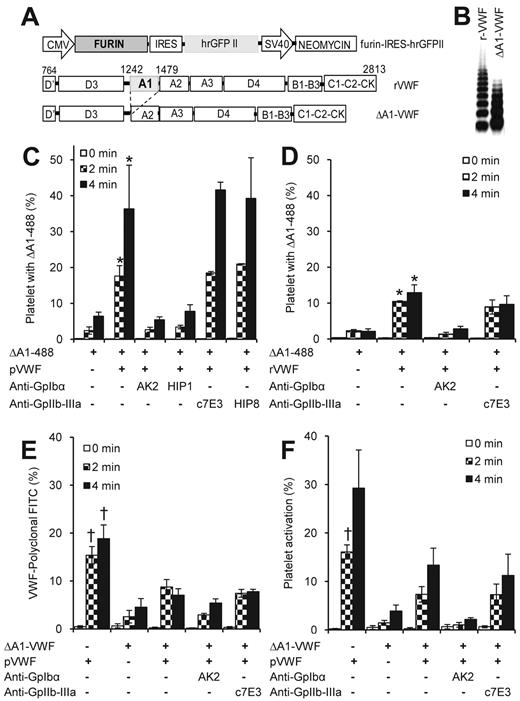
Augmented pVWF-488 binding on addition of unlabeled pVWF in Figure 1 may be the result of the self-association of VWF on platelet receptor GpIbα.12,13 To test this possibility, we expressed multimeric rVWF and VWF that lacked the A1 domain (ΔA1-VWF; Figure 2). To this end, a CHO cell line overexpressing furin was first created (Figure 2A). These cells are green because of the presence of the green fluorescent protein variant (hrGFP-II) that is driven by the internal ribosome entry site. Recombinant VWF expressed in these CHO-furin cells lacked the propeptide sequence (supplemental Figure 5A). Although full-length rVWF expressed all 2050 amino acids of the mature protein, ΔA1-VWF lacked amino acids from 1242 to 1479. Both proteins were expressed and purified from CHO cells as multimers with up to 5 to 8 protomer units (Figure 2B). As expected, ΔA1-VWF did not recognize a mAb directed against the VWF A1-domain (supplemental Figure 5B), and it also did not bind platelets via GpIbα (supplemental Figure 5C). Both proteins appear to be folded well because cleavage by metalloprotease ADAMTS13 required the addition of 1.5M urea to expose the buried Tyr1605-Met1606 cleavage site (supplemental Figure 5D). These proteins also bound FVIII via the D′D3 domain efficiently (supplemental Figure 5E). Finally, consistent with the literature,26-28 ΔA1-VWF displayed binding for type III collagen (supplemental Figure 5F). Thus, ΔA1-VWF displayed similar properties as rVWF except that it lacked A1-domain function.
Application of ΔA1-488 to monitor VWF self-association. (A) CHO-furin cells were generated by stably transfecting furin-internal ribosome entry site-hrGFP into CHO cells. Both recombinant full-length human VWF (rVWF) and VWF lacking the A1-domain (ΔA1-VWF; ie, amino acids 1242-1479) were expressed in these cells. (B) Western blot shows the multimer distribution of recombinant proteins. (C-D) ΔA1-VWF was labeled with Alexa-488 to produce ΔA1-488. ΔA1-488 bound platelets in the presence of both 5 μg/mL plasma (C, pVWF) and recombinant full-length VWF (D, rVWF). Shear rate is 9600/s. ΔA1-488 binding to platelets is the result of VWF self-association. Binding was blocked by mAbs against GpIbα but not GpIIb-IIIa. (E-F) A total of 5 μg/mL pVWF was shear mixed with 5 μg/mL ΔA1-VWF at 9600/s in the presence or absence of anti-GpIbα/GpIIb-IIIa mAbs for the indicated time. Two samples were withdrawn. The total VWF bound to platelets was measured in one sample using FITC-conjugated rabbit anti–human VWF polyclonal Ab (E). Platelet activation was quantified in the other using annexin-FITC. Both pVWF binding and cell activation were partially reduced by ΔA1-VWF in a GpIbα-dependent manner. Shear protocol in all panels is identical to Figure 1. *P < .05 with respect to all other treatments except blocking with GpIIb-IIIa mAbs. †P < .05 with respect to all other treatments.
Application of ΔA1-488 to monitor VWF self-association. (A) CHO-furin cells were generated by stably transfecting furin-internal ribosome entry site-hrGFP into CHO cells. Both recombinant full-length human VWF (rVWF) and VWF lacking the A1-domain (ΔA1-VWF; ie, amino acids 1242-1479) were expressed in these cells. (B) Western blot shows the multimer distribution of recombinant proteins. (C-D) ΔA1-VWF was labeled with Alexa-488 to produce ΔA1-488. ΔA1-488 bound platelets in the presence of both 5 μg/mL plasma (C, pVWF) and recombinant full-length VWF (D, rVWF). Shear rate is 9600/s. ΔA1-488 binding to platelets is the result of VWF self-association. Binding was blocked by mAbs against GpIbα but not GpIIb-IIIa. (E-F) A total of 5 μg/mL pVWF was shear mixed with 5 μg/mL ΔA1-VWF at 9600/s in the presence or absence of anti-GpIbα/GpIIb-IIIa mAbs for the indicated time. Two samples were withdrawn. The total VWF bound to platelets was measured in one sample using FITC-conjugated rabbit anti–human VWF polyclonal Ab (E). Platelet activation was quantified in the other using annexin-FITC. Both pVWF binding and cell activation were partially reduced by ΔA1-VWF in a GpIbα-dependent manner. Shear protocol in all panels is identical to Figure 1. *P < .05 with respect to all other treatments except blocking with GpIIb-IIIa mAbs. †P < .05 with respect to all other treatments.
We monitored the binding of ΔA1-VWF conjugated with Alexa-488 (termed ΔA1-488) to platelets in the presence of either pVWF (Figure 2C) or rVWF (Figure 2D). In both cases, whereas the binding of ΔA1-488 to platelets was low, this was augmented in the presence of unlabeled full-length p/rVWF. The low ΔA1-488 binding observed in the absence of full-length VWF is probably the result of residual VWF present in the diluted PRP used in this assay. ΔA1-488 binding in the presence of full-length p/rVWF was dependent on VWF-GpIbα interaction, and it was independent of platelet GpIIb-IIIa. These data are consistent with the proposition that VWF self-associates on platelet GpIbα on shear.
The self-association of VWF on platelets is a physical phenomenon that can proceed in the absence of platelet activation or other plasma components. In this regard, ΔA1-488 binding to formalin-fixed platelets was enhanced on addition of pVWF. This binding proceeded efficiently at shear rate of more than 5800/s, and it was abrogated by anti-GpIbα mAb (supplemental Figure 6).
In additional runs, unlabeled ΔA1-VWF was added in the presence of pVWF to determine its effect on platelet activation. Here, we observed a decrease in VWF binding to platelets (Figure 2E) and platelet activation (Figure 2F) on addition of ΔA1-VWF. Regardless of the reduced binding, significant platelet activation was noted in the presence of ΔA1-VWF. Overall, the studies demonstrate the utility of ΔA1-488 for the detection of protein self-association. Association between ΔA1-VWF and pVWF in suspension under shear may reduce/mask the A1-domain of pVWF. This may account for the reduced cell activation in studies with unlabeled ΔA1-VWF.
VWF self-association occurs in whole blood
The binding of ΔA1-488 to platelets was measured both in the presence of blood plasma and in whole blood (Figure 3). In some runs (Figure 3A), FITC-conjugated pVWF (pVWF-FITC) was sheared with platelets in the presence of VWF-deficient plasma. Platelets were labeled with CD31-PE, and annexin-PE-Cy5 was used to monitor cell activation. Here, consistent with the proposition that VWF self-association can take place in plasma, we observed augmented VWF-FITC binding to platelet GpIbα on addition of unlabeled pVWF. In other experiments, shearing PRP with ΔA1-488 also resulted in the binding of the fluorescent VWF-mutant to platelets (Figure 3B). Here, confocal microscopy images show that ΔA1-488 localized on only a small fraction of platelet GpIbα receptors, and this protein appeared in the form of clusters that would be expected if it is self-associated. Fluid shear is required for this self-association process because VWF clusters containing ΔA1-488 do not appear on the surface of platelets held under static conditions (supplemental Figure 7). ΔA1-488 also bound platelets in the milieu of whole blood (Figure 3C-D). Fluid shear both augmented the binding of ΔA1-488 to platelets and cell activation. Approximately 50% of platelets that were activated were bound to ΔA1-488, and this suggests an important role of VWF self-association in triggering SIPAct. Control cytometry dot plots for the whole blood studies are provided in supplemental Figure 8.
VWF self-association in the presence of plasma proteins and in whole blood. (A) A total of 30 × 106 platelets/mL was diluted in 100% VWF-deficient plasma, instead of HEPES buffer, along with 10 μg/mL FITC-conjugated pVWF (pVWF-FITC). The sample was sheared at 9600/s for 4 minutes, either in the presence or absence of 10 μg/mL unconjugated pVWF and anti-GpIbα blocking mAb HIP1. A total of 60 μg/mL 7E3 was present during all runs to prevent VWF interaction with GpIIb-IIIa. Samples withdrawn were incubated for 5 minutes with CD31-PE and annexin-PE/Cy5 before cytometry analysis. *P < .05 with respect to all other treatments. (B) A total of 5 μg/mL ΔA1-488 (green) was added to PRP and sheared at 9600/s for 2 minutes. Samples fixed in 0.5% paraformaldehyde were labeled with VM16d (anti-GpIbα mAb, red) and examined using confocal microscopy. (C) A total of 5 μg/mL ΔA1-488 was added to whole blood and subjected to shear at 4700/s. Samples withdrawn at 4 minutes was incubated with CD31 PerCP-eFluor 710 to identify platelets and annexin-PE to monitor cell activation. Cytometry dot plot shows ΔA1-488 bound to inactive (top left quadrant) and activated platelets (top right). (D) Distribution of active platelets either with or without ΔA1-488 bound. Percentage is based on total number of platelets read in the cytometer. *Platelet activation (top right plus bottom right) is higher at 4 minutes compared with 0 minutes at this shear rate (P < .05). †Platelet activation at 4 minutes at this shear rate is higher compared with 300/s (P < .05).
VWF self-association in the presence of plasma proteins and in whole blood. (A) A total of 30 × 106 platelets/mL was diluted in 100% VWF-deficient plasma, instead of HEPES buffer, along with 10 μg/mL FITC-conjugated pVWF (pVWF-FITC). The sample was sheared at 9600/s for 4 minutes, either in the presence or absence of 10 μg/mL unconjugated pVWF and anti-GpIbα blocking mAb HIP1. A total of 60 μg/mL 7E3 was present during all runs to prevent VWF interaction with GpIIb-IIIa. Samples withdrawn were incubated for 5 minutes with CD31-PE and annexin-PE/Cy5 before cytometry analysis. *P < .05 with respect to all other treatments. (B) A total of 5 μg/mL ΔA1-488 (green) was added to PRP and sheared at 9600/s for 2 minutes. Samples fixed in 0.5% paraformaldehyde were labeled with VM16d (anti-GpIbα mAb, red) and examined using confocal microscopy. (C) A total of 5 μg/mL ΔA1-488 was added to whole blood and subjected to shear at 4700/s. Samples withdrawn at 4 minutes was incubated with CD31 PerCP-eFluor 710 to identify platelets and annexin-PE to monitor cell activation. Cytometry dot plot shows ΔA1-488 bound to inactive (top left quadrant) and activated platelets (top right). (D) Distribution of active platelets either with or without ΔA1-488 bound. Percentage is based on total number of platelets read in the cytometer. *Platelet activation (top right plus bottom right) is higher at 4 minutes compared with 0 minutes at this shear rate (P < .05). †Platelet activation at 4 minutes at this shear rate is higher compared with 300/s (P < .05).
VWF self-association depends on the applied shear stress but not the shear rate
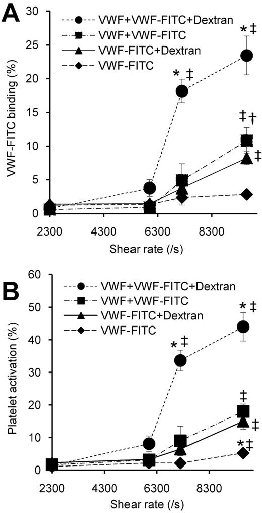
VWF binding to platelets25 and platelet activation13 are augmented by the applied shear stress. We tested whether VWF self-association is also shear-stress dependent by monitoring the enhanced binding of pVWF-FITC to platelets on addition of unlabeled pVWF (Figure 4). Whereas shear rate (G) was varied in some runs, media viscosity (μ) was increased in others by addition of dextran; 1.5% dextran doubles medium viscosity. Thus, the magnitude of applied shear stress (τ) at a given shear rate (τ = μ × G) is doubled for the Newtonian fluid used in this study. Here, VWF-FITC binding and platelet activation showed very similar dependence on both the applied shear rate and shear stress. VWF self-association, quantified as the extent of pVWF-FITC binding in the presence of unlabeled pVWF compared with that in the absence of pVWF, was augmented significantly above all other treatments on addition of dextran at shear rate more than 7200/s. The data suggest that VWF-self-association, like SIPAct, is a shear-stress–dependent phenomenon.
Shear stress dependence of VWF self-association. A total of 30 × 106 platelets/mL was subjected to a range of shear rates with 10 μg/mL pVWF-FITC and 60 μg/mL 7E3, either in the presence or absence of 10 μg/mL unlabeled pVWF. A total of 1.5% wt/vol dextran was added in some cases to double media viscosity. Platelets withdrawn at 4 minutes were incubated with CD31-PE and annexin-PE/Cy5 before cytometry analysis. (A) VWF-FITC binding to platelets. (B) Platelet activation. pVWF binding to platelets and platelet activation increased above a shear rate of 7200/s. Both VWF-FITC binding to platelets and VWF-self-association were shear stress-dependent processes. *P < .05 with respect to all other treatments at the given shear rate. ‡P < .05 with respect to 2300/s. †P < .05 with respect to pVWF-FITC alone at the same shear rate.
Shear stress dependence of VWF self-association. A total of 30 × 106 platelets/mL was subjected to a range of shear rates with 10 μg/mL pVWF-FITC and 60 μg/mL 7E3, either in the presence or absence of 10 μg/mL unlabeled pVWF. A total of 1.5% wt/vol dextran was added in some cases to double media viscosity. Platelets withdrawn at 4 minutes were incubated with CD31-PE and annexin-PE/Cy5 before cytometry analysis. (A) VWF-FITC binding to platelets. (B) Platelet activation. pVWF binding to platelets and platelet activation increased above a shear rate of 7200/s. Both VWF-FITC binding to platelets and VWF-self-association were shear stress-dependent processes. *P < .05 with respect to all other treatments at the given shear rate. ‡P < .05 with respect to 2300/s. †P < .05 with respect to pVWF-FITC alone at the same shear rate.
Drag force applied on platelet GpIbα triggers mechanotransduction
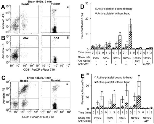
We hypothesize that the hydrodynamic force applied on platelet GpIbα after VWF self-association on the receptor may trigger mechanotransduction. This outside-in signaling then contributes to SIPAct. To test this possibility, beads bearing both immobilized VWF (pVWF-beads) and anti-GpIbα mAb AP-1 (anti-GpIbα-beads) were created (Figure 5). These beads were mixed with platelets at various shear rates.
Platelet mechanotransduction by beads bearing immobilized VWF and anti-GpIbα mAb. A total of 107 platelets/mL were mixed with beads bearing either pVWF (A-B,D) or mAbs AP-1 against GpIbα (C,E) at various shear rates. Platelet:bead ratio was approximately 5:1. Anti-GpIbα blocking mAb AK2 was present in panel B, whereas there were no blocking reagents in panels A and C. Samples withdrawn at indicated times were incubated with CD31 PerCP-eFluor 710 and annexin-PE for 5 minutes and analyzed using flow cytometry. Cytometry forward versus side scatter was used to gate either singlet beads (Ai,Bi,Ci) or singlet platelets (Aii,Bii,Cii). (D-E) Distribution of active platelets that were either free (Aii,Bii,Cii top right quadrant) or bound (Ai,Bi,Ci top right quadrant) to beads was quantified. Percentage is based on total number of platelets in the sheared mixture read in cytometer. Beads without platelets appear in bottom left quadrant of Ai, Bi, Ci. *Platelet activation is higher at 3 minutes compared with 0 minutes at these shear rates (P < .05).
Platelet mechanotransduction by beads bearing immobilized VWF and anti-GpIbα mAb. A total of 107 platelets/mL were mixed with beads bearing either pVWF (A-B,D) or mAbs AP-1 against GpIbα (C,E) at various shear rates. Platelet:bead ratio was approximately 5:1. Anti-GpIbα blocking mAb AK2 was present in panel B, whereas there were no blocking reagents in panels A and C. Samples withdrawn at indicated times were incubated with CD31 PerCP-eFluor 710 and annexin-PE for 5 minutes and analyzed using flow cytometry. Cytometry forward versus side scatter was used to gate either singlet beads (Ai,Bi,Ci) or singlet platelets (Aii,Bii,Cii). (D-E) Distribution of active platelets that were either free (Aii,Bii,Cii top right quadrant) or bound (Ai,Bi,Ci top right quadrant) to beads was quantified. Percentage is based on total number of platelets in the sheared mixture read in cytometer. Beads without platelets appear in bottom left quadrant of Ai, Bi, Ci. *Platelet activation is higher at 3 minutes compared with 0 minutes at these shear rates (P < .05).
Because the drag force applied by VWF immobilized on beads is substantially higher than free VWF in solution,29 we expected to observe enhanced platelet activation at lower shear rates in studies with pVWF-beads (Figure 5A-B,D). Consistent with this proposition, after 3 minutes of shear at 1863/s, 17% of the platelets were activated in the presence of pVWF-beads. Negligible platelet activation occurs at these shear rates in the absence of beads (Figure 4). Approximately two-thirds of the activated platelets were in solution, free of the pVWF-beads. This is probably the result of the labile nature of the VWF-GpIbα bond. The extent of platelet activation increased with the magnitude of applied shear rate, with significant levels of platelet activation being observed at shear rates down to 932/s (shear stress of ∼9 dyne/cm2). In control runs, both VWF-bead binding to platelets and cell activation were blocked by anti-VWF A1 domain mAb AVW-3 and anti-GpIbα mAb AK2.
In studies performed with anti-GpIbα-beads also, we observed enhanced platelet activation that was shear mediated. These beads triggered platelet activation at shear rates down to 233/s (Figure 5C,E). In this case, a majority of the activated platelets were associated with the beads unlike the previous case where they were in solution. This may be the result of the high affinity antigen-antibody interaction that creates a stronger molecular bond. Platelet binding to beads and cell activation were abrogated by soluble anti-GpIbα mAb AP-1.
Discussion
This study demonstrates that robust, shear-dependent, VWF self-association takes place on the platelet GpIbα receptor. VWF self-association occurs in the milieu of plasma and also in whole blood. Both the magnitude of VWF self-association and platelet activation vary as a function of the applied shear stress, but not shear rate. They proceed optimally above 60 dyne/cm2. The strong correlation between the shear requirements for VWF self-association and SIPAct13 suggests that VWF self-association is a necessary process that accompanies platelet activation. These overall conclusions were consistently observed regardless of the FITC/Alexa-488 labeling protocol used to detect VWF-binding or the annexin-fluorophore used to measure platelet activation. Taken together with prior publications, the results suggest 3 independent roles for fluid shear in augmenting: (1) the affinity of VWF-GpIbα binding, (2) VWF self-association on GpIbα, and (3) platelet mechanotransduction via GpIbα.
Shear dependent changes in VWF conformation enhances VWF-GpIbα binding
The application of fluid shear results in conformation changes in VWF. Such conformation changes in solution result in large-scale elongation/uncoiling of VWF30 and the exposure of protein hydrophobic domains21,31 at shear rates more than 5000/s (shear stress ∼ 50 dyne/cm2). Small-angle scattering studies show that smaller-length scale changes may occur at lower shear rates.21 Shear driven protein conformation change also enhances the binding of VWF to platelet GpIbα,25 and it exposes a cryptic proteolytic site that is hidden within the native A2-domain.32 Point mutations in VWF or GpIbα associated with von Willebrand disease, either von Willebrand disease type 2B or platelet-type von Willebrand disease, enhance VWF binding affinity and platelet activation at low shear rates.33 In this context, because mutations in GpIbα are unlikely to affect VWF self-association rates, the data suggest that the shear-driven binding of VWF to platelets is separable from protein self-association.
The precise conformation changes that promote VWF binding and proteolysis have not been determined. Choi et al34 suggest that shear application between 2000 and 5000/s trigger protein conformation changes such that several amino acids in the D3 and C domains of VWF undergo shear-induced disulfide bond formation. Others also suggest that the native VWF-D′D3 domain shields the VWF-A1 domain from binding platelet GpIbα.35 Finally, the proteolytic processing of VWF by ADAMTS13 under shear is enhanced by the binding of FVIII to the VWF-D′D3 domain.36 Thus, one possible model suggests that shear promotes the rearrangement of individual domains within the globular section of VWF, which contains both D′D3 and A1.21 This then enhances both VWF A1-domain binding affinity for platelet GpIbα and proteolysis of VWF-A2.
VWF self-association on platelets is a shear stress-dependent process
In addition to conformation changes in solution, studies that apply shear to VWF and then analyze protein size using light scattering show that VWF subjected to shear more than 2300/s undergoes aggregation or self-association.13 Although it has been suggested that VWF self-association may occur in the absence of shear,37 these authors present data from enzyme-linked immunosorbent assay studies where VWF is immobilized on plastic plates. Such immobilization itself may alter the native conformation of the protein and promote self-association. In our system, which contains platelets, however, it is fluid shear stress that drives the accumulation of more than one VWF molecule on a single platelet GpIbα receptor. This is particularly evident in studies where variation in media viscosity using dextran enhances the extent of self-association (Figure 4). In studies performed with PRP also, ΔA1-488 binding to platelets was only observed in the presence of shear (Figure 3B).
The mechanism of VWF self-association is not fully established. Savage et al12 demonstrated that, although platelets alone fail to bind surfaces coated with immobilized ΔA1-VWF, the adhesion of these cells to this substrate is enhanced if either VWF or ΔA3-VWF (VWF lacking the A3 domain) is present in solution. Thus, self-association does not involve the homotypic binding of VWF-A1 or -A3 domains. Ulricht et al37 suggest that multiple domain interactions probably contribute to VWF self-association. These authors demonstrate that proteolytically generated VWF fragments SpII (amino acids 1366-2050) and SpIII (amino acids 1-1365) in biotinylated forms are able to bind both immobilized SpII and SpIII in enzyme-linked immunosorbent assay. Finally, using a viscometer to apply shear and using MALDI-TOF/TOF for analysis, it has been shown that free thiols on the VWF surface may form new disulfide bonds on shear application.34 These Cys residues, which are susceptible to thiol-disulfide exchange, are located in the C-domains and also the D3 domain of plasma VWF. Overall, although there is partial information on the mechanism of VWF self-association, further development of ΔA1-488 and analogs of this reagent may reveal more precisely the molecular basis of this phenomenon. Such studies may reveal the extent to which VWF self-association in solution under shear precedes the accumulation of the molecular complex on platelet GpIbα. Alternatively, it can allow us to determine whether VWF recruited on GpIbα has an altered conformation that is more prone to self-association compared with the native protein.
VWF self-association contributes to shear-induced platelet activation
The overall size of GpIbα may be a key feature controlling its ability to transmit mechanical outside-in signals. Electron microscopy observations of GpIbα suggest that the globular head section of this glycoprotein resembles a small sphere of radius 4.5 nm and that this domain is attached to cells via a slender stalk of length 35 nm.38 Our microscopy images reveal that self-associated VWF bound to GpIbα resembles a clump, with only a few of the platelet GpIbα receptors being occupied by the aggregated protein (Figure 3B). Here, self-associated VWF effectively increases the “apparent” size of the GpIbα globular head section.
Based on our earlier work,29 we estimate the magnitude of force applied on platelet GpIbα under the experimental conditions described in this manuscript (Table 1). According to this theory, at a shear stress of 100 dyne/cm2 (shear rate of 10 000/s, 1 cp media viscosity), the maximum force applied on native GpIbα on platelets is small at approximately 0.1 pN. Attachment of a single VWF (radius ∼ 100 nm) to this receptor at the same shear stress increases the magnitude of applied shear force to 8 pN. Assuming that the self-associated VWF is 200 nm in size, we estimate that GpIbα bound to the self-associated protein complex experiences a peak force of 23 pN. A similar magnitude of force is applied when polystyrene beads with immobilized VWF or anti-GpIbα mAb are shear mixed with platelets at a low shear stress of 2 dyne/cm2. Attachment of GpIbα receptors located on adjacent platelets to a single VWF complex at 100 dyne/cm2 can increase the magnitude of applied force to approximately 490 pN. Although some of these force estimates are large compared with the measured VWF-GpIbα bond rupture force39 (11.4 pN), it is necessary to note that these are peak instantaneous forces and they may be distributed over multiple GpIbα receptors on cells. Compared with these forces that are applied to platelets in suspension, forces in the order of 15 to 100 pN are applied to platelet-substrate bonds as these cells translocate on immobilized VWF at a shear rate of 2000/s.40 Overall, VWF self-association on platelets in solution can enhance the magnitude of the applied shear force on GpIbα by increasing the apparent/effective radius of gyration at the receptor's globular head section. Under this condition, the mechanical force applied on platelet GpIbα in solution can rival that of platelets translocating on a substrate with immobilized VWF. Finally, the study also demonstrates the utility of antibody-coated beads to study receptor-mediated mechanotransduction.
Estimated force applied on platelet GpIbα*
| . | Radius 1 (a1), μm . | Radius 2 (a2), μm . | Separation distance (d), μm . | Shear stress, dyne/cm2 . | Peak force F, pN† . |
|---|---|---|---|---|---|
| GpIbα alone | 1.5 | 0.0045 | 0.035 | 100 | 0.085 |
| GpIbα with single VWF | 1.5 | 0.1 | 0.035 | 100 | 7.6 |
| GpIbα with self-associated VWF | 1.5 | 0.2 | 0.035 | 100 | 23.2 |
| GpIbα pulled by second platelet | 1.5 | 1.5 | 0.035 to 1 | 100 | 430 to 495 |
| GpIbα pulled by polystyrene bead | 1.5 | 3 | 2 | 16 |
The force applied between two spheres with radius a1 and a2 separated by a distance d was calculated.29 Applied shear stress is 100 dyne/cm2 (ie, shear rate = 10 000/s and medium viscosity = 1 cp) in all cases except for the case of platelet-bead interactions where shear stress was 2 dyne/cm2. Platelets are modeled as sphere with radius a1 = 1.5 μm. a2 and d are varied according to measured molecular dimensions of GpIbα, VWF, platelets, and hypothetical size of self-associated VWF.
In all cases, it is assumed that VWF or protein complex is bound to a single GpIbα receptor.
Application of mechanical signals via GpIbα can trigger cell signaling events that lead to platelet activation and subsequent aggregation. In this regard, cell activation involves intracellular calcium elevation and a complex interaction of numerous molecular pathways including, but not limited to, the phosphoinositide-3-kinase-Akt, Src/phospholipase C, immunoreceptor tyrosine-based activation motif, adhesion and degranulation promoting adapter protein, mitogen-activated protein kinase, thromboxane A2, and adenosine diphosphate driven pathways.41 Together, these pathways contribute to the expression of phosphatidylserine on the outer leaflets of activated platelets and changes in the ligand-binding epitope of platelet receptor GpIIb-IIIa. Platelet receptors GpIbα and GpIIb-IIIa together mediate stable platelet aggregation under shear.13,42 In addition to these processes, it is suggested that shear-induced platelet-VWF interactions trigger GpIbα ectodomain shedding, and this may also contribute to regulating the kinetics of cell adhesion.43
In conclusion, we demonstrate that VWF self-associates on platelet GpIbα. Such self-association increases the multivalency of VWF, and this probably enhances the avidity of VWF-GpIbα interactions in the vasculature. High-binding avidities may be necessary for the sustained application of forces that contribute to SIPAct. In support of this, some clinical studies suggest that platelet activation under shear may contribute to the pathogenesis of acute myocardial infarction44 and cerebral ischemia.45 It is also suggested in an arterial stenosis model that SIPAct may act in synergy with vascular injury to regulate platelet aggregation in circulation.46 Alternatively, enhanced VWF binding via self-association to single platelets or bridging between 2 platelets may enhance VWF A2-domain proteolysis by ADAMTS13.8 In this regard, abnormal high shear stress in the vasculature, which is associated with aortic stenosis47 and pulmonary arterial hypertension,48 results in the loss of large VWF multimers. This can contribute to bleeding. Future studies in appropriate disease models will be necessary to delineate between the precise contributions of VWF self-association to platelet activation and its effect on protein multimer distribution.
An Inside Blood analysis of this article appears at the front of this issue.
The online version of this article contains a data supplement.
The publication costs of this article were defrayed in part by page charge payment. Therefore, and solely to indicate this fact, this article is hereby marked “advertisement” in accordance with 18 USC section 1734.
Acknowledgment
This work was supported by the National Institutes of Health (grants HL 77258 and HL 76211).
National Institutes of Health
Authorship
Contribution: K.M.D. designed and performed experiments and wrote the manuscript; I.S. designed and performed experiments and wrote the first draft of the manuscript; N.M. performed experiments; and S.N. designed research, performed experiments, coordinated project activities, and wrote the paper.
Conflict-of-interest disclosure: The authors declare no competing financial interests.
Correspondence: Sriram Neelamegham, 906 Furnas Hall, State University of New York, Buffalo, NY 14260; e-mail: neel@buffalo.edu.


This feature is available to Subscribers Only
Sign In or Create an Account Close Modal