A 16-year-old female with AIDS presented with 3 months' history of fever, loose stools, and significant weight loss. She had marked pallor, erythematous plaques on the face and neck, enlarged liver and spleen 2.0 cm, below the respective costal margins, and bilateral cervical lymphadenopathy. Both her parents had been suffering from AIDS and subsequently died of disseminated tuberculosis.
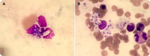
Her chest x-ray was normal and ultrasonography affirmed hepatosplenomegaly. HIV serology was positive and CD4 count was 0.012 × 10⁹/L. Complete blood counts showed pancytopenia with hemoglobin of 57 g/L, total leukocyte count of 1.1 × 10⁹/L, and platelet count of 18 × 109/L. The peripheral blood film (panel A) revealed monocytes and neutrophils with the presence of yeast-like intracellular organisms, 2 to 4 μm in diameter. These organisms had an eccentric chromatin and pseudocapsule, confirming to the morphology of Histoplasma capsulatum. There was evidence of histoplasmosis in 5% of the leukocytes. Bone marrow aspirate smear (panel B) and biopsy showed numerous histiocytes containing abundant intracellular organisms. These organisms stained bright pink on periodic acid-Schiff stain.
Treatment with amphotericin B was started immediately; however, the patient succumbed to the infection.
A high index of suspicion and a careful examination of the peripheral blood smear may disclose histoplasmosis.
Many Blood Work images are provided by the ASH IMAGE BANK, a reference and teaching tool that is continually updated with new atlas images and images of case studies. For more information or to contribute to the Image Bank, visit www.ashimagebank.org.


This feature is available to Subscribers Only
Sign In or Create an Account Close Modal