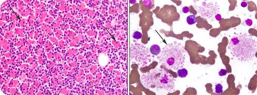
A 63-year-old male presented with progressive weakness, weight loss, and bone pains. Previous investigations showed normochromic normocytic anemia and a raised erythrocyte sedimentation rate. Further laboratory work-up revealed IgG κ monoclonal gammopathy in the serum. The IgG level was 31.90 g/L (normal, 6.94-16.18 g/L). Other laboratory tests showed hemoglobin 91.0 g/L, white blood cell count 8.9 × 10⁹/L, platelet count 191 × 109/L, serum creatinine 247.52 μmol/L, and normal serum albumin and calcium. Skeletal survey showed generalized osteoporosis and multiple lytic lesions throughout the skeleton including the skull, mandible, ribs, scapulae, and left proximal humerus. Bone marrow contained 36% plasma cells along with numerous globular and crystalline-laden histiocytes (see arrows). The histiocytes were positive for CD68 immunostain and plasma cells expressed positivity for CD138. A diagnosis of crystal storing histiocytosis (CSH) in multiple myeloma was made.
CSH is a rare morphologic finding characterized by accumulation of monoclonal immunoglobulin light chains within histiocytes. It is seen in B-cell lymphoproliferative disorders, most commonly in multiple myeloma, and is usually associated with IgG paraprotein and kappa light chain accumulation. CSH has also been seen in other tissues including kidneys, spleen, lymph nodes, skin, thyroid, lungs, and gastrointestinal tract. There is no intervention aimed specifically at CSH. Therapy is directed against the underlying hematological malignancy.
Many Blood Work images are provided by the ASH IMAGE BANK, a reference and teaching tool that is continually updated with new atlas images and images of case studies. For more information or to contribute to the Image Bank, visit www.ashimagebank.org.


This feature is available to Subscribers Only
Sign In or Create an Account Close Modal