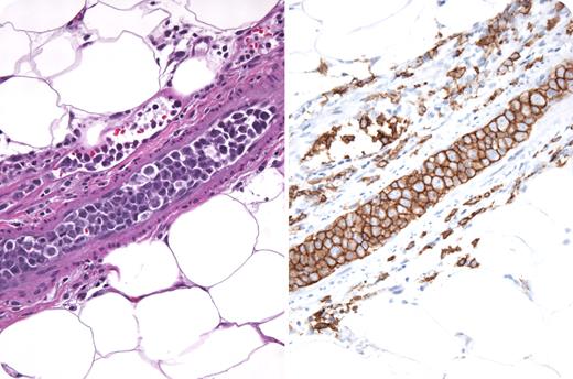
A 74-year-old woman presented with 6 months of fatigue, fevers, chills, dyspnea, weight loss, and blurry vision. Physical examination revealed only fever and mild splenomegaly. She had mild anemia, thrombocytopenia, lactic dehydrogenase that was 3 times normal, a markedly increased sedimentation rate, and hypoalbuminemia. CT/MRI of the head and body revealed no additional abnormalities. Bone marrow examination demonstrated moderate fibrosis, normal cytogenetics, and no dysplasia. She developed new skin nodules and a biopsy was performed. The photograph shows intravascular “plugs” composed of large atypical CD20 B cells with aberrant expression of CD5. Diagnosed as intravascular lymphoma (IVL), she received chemotherapy with rituximab (EPOCH-R) that resulted in a complete resolution of symptoms and disappearance of the skin nodules. IVL are rare, aggressive extranodal diffuse large cell lymphomas, characterized by intravascular malignant lymphoid tumor cells with a remarkable sparing of lymph nodes and nonvascular organ parenchyma. IVL are generally positive for CD19, CD20, CD22, CD79a, and occasionally CD5. Certain tumor cell surface molecules, such as CD11a/49d, may bind to CD 54/106 on endothelial cells and result in vascular compartmentalization. Clinical presentation is diagnostically challenging and nonspecific; nearly half of cases present with fever of unknown origin. Anemia, high lactic acid dehydrogenase levels, and high sedimentation rates are typical. CNS manifestations are also common and often fatal. More than half of cases are diagnosed postmortem; therefore, a high index of clinical suspicion is paramount. Median survival is 5.5 months; however, anthracycline-based regimens in combination with rituximab have proven successful. CNS-directed therapy is recommended for patients with neurologic symptoms.
A 74-year-old woman presented with 6 months of fatigue, fevers, chills, dyspnea, weight loss, and blurry vision. Physical examination revealed only fever and mild splenomegaly. She had mild anemia, thrombocytopenia, lactic dehydrogenase that was 3 times normal, a markedly increased sedimentation rate, and hypoalbuminemia. CT/MRI of the head and body revealed no additional abnormalities. Bone marrow examination demonstrated moderate fibrosis, normal cytogenetics, and no dysplasia. She developed new skin nodules and a biopsy was performed. The photograph shows intravascular “plugs” composed of large atypical CD20 B cells with aberrant expression of CD5. Diagnosed as intravascular lymphoma (IVL), she received chemotherapy with rituximab (EPOCH-R) that resulted in a complete resolution of symptoms and disappearance of the skin nodules. IVL are rare, aggressive extranodal diffuse large cell lymphomas, characterized by intravascular malignant lymphoid tumor cells with a remarkable sparing of lymph nodes and nonvascular organ parenchyma. IVL are generally positive for CD19, CD20, CD22, CD79a, and occasionally CD5. Certain tumor cell surface molecules, such as CD11a/49d, may bind to CD 54/106 on endothelial cells and result in vascular compartmentalization. Clinical presentation is diagnostically challenging and nonspecific; nearly half of cases present with fever of unknown origin. Anemia, high lactic acid dehydrogenase levels, and high sedimentation rates are typical. CNS manifestations are also common and often fatal. More than half of cases are diagnosed postmortem; therefore, a high index of clinical suspicion is paramount. Median survival is 5.5 months; however, anthracycline-based regimens in combination with rituximab have proven successful. CNS-directed therapy is recommended for patients with neurologic symptoms.
Many Blood Work images are provided by the ASH IMAGE BANK, a reference and teaching tool that is continually updated with new atlas images and images of case studies. For more information or to contribute to the Image Bank, visit www.ashimagebank.org.
National Institutes of Health


This feature is available to Subscribers Only
Sign In or Create an Account Close Modal