Abstract
Abstract 399
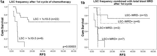
In acute myeloid leukemia (AML), relapses originate from the outgrowth of therapy surviving leukemic blasts know as minimal residual disease (MRD). Accumulating evidence shows that leukemia initiating cells or leukemic stem cells (LSCs) are responsible for persistence and outgrowth of AML. Monitoring LSCs during and after therapy might thus offer accurate prognostic information. However, as LSCs and hematopoietic stem cells (HSCs) both reside within the immunophenotypically defined CD34+CD38- compartment, accurate discrimination between LSCs and HSCs is required. We previously showed that within the CD34+CD38- stem cell compartment, LSCs can be discriminated from HSC by aberrant expression of markers (leukemia associated phenotype, LAP), including lineage markers like CD7, CD19 and CD56 and the novel LSC marker CLL-1 (van Rhenen, Leukemia 2007, Blood 2007). In addition, we reported that flowcytometer light scatter properties add to even better detection of LSCs, allowing LSCs detection in AML cases lacking LAP (ASH abstract 1353, 2008). Using this gating strategy, we determined LSC frequency in 64 remission bone marrow samples of CD34+ AML patients. A stem cell compartment was defined as a minimum of 5 clustered CD34+CD38- events with a minimal analyzed number of 500,000 white blood cells. After first cycle of chemotherapy, high LSC frequency (>1 × 10⁻³) clearly predicted adverse relapse free survival (RFS, figure 1a). LSC frequency above cut-off led to a median RFS of 5 months (n=9), while patients with LSC frequency below cut-off (n=22) showed a significantly longer median RFS of >56 months (p=0.00003). In spite of the relatively low number of patients, again a high LSC frequency (>2 × 10⁻⁴) after the second cycle and after consolidation therapy predicted worse RFS: after second cycle, median RFS was 6 months (n=9) vs. >43 months for patients with LSC frequency below cut-off (p=0.004). After consolidation, these figures were 6 months (n=7) vs. >32 months (n=6, p=0.03). Although total blast MRD (leukemic blasts as % of WBC) is known to predict survival (N.Feller et al. Leukemia 2004), monitoring LSCs as compared to total blast MRD has two major advantages: the specificity is higher (van Rhenen et al. Leukemia 2007) and well-known LSC makers like CLL-1, CD96 and CD123 can in principle be used for LSC monitoring, but not for total blast MRD detection since these markers are also expressed on normal progenitor cells. On the other hand, LSCs constitute only a small fraction of all leukemic blasts and therefore monitoring total blast MRD may have the advantage of a higher sensitivity. We thus tested the hypothesis that even more accurate prognostic information could be obtained by combining LSC frequency with total blast MRD. Total blast MRD after first cycle was predictive for survival with borderline significance (p=0.08): a cut-off of 0.3% resulted in two patient groups with median RFS of 9 months vs. >56 months. Figure 1b shows the result of the combined data of LSC and MRD frequency after first cycle therapy. We used the terms LSC+ and MRD+ for cell frequencies above cut-off and LSC- and MRD- for those below cut-off. We could clearly identify that apart from LSC+/MRD+ patients, LSC+/MRD- patients too have very poor prognosis, while MRD+/LSC- patients show an adverse prognosis as compared to LSC-/MRD- patients. These results from the first study on the in vivo fate of LSCs during and after therapy, strongly support the hypothesis that in CD34+ AML the leukemia initiating capacity originates from the CD34+CD38- population and is important for tumor survival and outgrowth. These results show that LSC frequency might be superior in predicting prognosis of AML patients in CR as compared to MRD total blast frequency, while the combination of both may offer the most optimal parameter to guide future intervention therapies.
This work was supported by Netherlands Cancer Foundation KWF.
No relevant conflicts of interest to declare.
Author notes
Asterisk with author names denotes non-ASH members.


This feature is available to Subscribers Only
Sign In or Create an Account Close Modal