Abstract
Abstract 3030
Poster Board II-1006
NK cells have therapeutic potential for a wide variety of human malignancies. The major obstacle for adoptive NK cell immunotherapy is obtaining sufficient cell numbers, as these cells represent a small fraction of peripheral white blood cells, expand poorly ex vivo, and have limited life spans in vivo. Common gamma-chain cytokines are important in NK cell activation, maturation, and proliferation. Others have described improved ex vivo expansion of NK cells using soluble cytokines, when cocultured with stimulated peripheral blood mononuclear cells (PBMC) or Epstein Barr Virus (EBV) lymphoblastioid cell lines, or with artificial antigen presenting cells (aAPC) engineered with costimulatory molecules and/or membrane-bound IL-15 (mIL-15). Expansion of NK cells by these methods has been limited by senescence from telomere shortening. To generate clinical-grade T cells for adoptive transfer, our group developed aAPC derived from K562 retrovirally transduced to express the costimulatory molecules CD86 and CD137L. These aAPC were produced as a master cell bank and further genetically modified to express membrane-bound cytokines. Since IL-21 signals via STAT3, and STAT3 is a known activator of telomerase transcription, we investigated whether NK cell expansion with mIL-21 would provide a sustained proliferative advantage over or in combination with mIL-15.
K562 aAPC were retrovirally transduced to express CD64, CD86, CD137L, CD19 (Clone 9), and mIL-15 (Clone 4). These clones were further modified by Sleeping Beauty integration of mIL-21 (Clone 9+IL-21 and Clone 4+IL-21). Freshly isolated PBMC from 5 donors were co-cultured with irradiated K562 aAPC (Clone 4, Clone 4+mIL-21, and Clone 9+mIL-21) at a ratio of 2:1 (aAPC:PBMC) in the presence of 50 IU/ml of rhIL-2. Half of the media was changed every two days and cells were re-stimulated with aAPC every seven days at ratio of 2:1. Cells were counted and phenotyped on day 0, 7, 14, and 21 for CD3, CD16, CD56, NKG2D, KIR (2DL1, 2DL2/3, and 3DL1), and NCR (NKp30, NKp44, NKp46). A preclinical SOP to expand PBMC from a 20 mL blood draw was established and additional donors of known HLA type were expanded with Clone 9+mIL-21 for up to 7 weeks. Cytotoxicity function against K562, 721.221, Raji, and AML targets was measured using the Calcien-AM assay (Invitrogen). Telomere length of expanded and fresh NK cells was measured with the FlouFish assay using the telomere specific FITC conjugated (C3TA2)3 PNA probe.
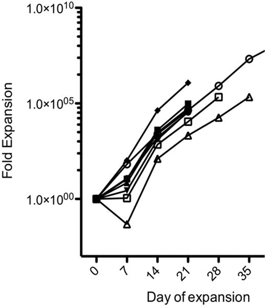
By day 14, aAPCs bearing mIL-21 induced greater total cell expansion than those with mIL-15 alone (188, 2900, and 2281-fold for Clone 4, Clone 4+mIL-21, and Clone 9+mIL-21, respectively). However, PBMC cultured without mIL-15 contained far fewer co-expanding T cells. Exponential expansion continued for up to 7 weeks without evidence of senescence when mIL-21 was present, reaching a mean of 91,566-fold expansion of the CD3−CD16/56+ population at 4 weeks. NK cells expanded with mIL-21 had increased expression of KIR and NCR, and expressed very high CD16 and NKG2D levels. These NK cells showed much higher cytotoxicity against all targets than fresh NK cells, retained KIR inhibition, and demonstrated enhanced killing via ADCC. Furthermore, telomere lengths of NK cells expanded with Clone 9+mIL-21 were longer than that of fresh NK cells or those expanded without mIL-21, perhaps explaining the continued expansion without senescence. Thus, NK cell expansion is improved using aAPCs expressing mIL-21 rather than mIL-15. We are currently establishing a GMP-grade working cell bank of Clone 9+mIL-21 for use in clinical trials.
Funding: Brenda and Howard Johnson Fund, UT MD Anderson Physician Scientist Program
No relevant conflicts of interest to declare.
Author notes
Asterisk with author names denotes non-ASH members.


This feature is available to Subscribers Only
Sign In or Create an Account Close Modal