

Abstract
Abstract 2759
Poster Board II-735
A subpopulation of cells with characteristics of cancer stem cells can be identified by the side population (SP) phenotype that is based on the efflux of the fluorescent dye Hoechst 33342 and detected by flow cytometric analysis. We previously reported the identification of SP cells in 4 human myeloma cell lines (HMCL) and 18 of 21 bone marrow samples from patients with myeloma in which the percentage of SP cells ranged from 0 to 4.9% (median=0.3; mean=0.8; SD=1.2). We now report the mechanism underlying the Hoechst 33342 efflux in SP cells, which may be a contributor to the drug resistance in myeloma cancer stem cells (MCSC). SP and main population (MP) subsets of HMCL (KMS-11 and RPMI-8226), based on Hoechst-33342 staining, were analyzed or sorted by BD FACSAria™ II using a blue and red dual-wavelength analysis after UV excitation. In this study, we examined the activities of three main drug transporter classes associated to SP phenotype – ABCB1 (MDR1), ABCG2 (BCRP1) and ABCC (MRP). Inhibition of the SP phenotype using PSC833, Ko143, and sulfinpyrazone (ABCB1, ABCG2, and ABCC inhibitors respectively) were first tested. Only the Ko143 and sulfinpyrazone were able to inhibit the entire SP phenotype in HMCL. Next, the activity of each transporter in unfractionated, sorted-SP and sorted-MP cells was examined. Relative cellular accumulation of fluorescent substrate for each transporter, namely rhodamine123 1 μg/mL, mitoxantrone 20 μM, and fluo-3 am 1 μg/mL, was determined. Experiments were conducted in the presence or absence of specific inhibitors. There were significant increases of mitoxantrone (P=0.0033** and P=0.0262*) and fluo-3 am (P=0.0006*** and P=0.0152*) in unfractionated RPMI-8226 and KMS-11 in the presence of Ko143 and sulfinpyrazone respectively. In the presence of sulfinpyrazone, benzbromarone, or dipyridamole (ABCC inhibitors), there was a significant increase of fluo-3 am in both SP and MP cells (P<0.05) sorted from KMS-11, however, only the sorted-SP cells showed significant increase of mitoxantrone in the presence of Ko143 (P=0.0462*) (Figure 1A). This suggests that while both ABCC and ABCG2 activity were significant in HMCL, the ABCC activity was similar in both SP and MP cells, whereas the ABCG2 activity was higher in the SP cells. The addition of PSC833 did not increase the accumulation of rhodamine 123 in the unfractionated, sorted-SP, or sorted-MP cells. This confirmed that there is negligible ABCB1 activity in HMCL. Consistent with the functional assay, western blot results revealed no ABCB1 expression in both SP and MP cells from KMS-11. There was a moderate ABCC1 expression in both fractions, and more ABCG2 in SP than MP cells (Figure 1B). This report suggests that while ABCG2 and ABCC activity are present in myeloma cells, ABCG2 is the main contributor to defining the SP by Hoechst efflux and perhaps the molecular determinant of MCSC. However, whether the stem cell related characteristic differences between SP and MP cells are solely because of these transporters is far from conclusive. Understanding these principles will allow investigators to develop therapeutic approaches that capitalize on the uniqueness of MCSC, thereby targeting the malignant cell and sparing normal cells. In conclusion, this report elucidates a distinct drug transporter expression in myeloma SP cells that could be a crucial target in cancer drug development and future clinical trials in patients with myeloma.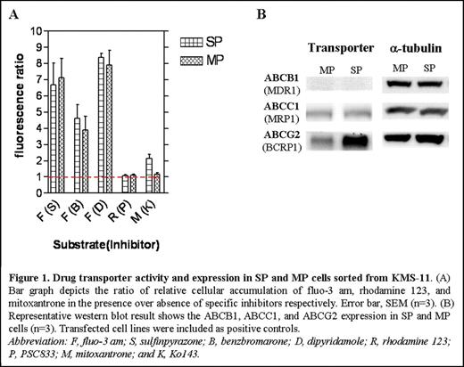

Disclosures:
No relevant conflicts of interest to declare.

This feature is available to Subscribers Only
Sign In or Create an Account Close Modal