Abstract
Local inflammation during cutaneous leishmaniasis is accompanied by accumulation of CD11b+ cells at the site of the infection. A functional role for these monocytic cells in local angiogenesis in leishmaniasis has not been described so far. Here, we show that CD11b+ cells express high levels of the myeloid differentiation antigen carcinoembryonic antigen-related cell adhesion molecule 1 (CEACAM1). In experimental cutaneous leishmaniasis in C57BL/6 wild-type (B6.WT) and B6.Ceacam1−/− mice, we found that only B6.Ceacam1−/− mice develop edemas and exhibit impairment of both hemangiogenesis and lymphangiogenesis. Because CEACAM1 expression correlates with functional angiogenesis, we further analyzed the role of the CD11b+ population. In B6.Ceacam1−/− mice, we found systemic reduction of Ly-6Chigh/CD11bhigh monocyte precursors. To investigate whether CEACAM1+ myeloid cells are causally related to efficient angiogenesis, we used reverse bone marrow transplants (BMTs) to restore CEACAM1+ or CEACAM1− bone marrow in B6.Ceacam1−/− or B6.WT recipients, respectively. We found that angiogenesis was restored by CEACAM1+ BMT only. In addition, we observed reduced morphogenic potential of inflammatory cells in Matrigel implants in CEACAM1− backgrounds or after systemic depletion of CD11bhigh macrophages. Taken together, we show for the first time that CEACAM1+ myeloid cells are crucial for angiogenesis in inflammation.
Introduction
Inflammatory cells such as CD11b+ monocytes have been in the focus of controversial discussions about their role in neovascularization.1-6 Bone marrow (BM) is a source for precursor cells that exhibit considerable plasticity because they can replenish tissue-resident pool leukocytes or merge or transdifferentiate into vascular structures.1-4 Functional correlations between the plasticity and angiogenic properties of specific myelomonocytic populations or macrophage precursors have been detailed in recent reports.5-9 Carcinoembryonic antigen-related cell adhesion molecule 1 (CEACAM1) is engaged in homotypic and heterotypic adhesion processes during cellular growth and proliferation or innate and inflammatory immune responses.10-12 Recently, we showed that CEACAM1 modulates angiogenesis and vascular remodeling.13 However, the involvement of CEACAM1 in angiogenesis has been described from an endothelial-centric view so far. It has remained unclear if CEACAM1-expressing progenitors from blood or BM may play a role in angiogenesis in inflammation.
To address this question we used the experimental model of cutaneous leishmaniasis, known to produce a severe local inflammation at the site of infection mainly caused by infiltrating CD11bhigh cells.14-16 After subcutaneous inoculation, the obligatory intracellular parasite Leishmania major is engulfed by macrophages, where it enters its replication cycle. In later phases of the infection, macrophages can eliminate the parasites after activation by IFN-γ producing T helper type 1 (Th1) cells.17-19 Contrary to the well-documented adaptive immune response in experimental leishmaniasis, the effect of local leukocyte turnover on angiogenesis in this model is unknown. Here, we addressed the question whether myeloid cells modulate angiogenesis in the model of cutaneous leishmaniasis and show that CEACAM1 expression on CD11bhigh cells is essential for angiogenesis in inflammation.
Methods
Mice
Six- to 8-week-old C57BL/6 wild-type mice (B6.WT; CD45.1+ or CD45.2+ on C57BL/6 background) and Ceacam1-deficient mice on C57BL/6 background (B6.Ceacam1−/−) mice were housed under standard conditions in the animal facilities of the Bernhard-Nocht-Institute for Tropical Medicine and the University Medical Center Hamburg-Eppendorf. Experiments were performed according to the guidelines for the care and the use of experimental animals and were approved by the University's ethical boards. B6.Ceacam1−/− mice were generated as described.20,21 Infections with L major (MHOM/IL/81/FE/BNI) Virulent parasites were propagated in blood agar plates, and 3 × 10⁶ promastigotes were subcutaneously injected into the right hind footpad of the mice as described.22
Matrigel implantation assays and cell preparations
Matrigel implantation assays containing Leishmania parasites (4 × 10⁶L major per implant) plus VEGF-C (R&D Systems, Wiesbaden-Nordenstadt, Germany; 500 ng/implant) were performed as described.13 Single-cell suspensions were obtained after enzyme digestion in Dispase (BD Biosciences, Heidelberg, Germany) and Collagenase D (Roche Diagnostics, Mannheim, Germany).
Immunohistochemistry and quantification of edematous areas and vessels
Matrigel implants or footpads were cryopreserved and embedded in OCT compound (Tissue Tek; Diatec, Hallstadt/Bamberg, Germany).
Ten-μm sections were fixed in ice-cold acetone and stained with hematoxylin/eosin (H&E) or the following antibodies: anti–LYVE-1 (lymphatic vessel endothelial hyaluronic acid receptor 1; Biomol, Lörrach, Germany), biotinylated anti–MECA-32, and anti-CD11b antibodies (BD Biosciences). As secondary antibodies, we used anti–rabbit TRITC- or anti–rat Cy5-labeled antibodies from Dianova (Hamburg, Germany), and Alexa-488–labeled streptavidin (BD Biosciences, Karlsruhe, Germany). Nuclei were stained with DAPI (Sigma-Aldrich, Deisenhofen, Germany). Slides were blinded and analyzed on an upright Zeiss Axioskop 2 plus microscope (Carl Zeiss, Jena, Germany), and an upright Leica DM5000B fluorescence microscope (Leica, Wetzlar, Germany) equipped with a Leica DFC 360 FX camera using HCX PL FLUOTAR 10×/0.3, PLAN APO 20×/0.9, HCX PL APO 40×/0.75, and HCX PL APO 100×/1.4-0.7 lenses. Images were acquired using Openlab software (Improvision, Coventry, United Kingdom) and Leica Application Suite software. Image processing was performed with Openlab software and Adobe Photoshop CS3 (Adobe Systems, San Diego, CA). For quantification of the edematous tissue, the DAPI− areas within the infiltrate of the inflamed footpads were calculated with the use of Adobe PhotoshopCS3. Similarly, lymphatic and blood vessel growth were evaluated by quantifying LYVE-1+ and MECA-32+ areas. For each parameter, slides were photographed in a meandering pattern. Per specimen (n = 6 each), at least 20 pictures per footpad were taken for computer-assisted processing.
Flow cytometry analyses
Either single-cell preparations from footpads, digested with collagenase D (Roche Diagnostics), 106 leukocytes or 2 × 10⁵ cells from Matrigel implants were analyzed with the monoclonal antibodies (mAbs) mentioned earlier; in addition, anti–Ly-6C (late monocyte precursor differentiation antigen) mAb (BMA Biomedicals, Augst, Switzerland), Alexa 488–conjugated anti-CEACAM1 (CC1; a kind gift of K. Holmes, University of Colorado Health Sciences Center, Aurora, CO), PerCP-Cy5.5–conjugated anti-CD11b (BD Biosciences), and PE-conjugated anti-CD45.1 (BD Biosciences) were used. Flow cytometry was performed with a FACSCalibur flow cytometer (BD Biosciences). Data were processed with CellQuestPro software (BD Biosciences). For quantification of cells isolated from footpads, latex beads (Sigma-Aldrich) served as internal standards.
Tube formation assay
Cells recovered from Matrigel plugs on day 7 were used in a tube-formation assay adapted from Maruyama et al.5 Matrigel (100 μL; BD Biosciences) was diluted with 100 μL endothelial cell medium EBM-2 (Cambrex, Milan, Italy), added to 4-chamber slides (Lab-Tek I; Nunc, Roskilde, Denmark) and allowed to gel for 30 minutes at 37°C. Different amounts of cells (5 × 10⁵, 2.5 × 10⁵, 1 × 10⁵, 0.5 × 10⁵) were seeded onto the gel in 500 μL EBM-2 containing 3% FCS. Tube formation was monitored on day 2 by counting arborizing structures manually.
Bone marrow transfer
B6.WT and B6.Ceacam1−/− mice were irradiated with 11 Gy. One day after irradiation, mice received 107 BM cells from appropriate donors. BMT was evaluated in flow cytometry analyses 60 days after transplantation to confirm reconstitution. In our experiments, CEACAM1+ BM from B6.WT mice expressing CD45.1 was transferred into B6.Ceacam1−/− recipients expressing CD45.2 (“rescue” BM transplantation). For the “un-rescue” BM transplantation, we used B6.Ceacam1−/− mice as donors for B6.WT recipients (CD45.1).
Generation of polyclonal anti-CEACAM1 antiserum P1
Rabbit polyclonal antiserum was raised by subcutaneous injection of purified murine CEACAM1 (4 injections with 1 mg/mL pure CEACAM1 in incomplete Freund adjuvants). Soluble CEACAM1 only containing the extracellular portion of murine CEACAM123 plus a hexahistidine tag was expressed in Sf21 cells and was affinity purifed. Purity was greater than 95% according to SDS–polyacrylamide gel electrophoresis and Coomassie staining. The anti–CEACAM1-antiserum “P1” was harvested after the fourth immunization by terminal bleeding. Potency and specificity of the antiserum were characterized by Western blotting and in immune histochemistry.
Proliferation assay and quantification of interferon-γ
Lymph node cells (3 × 10⁵) from naive or infected mice were cultured in 200 μL either unstimulated or incubated with ConA (2 μg/mL; Sigma-Aldrich) or L-Ag (derived from 9 × 10⁵L major parasites) in 96-well culture dishes at 37°C and 5% CO2 for 3 days. The amount of IFN-γ in supernatants was quantified with DuoSet ELISA (enzyme-linked immunosorbent assay) Development system according to the manufacturer's instructions (R&D Systems). Proliferation was measured by [H3]-thymidine incorporation (0.02 μCi [740 Bg/well]; Amersham/GE Healthcare, München, Germany) for 18 hours after stimulation.24
Quantification of anti-L major–specific immunoglobulins by ELISAs
ELISAs for the quantification of L major–specific immunoglobulins in mouse serum were performed as described.24
Depletion of CD11bhigh macrophage precursors
B6.WT mice received liposomes either loaded with PBS or clodronate (Cl2MDP) on the day before Matrigel implantation, and on days 2 and 5 after Matrigel injections. Flow cytometry analyses to monitor CD11b+ population dynamics were performed on days 2, 5, and 7 in peripheral blood. Cl2MDP (or clodronate) was a gift of Roche Diagnostics, Mannheim, Germany. It was encapsulated in liposomes by N.v.R. as described earlier.25-27
Statistical analyses
Statistical analyses were performed with the Student t test.
Results
Cutaneous leishmaniasis provokes edema formation and impairment of local angiogenesis in B6.Ceacam1−/− mice
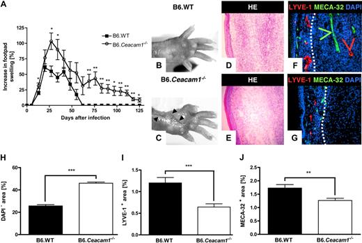
In our model of cutaneous leishmaniasis, we inoculated L major parasites into the right hind footpads of B6.WT and B6.Ceacam1−/− mice. After infection, footpad swelling was recorded weekly through the entire course of the infection and calculated relative to the diameter of the uninfected footpad. As shown in Figure 1A, footpad swelling reached a maximum 3 to 4 weeks after infection. In B6.Ceacam1−/− mice, footpad swelling was significantly more pronounced compared with B6.WT controls and did not reach normal levels until 125 days after infection (Figure 1A). In contrast, no differences between the infected and uninfected footpads were noticeable approximately 60 days after infection in B6.WT mice (Figure 1A). In addition to marked increases in footpad swelling, footpads from B6.Ceacam1−/− but not B6.WT mice exhibited ulcerations, documented by photographs taken on day 41 after infection (Figure 1B,C). Because increases in footpad swelling were most significant around day 21 after infection, we chose this time point for all subsequent experiments, if not stated otherwise. To further investigate the underlying causes for differences in the inflammatory phenotype, we performed histologic analyses. In H&E stains and detection of blood and lymphatic vessels by immunohistology, we found tissue compaction and extensive vascularization in infected footpads of B6.WT mice (Figure 1D,F). Newly formed lymphatic and blood vessels in the infected footpad of a B6.WT mouse are visible in the infiltrate (Figure 1F green and red arrowheads). In infected footpads of B6.Ceacam1−/− mice, cell-free spaces are visible in the H&E stains (Figure 1E) and in immunofluorescence (Figure 1G). Poor vessel formation was detectable in the infiltrate (Figure 1G). Blood and lymphatic vessels were mainly found in the skin (Figure 1G). The boundaries between the skin and the inflammatory infiltrate are indicated by white dotted lines (Figure 1F,G). Hence, the edemas in the B6.Ceacam1−/− mice result from the accumulation of liquid in tissues caused by impaired vascular drain of interstitial fluids. The edematous areas (quantified as areas devoid of DAPI-staining) in the inflamed footpads of B6.WT and B6.Ceacam1−/− mice are shown in Figure 1H. Although approximately 50% of total areas in the infected footpads are devoid of DAPI staining in B6.Ceacam1−/− mice, cell-free areas comprise up to approximately 35% in inflamed footpads of B6.WT mice only (Figure 1H). The initial impression of inefficient local angiogenesis in B6.Ceacam1−/− mice was corroborated by quantitative analyses of lymphatic and blood vessels. Because lymphatic vessels may be dilated under inflammatory conditions and exhibit variable diameters, we expressed lymphatic vessel densities in relation to the total area analyzed (Figure 1I). We found that LYVE-1+ areas or areas occupied by lymphatic vessels were significantly higher in B6.WT than in B6.Ceacam1−/− mice. Similarly, the area claimed by MECA-32+ structures representing blood vessels was significantly reduced in the infected footpads of B6.Ceacam1−/− mice compared with B6.WT animals (Figure 1J).
Course of cutaneous leishmaniasis in B6.Ceacam1−/− and B6.WT mice and characterization of local lymphatic and blood vessel formation. (A) Footpad swelling after infection with Leishmania major. Weekly recordings of footpad swelling are shown for B6.Ceacam1−/− (○) and B6.WT mice (■). Footpad swelling is expressed as the percentage of increase of the infected over the noninfected footpad, respectively. Data shown here summarize the mean (± SEM) from 6 animals each; the experiment was repeated 3 times. *P < .05, **P < .01. Photographs of footpads from a B6.WT (B) and a B6.Ceacam1−/− mouse (C) document ulcerations (indicated with  ) in the B6.Ceacam1−/− animal on day 41 after infection. (D-G) Representative histologic analyses of cross-sections of the infected footpads of B6.WT (D,F) and B6.Ceacam1−/− mice day 21 after infection (E,G). Cryostat sections were analyzed by H&E staining (D,E) and immune fluorescent labeling (F,G) of lymphatic vessels (anti–LYVE-1 antibody; shown in red), blood vessels (anti–Meca-32 antibody; shown in green), and nuclei (DAPI; shown in blue). The dotted white line indicates the borderline between skin and inflammatory infiltrate. Magnification × 200. (H-J) Quantification of edematous areas and lymph and blood vessel formation in cross-sections of infected footpads of B6.WT (■) and B6.Ceacam1−/− mice (□). Data shown here represent mean (± SEM) from at least 6 specimens. (H) Cell-free areas were determined by quantifying the DAPI-free areas, expressed relative to the total inflammatory area analyzed; ***P < .001. (I) Quantification of lymphatic vessels by calculating the percentage of LYVE-1+ areas in cross-sections of infected footpads; ***P < .001. (J) Quantification of MECA-32+ areas relative to the total areas analyzed and expressed as the percentage of MECA-32+ areas, **P < .01.
Course of cutaneous leishmaniasis in B6.Ceacam1−/− and B6.WT mice and characterization of local lymphatic and blood vessel formation. (A) Footpad swelling after infection with Leishmania major. Weekly recordings of footpad swelling are shown for B6.Ceacam1−/− (○) and B6.WT mice (■). Footpad swelling is expressed as the percentage of increase of the infected over the noninfected footpad, respectively. Data shown here summarize the mean (± SEM) from 6 animals each; the experiment was repeated 3 times. *P < .05, **P < .01. Photographs of footpads from a B6.WT (B) and a B6.Ceacam1−/− mouse (C) document ulcerations (indicated with  ) in the B6.Ceacam1−/− animal on day 41 after infection. (D-G) Representative histologic analyses of cross-sections of the infected footpads of B6.WT (D,F) and B6.Ceacam1−/− mice day 21 after infection (E,G). Cryostat sections were analyzed by H&E staining (D,E) and immune fluorescent labeling (F,G) of lymphatic vessels (anti–LYVE-1 antibody; shown in red), blood vessels (anti–Meca-32 antibody; shown in green), and nuclei (DAPI; shown in blue). The dotted white line indicates the borderline between skin and inflammatory infiltrate. Magnification × 200. (H-J) Quantification of edematous areas and lymph and blood vessel formation in cross-sections of infected footpads of B6.WT (■) and B6.Ceacam1−/− mice (□). Data shown here represent mean (± SEM) from at least 6 specimens. (H) Cell-free areas were determined by quantifying the DAPI-free areas, expressed relative to the total inflammatory area analyzed; ***P < .001. (I) Quantification of lymphatic vessels by calculating the percentage of LYVE-1+ areas in cross-sections of infected footpads; ***P < .001. (J) Quantification of MECA-32+ areas relative to the total areas analyzed and expressed as the percentage of MECA-32+ areas, **P < .01.
Because leishmaniasis is accompanied by a strong inflammatory response, we next characterized the humoral and cellular immune responses to elucidate whether differences in the immune responses might influence angiogenesis in CEACAM1+ and CEACAM1− backgrounds.
Adaptive immune response against L major is not affected by loss of CEACAM1 expression
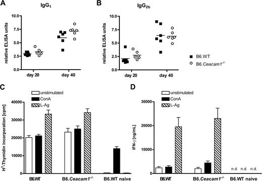
To evaluate the humoral and cellular immune responses against L major, we measured parasite-specific immunoglobulin production on days 20 and 40 after infection. As summarized in Figure 2A and B, comparable levels of anti-L major–specific IgG1 and IgG2b antibodies were detected in B6.WT and B6.Ceacam1−/− mice. Hence, CEACAM1 deficiency does not affect the B-cell response against L major parasites. To further analyze whether CEACAM1 deficiency results in alterations of the cellular immune response, we determined proliferation of whole-cell preparations from the popliteal lymph nodes of B6.WT and B6.Ceacam1−/− mice 21 days after infection. As shown in Figure 1C, proliferation of skin-draining lymph node cells is not reduced in B6.Ceacam1−/− mice. Although cell proliferation is commonly accepted as a read-out for T-cell priming and clonal expansion, cytokine secretion does not necessarily correlate with proliferation. Thus, we quantified IFN-γ in supernatants from these cell cultures indicative for an efficient Th1 response. We found comparable levels of IFN-γ in supernatants from nodal cells derived from B6.WT and B6.Ceacam1−/− mice that had been restimulated with L major antigen (L-Ag) or concanavalin A (ConA; Figure 2D). Lymph node cells from naive B6.WT mice were used as controls. In conclusion, CEACAM1 deficiency does not alter the proliferative response and IFN-γ production. Thus, we conclude that the protective Th1-type immune response against L major is not altered in the absence of CEACAM1. Therefore, the differences in the pathologic phenotype between B6.WT and B6.Ceacam1−/− mice are not a result of an inadequate adaptive immune response.
Characterization of the adaptive immune response toward L major in B6.Ceacam1−/− and B6.WT mice. (A,B) Quantification by enzyme-linked immunosorbent assay (ELISA) of L major–specific immunoglobulins in serum, IgG1 (A) and IgG2b (B) on days 20 and 40 after infection. The amounts of the L major–specific antibodies are expressed as relative ELISA units. Each symbol represents data from one B6.WT (■) or B6.Ceacam1−/− mouse (○; n = 6 each). Horizontal lines in (A) and (B) indicate the mean. (C) Proliferation of lymph node cells from naive B6.WT mice and B6.Ceacam1−/− and B6.WT mice on day 21 after infection after treatment with cell culture medium (□), ConA (■), and L-Ag (▧) expressed in counts per minute (cpm) after H3-thymidine incorporation. Data shown here summarize mean (± SEM) from 6 animals per group. (D) Quantification of IFN-γ production in supernatants from lymph node cells from naive B6.WT mice and from B6.WT and B6.Ceacam1−/− mice after infection with L major on day 21 after infection in response to medium (□), ConA (■), or L-Ag (▧). Data are summarized as mean (± SEM) from 6 mice per group (n.d. indicates not detectable).
Characterization of the adaptive immune response toward L major in B6.Ceacam1−/− and B6.WT mice. (A,B) Quantification by enzyme-linked immunosorbent assay (ELISA) of L major–specific immunoglobulins in serum, IgG1 (A) and IgG2b (B) on days 20 and 40 after infection. The amounts of the L major–specific antibodies are expressed as relative ELISA units. Each symbol represents data from one B6.WT (■) or B6.Ceacam1−/− mouse (○; n = 6 each). Horizontal lines in (A) and (B) indicate the mean. (C) Proliferation of lymph node cells from naive B6.WT mice and B6.Ceacam1−/− and B6.WT mice on day 21 after infection after treatment with cell culture medium (□), ConA (■), and L-Ag (▧) expressed in counts per minute (cpm) after H3-thymidine incorporation. Data shown here summarize mean (± SEM) from 6 animals per group. (D) Quantification of IFN-γ production in supernatants from lymph node cells from naive B6.WT mice and from B6.WT and B6.Ceacam1−/− mice after infection with L major on day 21 after infection in response to medium (□), ConA (■), or L-Ag (▧). Data are summarized as mean (± SEM) from 6 mice per group (n.d. indicates not detectable).
Ly-6Chigh/CD11bhigh cells that coexpress CEACAM1 increase systemically after infection with L major
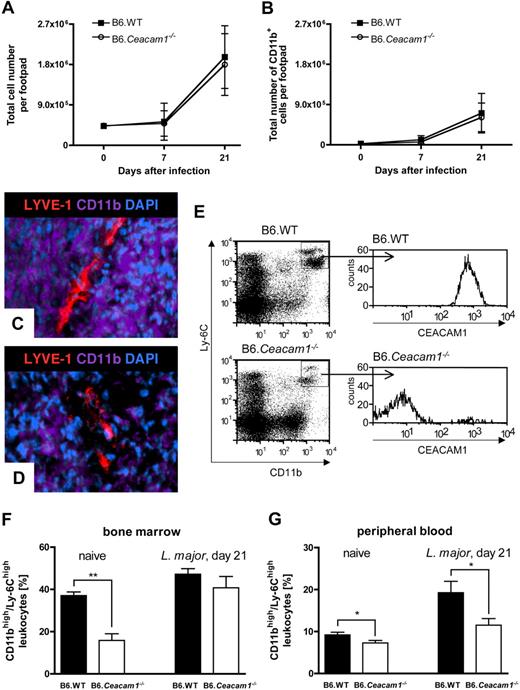
In addition to the adaptive immune response, we analyzed the macrophage populations at the site of the infection. During the early phases of leishmaniasis, we found a substantial increase in total cell counts in the footpads of both mouse lines (Figure 3A), accompanied by enhanced myelopoiesis and a massive influx of CD11b+ cells into the infected areas (Figure 3B).27
Characterization of the dynamics of the CD11b+ population in B6.Ceacam1−/− and B6.WT mice during leishmaniasis. (A,B) Quantification of cellular influx into infected footpads in B6.Ceacam1−/− and B6.WT mice by flow cytometry. Increase in total cell counts (A) and CD11bhigh cells (B) in the infected footpads after infection with L major on days 0, 7, and 21 after infection in B6.WT (■) and B6.Ceacam1−/− mice (○). (C,D) Representative images after immune fluorescence staining of CD11b+ cells (anti-CD11b antibody; shown in purple), lymphatic vessels (anti–LYVE-1 antibody, shown in red) in cross-sections of the infected footpads in a B6.WT mouse (C) and a B6.Ceacam1−/− mouse (D). Nuclei are stained with DAPI (blue). Magnification × 400. (E) Representative histograms show high CEACAM1 expression on the Ly-6Chigh/CD11bhigh population from peripheral blood of a B6.WT mouse (top histogram, top right square in the dot plot) but not on Ly-6Chigh/CD11bhigh population in B6.Ceacam1−/− mouse (bottom histogram). Note that the Ly-6Chigh/CD11bhigh population is diminished in the peripheral blood of a naive B6.Ceacam1−/− mouse (top right square in the bottom dot plot histogram). (F,G) Quantification of Ly-6Chigh/CD11bhigh monocyte precursors in the bone marrow (F) and peripheral blood (G) in naive and infected B6.WT (■) and B6.Ceacam1−/− mice (□). Note that naive B6.Ceacam1−/− mice harbor a significantly smaller Ly-6Chigh/CD11bhigh progenitor population in the bone marrow before infection compared with B6.WT animals, **P < .01. In peripheral blood, B6.Ceacam1−/− mice maintain a significantly reduced Ly-6Chigh/CD11bhigh population before and after infection with L major, P < .05. Data are presented as mean (± SEM) from at least 9 mice each.
Characterization of the dynamics of the CD11b+ population in B6.Ceacam1−/− and B6.WT mice during leishmaniasis. (A,B) Quantification of cellular influx into infected footpads in B6.Ceacam1−/− and B6.WT mice by flow cytometry. Increase in total cell counts (A) and CD11bhigh cells (B) in the infected footpads after infection with L major on days 0, 7, and 21 after infection in B6.WT (■) and B6.Ceacam1−/− mice (○). (C,D) Representative images after immune fluorescence staining of CD11b+ cells (anti-CD11b antibody; shown in purple), lymphatic vessels (anti–LYVE-1 antibody, shown in red) in cross-sections of the infected footpads in a B6.WT mouse (C) and a B6.Ceacam1−/− mouse (D). Nuclei are stained with DAPI (blue). Magnification × 400. (E) Representative histograms show high CEACAM1 expression on the Ly-6Chigh/CD11bhigh population from peripheral blood of a B6.WT mouse (top histogram, top right square in the dot plot) but not on Ly-6Chigh/CD11bhigh population in B6.Ceacam1−/− mouse (bottom histogram). Note that the Ly-6Chigh/CD11bhigh population is diminished in the peripheral blood of a naive B6.Ceacam1−/− mouse (top right square in the bottom dot plot histogram). (F,G) Quantification of Ly-6Chigh/CD11bhigh monocyte precursors in the bone marrow (F) and peripheral blood (G) in naive and infected B6.WT (■) and B6.Ceacam1−/− mice (□). Note that naive B6.Ceacam1−/− mice harbor a significantly smaller Ly-6Chigh/CD11bhigh progenitor population in the bone marrow before infection compared with B6.WT animals, **P < .01. In peripheral blood, B6.Ceacam1−/− mice maintain a significantly reduced Ly-6Chigh/CD11bhigh population before and after infection with L major, P < .05. Data are presented as mean (± SEM) from at least 9 mice each.
The inflammatory tissue largely consisted of CD11b+ cells, as detected in cross-sections of the inflamed footpads, with the use of fluorescently labeled anti-CD11b antibodies (Figure 3C,D shown in purple). In Figure 3C, CD11b+ cells are grouped around a large LYVE-1+ vessel in the footpad of a B6.WT mouse. In footpads from B6.Ceacam1−/− mice (Figure 3D), CD11b+ cells form a lose infiltrate that contains a discontinuous structure of LYVE-1+ cells (Figure 3D). Because both B6.WT and B6.Ceacam1−/− mice exhibit substantial influx of CD11b+ cells into the inflamed areas, but show differences in cellular distribution that coincide with edema formation in B6.Ceacam1−/− mice, we subjected the CD11b+ populations to more detailed analyses. As shown in qualitative analyses in Figure 3E, Ly-6Chigh/CD11bhigh monocytic precursors are detectable in both mouse lines, but, importantly, in B6.WT mice, this population is also highly positive for CEACAM1. Moreover, the Ly-6Chigh/CD11bhigh population appeared to be diminished in the peripheral blood of B6.Ceacam1−/− mice (Figure 3E). Quantitative analyses of the monocytic precursor populations in bone marrow (Figure 3F) and peripheral blood (Figure 3G) of naive and infected mice confirmed that B6.Ceacam1−/− mice exhibit a significant reduction in their Ly-6Chigh/CD11bhigh populations before and during infection with L major. Both mouse lines, however, equally respond to the infection with expansion of the Ly-6Chigh/CD11bhigh population both in bone marrow and peripheral blood (Figure 3F,G). This indicates that reduction in monocytic precursors is inherent to a CEACAM1− phenotype.
Taken together, we observed that B6.Ceacam1−/− mice exhibit significant differences in their innate immune response and their inflammatory phenotype as well as alterations in angiogenesis after infection with L major. However, we could not detect any differences in the humoral or T-cell responses between B6.WT and B6.Ceacam1−/− mice. Therefore, we decided to focus further functional analyses on the myeloid population positive for CEACAM1 and CD11b.
Reverse BM transplantation with CEACAM1+ and CEACAM1− BM donors shows an essential role for Ly-6Chigh/CD11bhigh/CEACAM1+ monocytic progenitors in angiogenesis
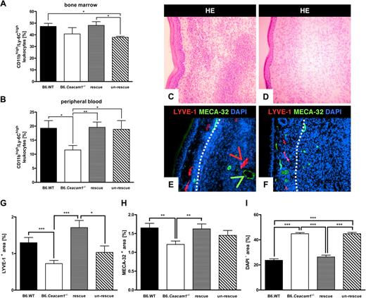
To evaluate the effect of CEACAM1+ myeloid cells on angiogenesis, we performed reverse BM transplantations with B6.WT donors (expressing CD45.1) for B6.Ceacam1−/− recipients (expressing CD45.2; named “rescue” experiment hereafter) and B6.Ceacam1−/− donors for B6.WT mice as recipients (named “un-rescue” experiment in the following text). After reconstitution, the mice were infected with L major as described before, including B6.WT and B6.Ceacam1−/− mice as controls. Ly-6Chigh/CD11bhigh progenitors were quantified in the BM and peripheral blood of infected mice on day 21 after infection, respectively (Figure 4A,B). Analyzing the Ly-6Chigh/CD11bhigh fraction in the BM, we showed that this population is significantly reduced after transfer of CEACAM1− BM into B6.WT recipients. Conversely, in the rescue experiment, the numbers of Ly-6Chigh/CD11bhigh cells were restored to comparable levels as in B6.WT mice (Figure 4A). Comparably, in peripheral blood, Ly-6Chigh/CD11bhigh monocyte precursors were replenished to levels in B6.WT mice after transfer of CEACAM1+ BM into B6.Ceacam1−/− mice (rescue BM transplantation; Figure 4B). However, no significant differences between Ly-6Chigh/CD11bhigh monocytic precursors were found between the rescue or un-rescue experiments. To evaluate the effects of the BM transplantations on angiogenesis, we analyzed histologic sections of the infected footpads after H&E and immune fluorescent labeling of blood and lymphatic vessels, respectively (Figure 4C-F). As shown in the H&E stain, the infiltrate contained rather high cellular densities after the rescue BM transplantation (Figure 4C), but exhibited large cell-free areas in footpads of mice that had undergone the un-rescue BM transplantation (Figure 4D). These findings are congruent with the staining patterns found in the B6.WT and B6.Ceacam1−/− mice, shown in Figure 1D through G. Here, the infected footpads from the rescued B6.Ceacam1−/− mice showed a similar pattern as in the B6.WT mice, and in footpads of the un-rescued B6.WT animals we found edema formation and reduction of angiogenesis comparable to our previous observations in B6.Ceacam1−/− mice (Figure 4E,F). Vascular densities and edematous areas were quantified as mentioned earlier, and data are summarized in Figure 4G-I. In the B6.Ceacam1−/− mice that had undergone the rescue transfer, LYVE-1+ areas in the footpad were significantly increased compared with the B6.Ceacam1−/− and un-rescued mice (Figure 4G). Contrary, lymphatic vessels areas were diminished in the B6.WT mice that had received CEACAM1− BM (un-rescue BM transplantation). Here, the lymphatic vascular areas were significantly smaller than in animals that had undergone the rescue transfers (Figure 4G). Similarly, blood vessel formation was restored in the rescued animals, and the areas claimed by MECA-32+ vessels were significantly increased compared with footpads of B6.Ceacam1−/− mice (Figure 4H). Likewise, after the un-rescue BM transplantation, the extent of vascularization was reduced. In agreement with these observations, we found that the edematous areas in the footpads of mice after the rescue BM transplantation were reduced by half compared with the edemas observed in footpads of B6.Ceacam1−/− animals (Figure 4I). In line with these findings, B6.WT mice that had received CEACAM1− BM (un-rescue BM transplantation) before infection exhibited cell-free areas to the same extent as B6.Ceacam1−/− animals (Figure 4I). In conclusion, the BM transplantation experiments show that the presence of CD11b+ cells with the potential to express CEACAM1 is crucial for angiogenesis in inflammation.
Analysis of the CEACAM1+/Ly-6Chigh/CD11bhigh progenitor population and local angiogenesis after reverse transfer of CEACAM1+ and CEACAM1− BM into recipient mice. (A,B) Summary of quantitative flow cytometric analyses of Ly-6Chigh/CD11bhigh populations in bone marrow (A) and peripheral blood (B) on day 21 after infection with L major after CEACAM1+ and CEACAM1− BMT into B6.Ceacam1−/− and B6.WT mice, respectively. Quantifications of Ly-6Chigh/CD11bhigh monocytic precursors after infection with L major and transfer of CEACAM1+ BM into CEACAM1− recipients (rescue,  ) and transfer of CEACAM1− BM into B6.WT recipients (un-rescue, ▧) are shown. Data from B6.WT and B6.Ceacam1−/− mice are depicted in ■ and □, respectively. Data are represented as means (± SEM) from at least 6 animals each, *P < .05; **P < .01. (C-F) Histologic analyses of cross-sections of infected footpads on day 21 after infection in H&E stainings (C,D) and immune fluorescence (E,F) after BM transplantation of CEACAM1+ BM into B6.Ceacam1−/− mice (rescue; C,E) and CEACAM1− BM into B6.WT mice (un-rescue; D,F). (E,F) Lymphatic vessels are shown in red (anti–LYVE-1 labeling) and blood vessels are colored in green (anti–MECA-32 labeling). Nuclei are shown in blue (DAPI). The white dotted line indicates the boundary between the skin and the inflammatory infiltrate. Magnification ×200. (G-I) Quantification of LYVE-1+ (G) and MECA-32+ areas (H) as well as cell-free spaces (I) in inflamed areas of infected footpads in control mice and after BM transplantation as indicated. *P < .05; **P < .01; ***P < .001.
Analysis of the CEACAM1+/Ly-6Chigh/CD11bhigh progenitor population and local angiogenesis after reverse transfer of CEACAM1+ and CEACAM1− BM into recipient mice. (A,B) Summary of quantitative flow cytometric analyses of Ly-6Chigh/CD11bhigh populations in bone marrow (A) and peripheral blood (B) on day 21 after infection with L major after CEACAM1+ and CEACAM1− BMT into B6.Ceacam1−/− and B6.WT mice, respectively. Quantifications of Ly-6Chigh/CD11bhigh monocytic precursors after infection with L major and transfer of CEACAM1+ BM into CEACAM1− recipients (rescue,  ) and transfer of CEACAM1− BM into B6.WT recipients (un-rescue, ▧) are shown. Data from B6.WT and B6.Ceacam1−/− mice are depicted in ■ and □, respectively. Data are represented as means (± SEM) from at least 6 animals each, *P < .05; **P < .01. (C-F) Histologic analyses of cross-sections of infected footpads on day 21 after infection in H&E stainings (C,D) and immune fluorescence (E,F) after BM transplantation of CEACAM1+ BM into B6.Ceacam1−/− mice (rescue; C,E) and CEACAM1− BM into B6.WT mice (un-rescue; D,F). (E,F) Lymphatic vessels are shown in red (anti–LYVE-1 labeling) and blood vessels are colored in green (anti–MECA-32 labeling). Nuclei are shown in blue (DAPI). The white dotted line indicates the boundary between the skin and the inflammatory infiltrate. Magnification ×200. (G-I) Quantification of LYVE-1+ (G) and MECA-32+ areas (H) as well as cell-free spaces (I) in inflamed areas of infected footpads in control mice and after BM transplantation as indicated. *P < .05; **P < .01; ***P < .001.
Lymphatic vessels coexpress CEACAM1 and LYVE-1 in inflammation
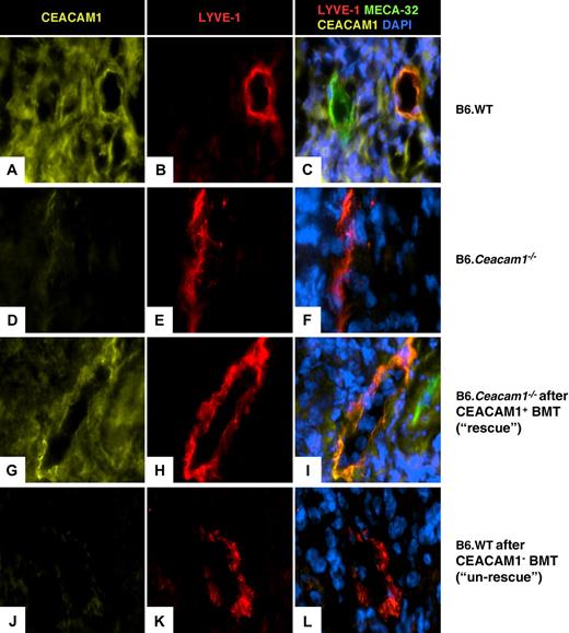
Because CEACAM1-expression on common myeloid cells infiltrating the site of infection with L major appears to be causally related to vessel formation under inflammatory conditions, we investigated expression of CEACAM1 in the footpads of infected mice before and after reverse BM transplantations. The 3 different panels (Figure 5) show single staining for CEACAM1 (yellow; left), LYVE-1 (red; middle), and the overlay plus MECA-32 (green) and DAPI (blue; right). In infected B6.WT mice, a highly CEACAM1+ cellular infiltrate is seen in infected footpads (Figure 5A) as well as in lymphatic vessels (Figure 5B). In the overlay (Figure 5C), coexpression of CEACAM1 on the lymphatic vessel as well as partial coexpression on a MECA-32+ blood vessel was detected. In the infected B6.WT mice, all of the newly formed lymphatic vessels were coexpressing CEACAM1 and LYVE-1. In footpads of B6.Ceacam1−/− mice (Figure 5D-F), only few LYVE-1+ structures are visible (Figure 5E), but they are negative for CEACAM1 as well as the infiltrate formed in response to the infection (anti-CEACAM1 staining, Figure 5D, and overlay, Figure 5F). Similarly to our observations in B6.WT mice, expression of CEACAM1 is detected in footpads of mice that had been subjected to the rescue BM transplantation with CEACAM1+ BM (Figure 5G), and most of the lymphatic vessels (Figure 5H) are also positive for CEACAM1 in the overlay (Figure 5I). Contrary, after the un-rescue BM transplantation with B6.Ceacam1−/− mice as donors and B6.WT as recipient mice, CEACAM1 expression in the infiltrate (Figure 5J) as well as on few lymphatic vessels was absent (Figure 5K and overlay, Figure 5L). In summary, our BM transplantation experiments show that vessel formation within the infiltrate is only efficient if CEACAM1 is expressed on myeloid cells.
Labeling of CEACAM1, LYVE-1, MECA-32, and nuclei in cross-sections of infected footpads. (A-C) Labeling of CEACAM1 (A, anti-CEACAM1 polyclonal antiserum P1; yellow) shows that the cellular infiltrate in the footpad is CEACAM1+. LYVE-1 is expressed on lymphatic vessels in the infiltrate (B, shown in red). In the overlay (C), coexpression of CEACAM1 and LYVE-1 on lymphatic vessels is shown in B6.WT mice. Blood endothelium (anti–MECA-32 labeling; green) also expresses CEACAM1 (C). In footpads from B6.Ceacam1−/− mice, no CEACAM1 labeling is present (D), and LYVE-1+ structures (E) are CEACAM1− (F). (G-I) After BM transplantation of CEACAM1+ BM into B6. Ceacam1−/− mice (rescue), CEACAM1 labeling is restored in the infected footpads (G), and LYVE-1+ structures (H) coexpress CEACAM1 (I). In the un-rescue experiment after CEACAM1− BM transplantation into B6.WT mice, CEACAM1-expressing cells are absent in the infiltrate (J) and lymphatic vessels (K,L). Representative photographs from 6 animals each are shown; magnification ×630.
Labeling of CEACAM1, LYVE-1, MECA-32, and nuclei in cross-sections of infected footpads. (A-C) Labeling of CEACAM1 (A, anti-CEACAM1 polyclonal antiserum P1; yellow) shows that the cellular infiltrate in the footpad is CEACAM1+. LYVE-1 is expressed on lymphatic vessels in the infiltrate (B, shown in red). In the overlay (C), coexpression of CEACAM1 and LYVE-1 on lymphatic vessels is shown in B6.WT mice. Blood endothelium (anti–MECA-32 labeling; green) also expresses CEACAM1 (C). In footpads from B6.Ceacam1−/− mice, no CEACAM1 labeling is present (D), and LYVE-1+ structures (E) are CEACAM1− (F). (G-I) After BM transplantation of CEACAM1+ BM into B6. Ceacam1−/− mice (rescue), CEACAM1 labeling is restored in the infected footpads (G), and LYVE-1+ structures (H) coexpress CEACAM1 (I). In the un-rescue experiment after CEACAM1− BM transplantation into B6.WT mice, CEACAM1-expressing cells are absent in the infiltrate (J) and lymphatic vessels (K,L). Representative photographs from 6 animals each are shown; magnification ×630.
CEACAM1-deficient macrophages fail to trigger angiogenesis in vitro
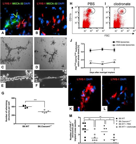
From our experiments conducted here, we cannot deduce whether CEACAM1 expression on CD11b+ macrophage precursors contributes directly or indirectly to angiogenesis in inflammation. Therefore, we sought for a suitable in vitro setting that allows mimicking leishmaniasis and also studying macrophage behavior. We performed Matrigel implantation assays with growth factor–enriched Matrigel also containing L major parasites. In this model, we intended to study formation of cellular assemblies and neovascularization in B6.WT and B6.Ceacam1−/− mice. The implants were retrieved 7 days after implantation and analyzed for the presence of vessel-like structures. In Figure 6A and B, immunohistochemical analyses of the implants are shown. In Figure 6A, an implant retrieved from a B6.WT mouse is shown. Here, we found MECA-32+ structures (shown in green) and elongated, lumenizing LYVE-1+ structures (red). In specimens from B6.Ceacam1−/− mice, we only found single LYVE-1+ cells and neither lymphatic nor blood vessel-like structures (Figure 6B). This was also confirmed in H&E stainings and transmission electron microscopy analyses, with lumen-forming structures in Matrigel implants only present in implants retrieved from B6.WT mice (Figure S1, available on the Blood website; see the Supplemental Materials link at the top of the online article).
Analysis of morphogenic properties of CEACAM1+ and CEACAM1− inflammatory cells in vitro and in vivo. (A,B) Matrigel implants were retrieved on day 7, and cross-sections were analyzed for MECA-32+ (shown in green) and LYVE-1+ (shown in red) structures. In implants from B6.WT mice (A), lumenizing MECA-32+ and LYVE-1+ structures are visible (arrows), whereas implants from B6.Ceacam1−/− mice only contain single LYVE-1+ cells and no MECA-32+ structures (B; n = 6, magnification × 630). (C-F) Matrigel tube formation assays using implant-derived cells from B6.WT (C,E) and B6.Ceacam1−/− mice (D,F) were documented 2 days after seeding 5 × 10⁵ cells/well; magnification × 50 (C,D) and × 200 (E,F). (G) Quantification of arborizing structures from 1 representative experiment of 4. Cells from B6.WT mice are shown in ■, those from B6. Ceacam1−/−-derived cells are shown in ○. ***P < .001. Horizontal lines indicate the mean. (H-L) Analysis of CD11bhigh monocytes and their morphogenic properties after treatment with PBS- or clodronate-loaded liposomes: Dot plot histograms of CD11bhigh cells in gate R3 after treatment with control liposomes (H) or clodronate-loaded liposomes (I). (J) Quantification of CD11bhigh populations in peripheral blood of B6.WT mice after treatment with PBS-loaded liposomes (■) or clodronate-loaded liposomes (▵). Data shown here present the mean (± SEM) of 6 animals each, with samples analyzed on days 2, 5, and 7 during Matrigel implantation; ***P < .001. (K,L) Cross-sections of Matrigel implants retrieved from B6.WT mice undergoing treatment with PBS-loaded liposomes (K) and clodronate-loaded liposomes (L). Cross-sections were stained for LYVE-1 (shown in red) and DAPI (blue), n = 6 specimens each, magnification × 1000. (M) Quantification of LYVE-1+ structures in implants from B6.WT (■) or B6.Ceacam1−/− mice (○), and B6.WT mice treated with PBS-loaded liposomes (▲) or clodronate-loaded liposomes (◇); **P < .01, *P < .05. Each symbol represents data from 1 implant. Horizontal lines indicate the mean. Experiments were performed with at least 5 animals.
Analysis of morphogenic properties of CEACAM1+ and CEACAM1− inflammatory cells in vitro and in vivo. (A,B) Matrigel implants were retrieved on day 7, and cross-sections were analyzed for MECA-32+ (shown in green) and LYVE-1+ (shown in red) structures. In implants from B6.WT mice (A), lumenizing MECA-32+ and LYVE-1+ structures are visible (arrows), whereas implants from B6.Ceacam1−/− mice only contain single LYVE-1+ cells and no MECA-32+ structures (B; n = 6, magnification × 630). (C-F) Matrigel tube formation assays using implant-derived cells from B6.WT (C,E) and B6.Ceacam1−/− mice (D,F) were documented 2 days after seeding 5 × 10⁵ cells/well; magnification × 50 (C,D) and × 200 (E,F). (G) Quantification of arborizing structures from 1 representative experiment of 4. Cells from B6.WT mice are shown in ■, those from B6. Ceacam1−/−-derived cells are shown in ○. ***P < .001. Horizontal lines indicate the mean. (H-L) Analysis of CD11bhigh monocytes and their morphogenic properties after treatment with PBS- or clodronate-loaded liposomes: Dot plot histograms of CD11bhigh cells in gate R3 after treatment with control liposomes (H) or clodronate-loaded liposomes (I). (J) Quantification of CD11bhigh populations in peripheral blood of B6.WT mice after treatment with PBS-loaded liposomes (■) or clodronate-loaded liposomes (▵). Data shown here present the mean (± SEM) of 6 animals each, with samples analyzed on days 2, 5, and 7 during Matrigel implantation; ***P < .001. (K,L) Cross-sections of Matrigel implants retrieved from B6.WT mice undergoing treatment with PBS-loaded liposomes (K) and clodronate-loaded liposomes (L). Cross-sections were stained for LYVE-1 (shown in red) and DAPI (blue), n = 6 specimens each, magnification × 1000. (M) Quantification of LYVE-1+ structures in implants from B6.WT (■) or B6.Ceacam1−/− mice (○), and B6.WT mice treated with PBS-loaded liposomes (▲) or clodronate-loaded liposomes (◇); **P < .01, *P < .05. Each symbol represents data from 1 implant. Horizontal lines indicate the mean. Experiments were performed with at least 5 animals.
To investigate whether implant-derived inflammatory cells are able to form cellular assemblies or to adopt a tubelike shape, we performed top-Matrigel tube formation assays (Figure 6C-G; Figure S2). As shown in Figure 6C and enlargement in Figure 6E, implant-derived cells from B6.WT mice form large cell clusters and thick tubelike branches. In contrast, cells from B6.Ceacam1−/− mice only form smaller colonies and thin tubular protrusions (Figure 6D,F). Arborizing structures are quantified in Figure 6G, indicating that implant-derived cells from B6.Ceacam1−/− mice showed a significantly reduced potential for tube formation compared with B6.WT control experiments. These findings were also corroborated by counting the numbers of junctions with 3 or more branches in arborizing entities, using different cell numbers seeded and by evaluation of different time points after seeding (Document S1; Figures S2, S3).
To confirm that CD11b+ macrophages may directly or indirectly assist vessel formation under inflammatory conditions in our model system, we performed Matrigel implantation assays under macrophage depletion. Clodronate-loaded liposomes have been reported to interfere with lymphangiogenesis in vivo.28 We used clodronate-loaded liposomes and PBS-loaded liposomes as controls that were injected on the day before and on days 2 and 5 after Matrigel implantation. As shown in Figure 6H-J, the CD11bhigh monocyte population was diminished significantly by treatment with clodronate-loaded liposomes, not by PBS-loaded liposomes throughout the entire time course. Sections of Matrigel implants were analyzed for the presence of vessel-like structures on day 7 after implantation. In Matrigel implants from mice that had received PBS-loaded liposomes, we found single LYVE-1+ cells, but we also observed cell clusters that resembled anastomosing vessel-like structures (Figure 6K). Only a few MECA-32+ structures were found (data not shown). In implants from mice that were subjected to clodronate-mediated monocyte depletion, we only found single cells that expressed LYVE-1, but we did not detect any extensive cell clusters or vessel-like structures (Figure 6L). Total numbers of LYVE-1+ lumenizing structures in cross-sections of Matrigel implants are compared in Figure 6M. Here, we show that depletion of CEACAM1+/CD11b+ monocytic cells or absence of CEACAM1 equally affects the formation of lumenizing LYVE1+ structures in Matrigel implants. From these data, we conclude that reduction of the CD11bhigh monocyte population in peripheral blood concurs with the reduction in cellular clustering in the Matrigel implants. Because CD11bhigh cells coexpress CEACAM1 on high levels (Figure 3E), a depletion of CD11b+ monocytes also includes depletion of CEACAM1+ monocytes that are potentially involved in angiogenesis. In addition, our observations about cellular clustering and structure formation in implants from B6.WT mice that were subjected to clodronate treatment are comparable to implants from B6.Ceacam1−/− mice (Figure 6B,L). In conclusion, these in vitro data showed that CEACAM1 and CD11b double-positive myeloid cells are pivotal for vessel formation.
Discussion
In our report, we used the experimental model of cutaneous leishmaniasis to study inflammatory lymphangiogenesis and hemangiogenesis in CEACAM1+ and CEACAM1− mouse backgrounds. Both mouse lines mounted an efficient adaptive immune response to L major. Still, the B6.Ceacam1−/− mice exhibited a different inflammatory phenotype in that they responded to the local inflammation provoked by L major with extended and increased footpad swelling and edema formation. These edemas resulted from accumulation of liquid in tissues caused by the impaired vascular drain of interstitial fluids. Congruently, we found inefficient hemangiogenesis and lymphangiogenesis in the inflamed footpads of B6.Ceacam1−/− mice. Because leishmaniasis triggers myelopoiesis and local accumulation of CD11b+ cells, we analyzed infiltrating cell populations in greater detail.27 We found that in footpads of the B6.Ceacam1−/− mice, local densities of inflammatory leukocytes were reduced, although the total numbers of CD11b+ cells in the footpads of B6.WT or B6.Ceacam1−/− mice were comparable. In B6.WT mice, these CD11b+ inflammatory cells within the infiltrate and their Ly-6Chigh/CD11bhigh progenitors express high levels of CEACAM1. Importantly, these Ly-6Chigh/CD11bhigh monocyte progenitors were diminished quantitatively in the BM and peripheral blood of B6.Ceacam1−/− mice before and after infection with L major. The quantitative differences in the CD11bhigh/Ly-6Chigh population were maintained throughout the course of the infection, indicating that CEACAM1 deficiency might reduce myeloid progenitor differentiation potential and maturation. Thus, CEACAM1-deficient mice respond to leishmaniasis with inefficient replenishment of myeloid precursors. This result reinforces the role of CEACAM1 as a differentiation antigen on myeloid cells, but it also corroborates previous reports that not only maturation and monocyte survival but also presentation of CD11b on the cell surface are affected by CEACAM1 expression.29-31
Therefore, it is most likely that, besides its endothelial expression, CEACAM1 expression on inflammatory CD11b+ myeloid cells is of functional relevance during neovascularization. CEACAM1 expression becomes up-regulated on the leukocyte surface once cells have been activated by pathogens or engagement of stimulatory receptors on their surface.11,32,33 For the first time, we describe a causal relation between CEACAM1 expression on inflammatory cells and local angiogenesis during an inflammatory response. In addition, we found that lymphatic vessels in the inflamed footpads coexpressed LYVE-1 and CEACAM1, whereas most lymphatic vessels in the skin of naive mice did not. This indicates that lymphatic vessels in inflammation express different surface markers than do “quiescent” steady state lymphatics. In addition, CEACAM1 expression on blood endothelium in mice or humans is not homogenous and shows some variations34 (A.K.H., unpublished data, August 2008).
A definite functional role for CD11b+ myeloid cells in neovascularization during hypoxia and tumor progression has been verified about their potential to produce proangiogenic cytokines or proteinases.28,35-37 However, it has been in the focus of controversial discussions how and if myeloid cells contribute physically to the formation of new blood and lymphatic vessels.
We cannot deduce from our data that myeloid cells positive for CD11b and CEACAM1 either fuse or integrate or transdifferentiate into lymphatic vessels and that they constitute definite lymphatic or blood endothelial cell precursors. Whether CEACAM1 expression on inflammatory lymphatic endothelial cells designates their origin, and whether it is indicative or supportive of progenitor integration will need further investigation. Different reports describe incorporation of Tie2+ or CD11b+ macrophages into newly formed vessels or coexpression of macrophage and lymphatic markers during early stages of vessel formation and in experiments ranging from 3 to 7 days.1,3,5,35,38,39 Still, these integration events were often documented only for singular cells in tissue sections, which either emphasizes that they are rather rare events or only occur in a specific time window. However, we cannot exclude the involvement of homophilic interactions between CEACAM1 molecules on leukocytes and blood or lymphatic endothelium, leading to extravasation or close endothelial interaction and subsequent incorporation into the vasculature, because CEACAM1-derived peptides enhance adhesion of neutrophilic granulocytes to immobilized human endothelial cells.40 Furthermore, it is not known if CEACAM1-mediated adhesion influences autocrine or juxtacrine signaling processes during inflammation or whether high cellular densities are required to create an angiogenic milieu. We have previously described that CEACAM1 is implicated in hemangiogenesis and vascular remodeling and that CEACAM1 expression enhances tissue recovery and capillary formation after femoral artery ligation.13,41 These reports and observations by others suggest that CEACAM1-dependent angiotrophic effects are elicited when the endothelium is challenged, eg, in inflammation, hypoxia, or tumor growth.13,41-43 In addition, CEACAM1 expression changes in lymphatic endothelial cells after infection with Kaposi-associated herpes virus or alterations in growth factor homeostasis in their environment.44,45 In turn, CEACAM1 expression on endothelial cells appears to influence lymphatic lineage marker expression, and it has been described itself as a lymphatic marker, which was identified in the context of comparative gene expression profiling of lymphatic or blood endothelia.45,46 However, CEACAM1 transcription levels did not respond to virally induced expression of prospero homeobox-related protein 1.47 This raises the question of whether CEACAM1 may be an upstream regulator of lymphatic lineage-specific signaling pathways or could be involved in progenitor commitment. The observation that CEACAM1 is expressed on blood endothelia in vivo, however, challenges this hypothesis.
To further explore a causative role for CEACAM1 expression on myeloid cells and the formation of new blood vessels and lymphatic vessels, we performed BM transplantation from CEACAM1+ donors into CEACAM1− recipients. Repeatedly, we analyzed angiogenesis after infection with L major. Strikingly, angiogenesis in the inflamed footpads was restored in the chimeras, and edemas were absent. These data were also corroborated with reverse BM transplantation of CEACAM1− BM into CEACAM1+ recipients, which resulted in the absence of local angiogenesis. This provides evidence that CEACAM1+ common myeloid progenitors are crucial for efficient inflammatory angiogenesis and that CEACAM1 is expressed either in newly formed lymphatics or in vessels embedded in inflammatory tissues. Under all conditions examined, blood vessels in the mice express CEACAM1 heterogeneously both under physiologic and pathologic conditions, congruent with human tissues.34,48,49
To further extend our investigations on monocyte/macrophage contribution to angiogenesis in vitro, we performed tube formation assays and Matrigel implantations with and without clodronate liposome–induced macrophage depletion. We found that in B6.Ceacam1−/− mice neoformation of vessel-like structures was impaired and that CEACAM1-deficient inflammatory cells exhibit reduced aggregation and tube-forming capacities in vitro. Interestingly, Matrigel implant-residing cells also showed impaired capabilities in the formation of cell aggregates and anastomosing structures under clodronate treatment, phenocopying the results we obtained in B6.Ceacam1−/− mice. Because clodronate liposomes specifically deplete CD11bhigh macrophage precursors positive for CEACAM1, we also depleted a CEACAM1+ population. Therefore, we suggest that expression of both CEACAM1 and CD11b is required on inflammatory cells to catalyze angiogenic processes (Figure 3B).27
In conclusion, we support the data that CEACAM1 is expressed on lymphatic endothelia under specific conditions.44,45,50 Here, we show for the first time that CEACAM1+/CD11b+ cells control angiogenesis during inflammation and that both blood and lymphatic vessel formation are affected by the loss of CEACAM1+/CD11bhigh cells. Further studies will need to be conducted to understand the detailed mechanisms of how vascular formation is controlled by CEACAM1+ common myeloid precursor cells.
An Inside Blood analysis of this article appears at the front of this issue.
The online version of this article contains a data supplement.
The publication costs of this article were defrayed in part by page charge payment. Therefore, and solely to indicate this fact, this article is hereby marked “advertisement” in accordance with 18 USC section 1734.
Acknowledgments
We thank Christa Frenz and Krimhild Scheike (University Medical Center Hamburg-Eppendorf) and Alexandra Veit, Ulricke Richardt, and Dr Christel Schmetz (Bernhard-Nocht-Institute, Hamburg) for expert technical assistance and support with transmission electron microscopy. We also thank Dr Sven Mostböck (University of Regensburg) for critical review of the manuscript.
This work was supported by the Priority Program of the German Research Foundation (DFG) SPP1190: the tumor-vessel interface (A.K.H. and C.W.), by DFG grant IT-13/3 (A.K.H. and W.D.I.), and by the Jung Foundation of Science and Research, Hamburg (U.R.).
The work presented in this manuscript was performed at the Department of Immunology, Bernhard-Nocht-Institute for Tropical Medicine and the Institute of Clinical Chemistry, University Medical Center Hamburg-Eppendorf (both Hamburg, Germany).
Authorship
Contribution: A.K.H. and U.R. designed and performed the research and wrote the manuscript; T.B. and N. Brewig performed the research; N.v.R. provided liposomes for macrophage depletion; P.L. performed computer-assisted vessel quantification and analyzed data; U.S., B.F., W.D.I., and C.W. analyzed and interpreted data; and N. Beauchemin provided B6.Ceacam1−/− mice and analyzed data.
Conflict-of-interest disclosure: The authors declare no competing financial interests.
Correspondence: Andrea K. Horst, Institute of Clinical Chemistry, Diagnostic Center, University Medical Center Hamburg Eppendorf, Martinistrasse 52, D-20246 Hamburg, Germany; e-mail: ahorst@uke.uni-hamburg.de; or Uwe Ritter, Department of Immunology, University of Regensburg, Franz-Josef-Strauss-Allee 11, D-93053 Regensburg, Germany; e-mail: uwe.ritter@klinik.uni-regensburg.de.
References
Author notes
*A.K.H. and T.B. contributed equally to this study.

This feature is available to Subscribers Only
Sign In or Create an Account Close Modal