Abstract
Inhibitors of cyclin-dependent kinases (Cdks) have been reported to have activities in chronic lymphocytic leukemia cells by inhibiting Cdk7 and Cdk9, which control transcription. Here we studied the novel Cdk inhibitor SNS-032, which exhibits potent and selective inhibitory activity against Cdk2, Cdk7, and Cdk9. We hypothesized that transient inhibition of transcription by SNS-032 would decrease antiapoptotic proteins, resulting in cell death. SNS-032 effectively killed chronic lymphocytic leukemia cells in vitro regardless of prognostic indicators and treatment history. This was associated with inhibition of phosphorylation of RNA polymerase II and inhibition of RNA synthesis. Consistent with the intrinsic turnover rates of their transcripts and proteins, antiapoptotic proteins, such as Mcl-1 and X-linked inhibitor of apoptosis protein (XIAP), were rapidly reduced on exposure to SNS-032, whereas Bcl-2 protein was not affected. The initial decrease of Mcl-1 protein was the result of transcriptional inhibition rather than cleavage by caspase. Compared with flavopiridol and roscovitine, SNS-032 was more potent, both in inhibition of RNA synthesis and at induction of apoptosis. SNS-032 activity was readily reversible; removal of SNS-032 reactivated RNA polymerase II, which led to resynthesis of Mcl-1 and cell survival. Thus, these data support the clinical development of SNS-032 in diseases that require short-lived oncoproteins for survival.
Introduction
Chronic lymphocytic leukemia (CLL) is characterized by the gradual accumulation of small, mature lymphocytes, with typical B-cell markers.1 Several lines of evidence suggest that the survival advantage of CLL lymphocytes is the result of the overexpression of antiapoptotic proteins of the Bcl-2 family.2-4 The Bcl-2 family consists of both antiapoptotic and proapoptotic proteins that share sequence homology within conserved Bcl-2 homology (BH) domains.5 Bcl-2 and Mcl-1 are antiapoptotic proteins that lend a survival advantage to CLL. They act by binding to proapoptotic proteins to prevent them from disrupting the mitochondrial outer membrane, an action that initiates apoptosis. On the other hand, X-linked inhibitor of apoptosis protein (XIAP) inhibits the activity of caspases 3, 7, and 9, preventing them from the induction of cell death.6 The mitochondria of the CLL cells are “primed” with death signals, and the cells require the continuous expression of antiapoptotic protein to maintain their survival.7,8
In such a biologic context, agents that aim at antagonizing or diminishing the antiapoptotic proteins cause the release of pro-death signals to commit cells to apoptosis. This has been a focus of new therapeutics in CLL. One such approach uses small molecular BH3 mimetics designed to interfere with interactions of antiapoptotic and proapoptotic proteins at the BH3 domain. These compounds, including ABT-737,3 GX15-070,9 Gossypol/AT-101,10,11 and TW-37,12 have shown impressive activity in vitro and are currently under investigation in clinical trials. A second approach is aimed at decreasing the expression level of Bcl-2. For example, Oblimersen (Genasense, Genta, Berkeley Heights, NJ) is an antisense oligonucleotide designed to target human Bcl-2 mRNA and reduce Bcl-2 expression.13 In addition, clinical trials are ongoing with AS1411 (Antisoma Research, London, United Kingdom), a nucleic acid aptamer that competes with Bcl-2 mRNA for binding to nucleolin, an action that destabilizes Bcl-2 mRNA and reduces its protein expression.14
A third approach uses transient exposure to inhibitors of cyclin-dependent kinases (Cdks) required for transcription, thereby selectively affecting short-lived antiapoptotic proteins.15-17 Although Cdk family members commonly regulate cell cycle events, some members are associated with transcription control. In particular, Cdk7 and Cdk9 have major roles in the initiation and elongation steps in transcription. For instance, Cdk7 is an integral component of the transcription factor TFIIH,18 which phosphorylates the Ser-5 in the heptad repeats of the C-terminal domain (CTD) of RNA polymerase II (Pol II), to facilitate transcription initiation. Cdk9, a portion of the elongation factor P-TEFb,19,20 performs a complementary function by phosphorylating Ser-2 in the CTD of RNA Pol II, which is required for transcript elongation. Although the prolonged inhibition of Cdk9 and Cdk7 will eventually affect all transcripts produced by RNA Pol II and subsequently their proteins, the immediate effect will be on those transcripts and proteins with inherently rapid turnover rates,21 such as Mcl-1 and XIAP. In such a context, inhibiting transcription would decrease Mcl-1 and XIAP expression, thus releasing their ability to block primed cells from initiating apoptosis.
This provided a rationale for using Cdk7 and Cdk9 inhibitors in CLL as well as other diseases that depend on such intrinsically labile proteins for survival. As such, both flavopiridol and roscovitine have shown in vitro activity in inhibiting RNA Pol II and reduced expression of Mcl-1 and XIAP, and efficiently induced apoptosis in CLL and multiple myeloma cells in vitro.15,16 Because the majority of the CLL cells are not cycling, the inhibition on the cell cycle regulating Cdks may not play a major role in their mechanism of action in CLL. Although disappointing in its initial clinical trials, flavopiridol administered on a pharmacokinetically derived dose schedule aimed at sustaining LC50 plasma levels for 4 hours has shown a 45% partial response rate in phase 1 trials in relapsed, high-risk CLL22 and a 48% response in a recently reported phase 2 trial.23
Here we present investigations of SNS-032, a novel and selective Cdk inhibitor that has emerged in clinical trials. SNS-032 (formerly known as BMS-387032) was originally synthesized by Bristol-Myers Squibb Pharmaceutical Research Institute (Stamford, CT) in an effort to generate a selective inhibitor of Cdk2. This compound was chosen for clinical development based on its strong and selective inhibitory activity for Cdk2 (IC50 = 38 nM) over Cdk1 and Cdk4 (IC50 = 480 and 925 nM, respectively) and a panel of unrelated kinases, its moderately low protein binding (63%), and its efficacious antitumor activity.24 Three phase 1 studies toward metastatic refractory solid tumors or lymphoma showed that this drug was well tolerated at 3 different dose schedules.25-27 Only after the compound was licensed by Sunesis Pharmaceuticals (South San Francisco, CA) was it realized that SNS-032 was also potent against Cdk 9 (IC50 = 4 nM) and Cdk7 (IC50 = 62 nM), the Cdks that are involved in the regulation of transcription.28 A phase 1 dose-escalation trial of SNS-032 was conducted by Sunesis Pharmaceuticals given weekly as a 1-hour intravenous infusion to patients with advanced solid tumors.29 The drug was well tolerated in a total of 21 patients enrolled in this study; 3 (15%) of them had stable disease. Another phase 1 multicenter trial of SNS-032 is currently ongoing in patients with advanced B-lymphoid malignancies, including CLL and multiple myeloma, using a pharmacokinetically derived dose schedule that targets the LC90 (115 ng/mL, 300 nM)30 for 6 hours.31 The current investigation tested the hypothesis that transiently inhibiting the synthesis of transcripts of antiapoptotic proteins that are necessary for CLL survival will result in the decrease in these proteins and cause the relatively rapid onset of cell death. Our results emphasized the importance of continued suppression of Mcl-1 expression to the initiation of apoptosis in CLL cells.
Methods
Patients
Samples from 51 CLL patients were used in this study. The median age of the patients was 63 years (range, 42-83 years) with 31 male patients and 20 female patients. Their median white blood cell count was 43 000/μL (range, 9500-235 000/μL). The median lymphocyte percentage was 89% (range, 63%-99%). Detailed patient characteristics are summarized in Table 1. Approval was obtained from the University of Texas M. D. Anderson Cancer Center Institutional Review Board for this investigation, and all patients agreed to participate and provided informed consent for use of their cells for in vitro studies, in accordance with the Declaration of Helsinki.
Characteristics of the CLL patients
| Patient no. . | Rai stage . | B2M, mg/L . | IgVH mutation* . | ZAP-70† . | No. of prior treatments . | Fludarabine-resistant . | Percentage TP53 del‡ . | Percentage ATM del‡ . |
|---|---|---|---|---|---|---|---|---|
| 1 | II | 3.2 | NA | − | 0 | NT | NA | NA |
| 2 | I | 3.5 | Unmutated | − | 0 | NT | <5 | 88 |
| 3 | I | 2.1 | Mutated | + | 1 | NT | 82.5 | 22 |
| 4 | II | 4.3 | Unmutated | + | 0 | NT | <5 | <5 |
| 5 | I | 5.6 | Mutated | NA | 2 | No | <5 | <5 |
| 6 | I | 1.8 | Unmutated | + | 0 | NT | <5 | 38 |
| 7 | I | 3 | Unmutated | + | 0 | NT | 83.5 | <5 |
| 8 | I | 4.2 | NA | NA | 0 | NT | NA | NA |
| 9 | II | 2.2 | Unmutated | NA | 1 | No | NA | NA |
| 10 | I | 6.2 | Unmutated | − | 0 | NT | 11.5 | <5 |
| 11 | II | 3.6 | Unmutated | + | 0 | NT | <5 | <5 |
| 12 | 0 | 2.2 | Mutated | − | 1 | NT | <5 | <5 |
| 13 | I | 1.7 | Unmutated | + | 0 | NT | <5 | <5 |
| 14 | II | 3.8 | NA | + | 0 | NT | <5 | <5 |
| 15 | I | 2.1 | NA | − | 0 | NT | <5 | <5 |
| 16 | 0 | 1.5 | NA | − | 0 | NT | <5 | <5 |
| 17 | I | 2.2 | Mutated | + | 0 | NT | <5 | <5 |
| 18 | I | 2.7 | Unmutated | NA | 0 | NT | <5 | <5 |
| 19 | I | 2.1 | Mutated | − | 0 | NT | <5 | <5 |
| 20 | 0 | 1.7 | NA | NA | 0 | NT | <5 | <5 |
| 21 | I | 2.2 | NA | NA | 0 | NT | NA | NA |
| 22 | III | 4.3 | Mutated | − | 0 | NT | <5 | <5 |
| 23 | I | 6.4 | Unmutated | + | 1 | NT | <5 | <5 |
| 24 | I | 3.4 | Unmutated | + | 2 | NT | <5 | <5 |
| 25 | I | 4.7 | Unmutated | + | 2 | NT | <5 | <5 |
| 26 | I | 2.6 | NA | + | 0 | NT | <5 | <5 |
| 27 | II | 4.1 | Mutated | − | 0 | NT | <5 | <5 |
| 28 | IV | 4.9 | Mutated | − | 1 | No | 88 | <5 |
| 29 | IV | 3.8 | NA | NA | 1 | No | <5 | 60 |
| 30 | I | 2.7 | Mutated | − | 0 | NT | <5 | <5 |
| 31 | IV | 3.3 | Mutated | − | 1 | NT | <5 | <5 |
| 32 | 0 | 2 | Unmutated | + | 0 | NT | <5 | <5 |
| 33 | 0 | 2.9 | Mutated | − | 0 | NT | 79.5 | <5 |
| 34 | I | 6 | NA | + | 0 | NT | 76.5 | 8.5 |
| 35 | IV | 7.4 | Unmutated | + | 3 | No | 96.5 | <5 |
| 36 | IV | 2.6 | Mutated | − | 0 | NT | <5 | <5 |
| 37 | 0 | 7.3 | Mutated | NA | 1 | NT | <5 | <5 |
| 38 | I | 7.5 | NA | NA | 0 | NT | NA | NA |
| 39 | 0 | 4.5 | Mutated | − | 1 | NT | 14 | <5 |
| 40 | IV | 8.1 | Unmutated | + | 2 | NT | <5 | <5 |
| 41 | I | 1.8 | Mutated | NA | 0 | NT | <5 | <5 |
| 42 | II | 2.4 | Mutated | + | 0 | NT | <5 | <5 |
| 43 | III | 3.2 | Unmutated | + | 0 | NT | <5 | 94.5 |
| 44 | II | 2.9 | Unmutated | − | 0 | NT | <5 | <5 |
| 45 | III | 3.4 | Unmutated | + | 2 | No | <5 | 61 |
| 46 | II | 2.1 | Mutated | − | 0 | NT | <5 | <5 |
| 47 | IV | 6.3 | Unmutated | + | 1 | Yes | <5 | <5 |
| 48 | I | 4.4 | Unmutated | + | 5 | Yes | NA | NA |
| 49 | II | 8.6 | NA | − | 3 | Yes | NA | NA |
| 50 | 0 | 2.1 | Mutated | − | 0 | NT | 8.5 | <5 |
| 51 | 0 | 3.4 | Unmutated | NA | 0 | NT | <5 | 61 |
Rai stage and B2M levels were assessed at the day of sample acquisition.
B2M indicates β-2-microglobulin; NA, information not available; NT, no fludarabine treatment; −, negative; and +, positive.
IgVH gene with less than 98% homology with the corresponding germline gene was considered mutated.
ZAP-70 expression was detected with either fluorescence in situ hybridization or flow cytometry.33 Sample with more than 20% of CLL cells expressing ZAP-70 is considered ZAP-70+.
TP53 or ATM gene was detected using fluorescence in situ hybridization of bone marrow cells using fluorescent probes designed to detect the 17p13.1 (TP53 gene) region of chromosome 17 or the 11q22.3 (ATM gene) region of chromosome 11. A total of 200 cells were analyzed for each probe. Numbers represent percentage of cells that are abnormal in at least one of the alleles of the gene loci; a percentage more than 5% was reported.
Isolation of CLL lymphocytes
Peripheral blood samples from the CLL patients were collected in heparin vacutainer tubes and centrifuged at 430g for 15 minutes to separate the plasma. The plasma (upper layer) was removed and saved for cell culture. The lower layer was diluted with phosphate-buffered saline (PBS), and the mononuclear cells were isolated by Ficoll density-gradient centrifugation. The isolated CLL cells were cultured at 107 cells/mL in RPMI 1640 medium (Sigma-Aldrich, St Louis, MO) containing 10% of autologous plasma.
Materials
SNS-032 was provided by Sunesis Pharmaceuticals. It was dissolved in dimethyl sulfoxide (DMSO) at 10 mM and stored at −20°C in small aliquots. [3H]Uridine (50 Ci/mmol) was purchased from Moravek Biochemicals (Brea, CA). Flavopiridol was obtained from the Drug Synthesis and Chemistry Branch, Division of Cancer Treatment, National Cancer Institute (Bethesda, MD). It was dissolved in DMSO at 10 mM and stored at −70°C in small aliquots. Roscovitine was purchased from LKT Laboratories (St Paul, MN). It was dissolved in DMSO at 10 mM and stored at −20°C in small aliquots. Annexin V-fluorescein isothiocyanate (FITC) Apoptosis Detection Kit was purchased from BD Biosciences (San Jose, CA). Propidium iodide (PI) solution (1 mg/mL) and tetramethyl rhodamine methyl ester (TMRM) were purchased from Sigma-Aldrich. ZVAD-FMK (methyl ester) was purchased from MP Biomedicals (Solon, OH). MG-132 was from EMD Biosciences (San Diego, CA).
Quantitation of cell death
Cell death in normal lymphocytes or CLL cells was evaluated by flow cytometry analysis using annexin V and PI double staining. After the drug treatment, CLL cells (106 cells) were washed with PBS and resuspended in 200 μL binding buffer with 5 μL annexin-FITC and incubated for 15 minutes in the dark at room temperature. After staining, 300 μL binding buffer with 5 μL of 50 μg/mL PI was added to each tube. Samples were analyzed immediately with a BD Biosciences FACSCalibur flow cytometer (BD Biosciences). Data acquisition and analysis were performed by the CellQuest program (BD Biosciences). Cells staining positive for either annexin V or PI were considered dead cells.
Mitochondrial membrane potential
Change in mitochondrial membrane potential was measured by flow cytometry using the fluorescent cation TMRM and annexin V double staining. After the drug treatment, CLL cells (106 cells) were washed with PBS and resuspended in 200 μL binding buffer with 5 μL annexin-FITC and 200 nM TMRM and incubated for 15 minutes in the dark at room temperature. After staining, 300 μL binding buffer was added to each tube. Samples were analyzed immediately with BD Biosciences FACSCalibur flow cytometer.
Measuring the RNA synthesis
RNA synthesis was measured by quantitating incorporation of [3H]uridine into the perchloric acid–insoluble materials. Briefly, after incubation with the drugs, CLL cells were labeled for 1 hour with [3H]uridine (10 μCi/mL). The cells were then washed twice with 10 mL of ice-cold PBS and then lysed while vortexing with 0.5 mL H2O and 0.5 mL 0.8 N perchloric acid. After centrifugation, the pellet was washed once with 1 mL 0.4 N perchloric acid and dissolved in 1 mL H2O with 50 μL of 10 N KOH overnight. The supernatant was then transferred to scintillation vials to quantitate radioactivity.
Immunoblotting
CLL cells were lysed as described previously.15 Cell lysate proteins (20 μg) were separated by sodium dodecyl sulfate–polyacrylamide gel electrophoresis and then electrotransferred to a nitrocellulose membrane (GE Osmonics, Minnetonka, MN). The membranes were blocked for 1 hour in PBS containing 5% nonfat dried milk and then incubated with primary antibodies for 3 hours, followed by incubation with secondary antibodies conjugated with fluorescent dyes for 1 hour. The blots were scanned by an Odyssey Infrared Imaging system (LI-COR Biosciences, Lincoln, NE) to obtain images and quantitations. The antibodies to Mcl-1 (S-19), Bcl-2, (100) Cdk7 (C-4), and Cdk9 (C-20) were purchased from Santa Cruz Biotechnology (Santa Cruz, CA). The antibody to poly(ADP-ribose) polymerase (PARP) was from BIOMOL International (Plymouth Meeting, PA). XIAP antibody was from BD Biosciences PharMingen (San Diego, CA). Antibodies for total RNA Pol II (8WG16) and phosphorylated CTD at Ser2 (H5) or Ser5 (H14) were purchased from Covance Research Products (Emeryville, CA). Alexa Fluor-680 goat anti–mouse IgG was purchased from Invitrogen (Carlsbad, CA). IRDye 800CW goat anti–rabbit IgG was from LI-COR Biosciences.
RNA isolation and real-time quantitative polymerase chain reaction
Total cellular RNA was isolated from the primary CLL cells using the RNeasy mini kit (QIAGEN, Valencia, CA) with DNase digestion to completely remove the genomic DNA. Total RNA (20-50 ng) was used for the one-step real-time polymerase chain reaction (PCR) reaction in the TaqMan One-Step RT-PCR Master Mix (Applied Biosystems, Foster City, CA). Each PCR reaction was carried out in a 25-μL volume on 96-well optical reaction plate for 30 minutes at 48°C for reverse transcription reaction, followed by 10 minutes at 95°C for initial denaturing, then followed by 40 cycles of 95°C for 15 seconds and 60°C for 2 minutes in the 7900HT Sequence Detection System (Applied Biosystems). The relative gene expression was analyzed by the Comparative Ct method using 18s ribosomal RNA as endogenous control, after confirming that the efficiencies of the target and the endogenous control amplifications were approximately equal. All the primers and probes and reverse-transcribed PCR buffers were purchased from Applied Biosystems.
Statistical analysis
Statistical analysis was carried out by the Student t test using the GraphPad Prism software (GraphPad Software, San Diego, CA). A P value less than .05 was considered to be statistically significant.
Results
SNS-032 induced apoptosis in CLL cells regardless of the prognostic factors and treatment history
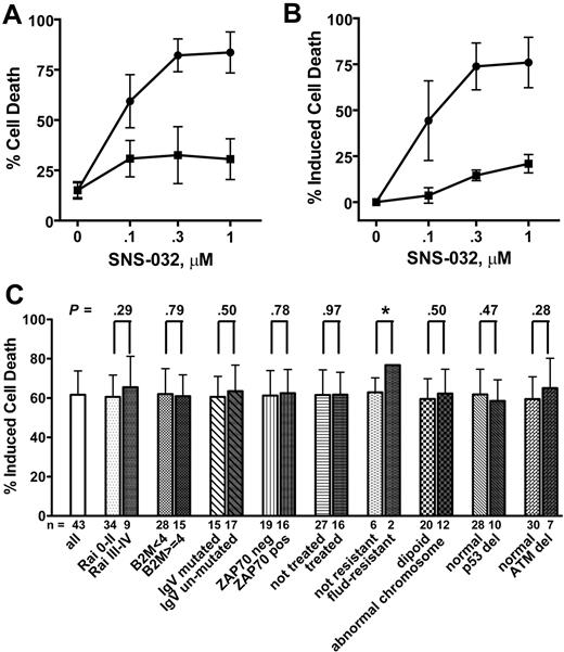
The cytotoxicity of SNS-032 was evaluated using annexin V/PI staining followed by flow cytometry after both a short (6 hours) and a longer incubation period (24 hours). The 6-hour time point was chosen because the SNS-032 is administered as a 15-minute loading dose followed by a 6-hour continuous infusion in the current clinical trial.31 CLL cells from 6 patient samples incubated with a range of SNS-032 concentrations (0.1-1 μM) for 6 hours induced cell death by 16% to 18% relative to untreated CLL cells (15.1% ± 3.7%, mean ± SD) (Figure 1A). In contrast, after 24 hours, cell death increased significantly in a concentration-dependent manner, which reached a maximum at 0.3 μM (82.2% ± 8.2%, mean ± SD), whereas the viabilities of the time-matched control cells were well maintained (15.1% ± 4.3% cell death, data not shown). A recent report of the phase 1 clinical trial of SNS-032 in B-cell malignancies showed that plasma SNS-032 more than 0.3 μM was well maintained during the infusion in patients who received total doses of 75 mg/m2.31 The induction of apoptosis was selective for CLL cells, as 0.3 μM SNS-032 only induced 14.5% plus or minus 2.9% (mean ± SD) cell death in mononuclear cells isolated from healthy donors, compared with a 73.8% plus or minus 12.8% cell death inductions in a set of 8 CLL samples (Figure 1B). Little variation was observed within the 6 samples used in Figure 1A, although the prognostic indicators of each patient varied. As shown in Table 1 (patients 1-4, 10, and 12), 2 of the 6 patients had received prior treatment, 3 had chromosomal abnormalities, 3 had unmutated immunoglobulin heavy-chain variable-region genes (IgVH),32 and 2 expressed high 70-kD ζ-associated protein (ZAP-70),33 indicating aggressive disease. Thus, these data suggested that SNS-032 may be toxic to CLL cells regardless of patient characteristics. To extend this observation, we compared 0.3 μM SNS-032–induced cell death after 24-hour incubation in 43 CLL samples. The net induced apoptosis averaged 61.6% plus or minus 12.1% (mean ± SD) after subtracting the cell death in time-matched controls (15.6% ± 8.9%; Figure 1C). There was no significant difference between cells from patients with either favorable or poor prognosis features, including Rai stage,34 β2-microglobulin levels,35 IgVH mutation status,32 ZAP-70 expression,33 previous treatment history, and chromosomal abnormalities. There was no evidence of a loss of potency in samples derived from fludarabine-resistant patients. In a group of samples analyzed by fluorescence in situ hybridization that had deletion of 17p, indicative of TP53 gene deletion (range, 8.5%-94.5%), there was no significant difference in cell death compared with the rest of the group that had normal TP53 gene loci. Similar results were observed for patients with del 11q, diagnostic of ATM gene deletion (range, 8.5%-94.5%). These results indicate that SNS-032 may induce apoptosis by a mechanism that is independent of p53 and ATM expression in the CLL cells.
SNS-032 potently induced apoptosis in CLL cells. (A) Induction of apoptosis in CLL cells isolated from 6 patients at 6-hour (■) and 24-hour (●) exposure to SNS-032. Cell death was measured by annexin V/PI double staining and quantitated by flow cytometry analysis (mean ± SD). (B) SNS-032 is selective against CLL cells. Cell death induced by SNS-032 was compared between the averages of 8 CLL cells (●) to mononuclear cells isolated from 3 healthy donors (■). (C) SNS-032 induced similar cell death regardless of patient characteristics and treatment history. Cell death induced by 0.3 μM SNS-032 after 24-hour incubation was compared in CLL cells from patients with favorable or poor prognostic factors. A P value of less than .05 was considered significant. Induced cell death was calculated as the difference between cell death in drug-treated samples minus that of time-matched controls. DMSO alone at highest concentration used here (0.01%) did not show toxicity to the CLL cells. *Sample number was too small to give a reliable P value.
SNS-032 potently induced apoptosis in CLL cells. (A) Induction of apoptosis in CLL cells isolated from 6 patients at 6-hour (■) and 24-hour (●) exposure to SNS-032. Cell death was measured by annexin V/PI double staining and quantitated by flow cytometry analysis (mean ± SD). (B) SNS-032 is selective against CLL cells. Cell death induced by SNS-032 was compared between the averages of 8 CLL cells (●) to mononuclear cells isolated from 3 healthy donors (■). (C) SNS-032 induced similar cell death regardless of patient characteristics and treatment history. Cell death induced by 0.3 μM SNS-032 after 24-hour incubation was compared in CLL cells from patients with favorable or poor prognostic factors. A P value of less than .05 was considered significant. Induced cell death was calculated as the difference between cell death in drug-treated samples minus that of time-matched controls. DMSO alone at highest concentration used here (0.01%) did not show toxicity to the CLL cells. *Sample number was too small to give a reliable P value.
Inhibition of RNA synthesis by SNS-032 in CLL cells
Treatment of CLL cells from the same group of 6 patient samples in Figure 1A with SNS-032 for 6 or 24 hours was associated with a decrease in phosphorylation of Ser2 and Ser5 of the CTD of RNA Pol II that appeared to be both concentration- and time-dependent (Figure 2A). SNS-032 decreased the ratios of pSer2/total RNA Pol II and pSer5/total RNA Pol II in a concentration-dependent manner (Figure 2B) that was remarkably consistent among samples. There was greater inhibition on Ser2 phosphorylation than that of Ser5, consistent with a lower IC50 for the inhibition of Cdk9 compared with Cdk7 (4 nM vs 62 nM).28 This was associated with inhibition of RNA synthesis, measured by tritiated uridine incorporation in the same group of 6 patient samples, which decreased in a concentration-dependent manner after SNS-032 treatment (Figure 2C). There was no apparent time dependence, indicating that the phosphorylation of RNA Pol II was inhibited rapidly after addition of SNS-032. Indeed, 0.3 μM SNS-032 reduced 88.3% of [3H]uridine incorporation after 1 hour (average of 2 patient samples, each measured in triplicate; data not shown). The protein levels of Cdk7 and Cdk9 were stable after 6 hours of SNS-032 exposure and declined at 24 hours (Figure 2A). The discordance between Cdk7 and Cdk9 protein levels and the Pol II phosphorylation status at 6 hours (Figure 2B) indicated that reducing of Cdk7 and Cdk9 levels could not account for the decreased RNA Pol II phosphorylation. Rather, the rapid inhibition on the activity of Cdk7 and Cdk9 led to the inactivation of RNA Pol II.
SNS-032 inhibited RNA synthesis in the CLL cells. CLL cells were incubated with 0.1, 0.3, and 1 μM SNS-032 for 6 and 24 hours, the phosphorylation of RNA Pol II and Cdk7, Cdk9 expression levels were analyzed by immunoblotting, using antibodies toward the phosphorylated Ser2 or Ser5 sites of the CTD, as well as total RNA Pol II, total Cdk7, and Cdk9. (A) A representative immunoblot from patient 4. (B) Inhibition of phosphorylation of RNA Pol II at Ser2 (■) and Ser5 (●) sites and protein levels of Cdk7 (▲) and Cdk9 (▼) after 6-hour (left) and 24-hour (right) incubation with SNS-032 from the 6 patient samples in Figure 1A. Levels of phosphorylation were quantified from the blots, normalized to total Pol II, and then expressed as a percentage of time-matched controls (mean ± SD). Levels of Cdk7 and Cdk9 were normalized to actin and expressed as percentage of time-matched controls (mean ± SD). (C) Inhibition of [3H]uridine incorporation by SNS-032 in CLL cells. The inhibition of [3H]uridine incorporation was measured in the same set of CLL samples at 6 hours (■) and 24 hours (●) in SNS-032. Data are percentage of time-matched controls (mean ± SD; n = 6 samples, each performed in triplicate). The average dpm value for untreated samples was 109 740 plus or minus 59 359 (mean ± SD).
SNS-032 inhibited RNA synthesis in the CLL cells. CLL cells were incubated with 0.1, 0.3, and 1 μM SNS-032 for 6 and 24 hours, the phosphorylation of RNA Pol II and Cdk7, Cdk9 expression levels were analyzed by immunoblotting, using antibodies toward the phosphorylated Ser2 or Ser5 sites of the CTD, as well as total RNA Pol II, total Cdk7, and Cdk9. (A) A representative immunoblot from patient 4. (B) Inhibition of phosphorylation of RNA Pol II at Ser2 (■) and Ser5 (●) sites and protein levels of Cdk7 (▲) and Cdk9 (▼) after 6-hour (left) and 24-hour (right) incubation with SNS-032 from the 6 patient samples in Figure 1A. Levels of phosphorylation were quantified from the blots, normalized to total Pol II, and then expressed as a percentage of time-matched controls (mean ± SD). Levels of Cdk7 and Cdk9 were normalized to actin and expressed as percentage of time-matched controls (mean ± SD). (C) Inhibition of [3H]uridine incorporation by SNS-032 in CLL cells. The inhibition of [3H]uridine incorporation was measured in the same set of CLL samples at 6 hours (■) and 24 hours (●) in SNS-032. Data are percentage of time-matched controls (mean ± SD; n = 6 samples, each performed in triplicate). The average dpm value for untreated samples was 109 740 plus or minus 59 359 (mean ± SD).
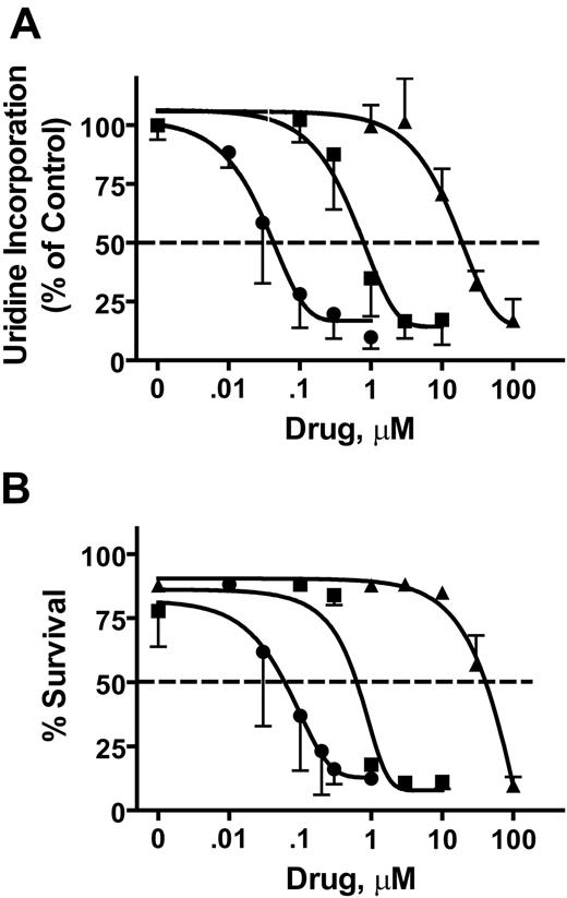
Both flavopiridol and roscovitine are inhibitory to Cdk9 and Cdk7 and affect Pol II phosphorylation status. In direct comparisons on CLL cells, SNS-032 was approximately 10- to 20-fold more potent than flavopiridol and approximately 400- to 700-fold more potent than roscovitine in the inhibition of RNA synthesis (Figure 3A) and induction of cell death (Figure 3B). Consistent with IC50 values against Cdk9 and Cdk7,36 these data indicate that SNS-032 is a more potent agent in these studies.
Comparison of the activity of SNS-032, flavopiridol, and roscovitine in CLL cells. The inhibition of uridine incorporation after 6 hours (A) and inhibition of cell survival measured by annexin V/PI staining after 24 hours (B) were compared among SNS-032 (●), flavopiridol (■), and roscovitine (▲) with concentrations that ranged between 0.01 and 100 μM. Data (mean ± SD) represent measurements from 3 to 5 individual patient samples.
Comparison of the activity of SNS-032, flavopiridol, and roscovitine in CLL cells. The inhibition of uridine incorporation after 6 hours (A) and inhibition of cell survival measured by annexin V/PI staining after 24 hours (B) were compared among SNS-032 (●), flavopiridol (■), and roscovitine (▲) with concentrations that ranged between 0.01 and 100 μM. Data (mean ± SD) represent measurements from 3 to 5 individual patient samples.
SNS-032 decreased the mRNA and protein levels of antiapoptotic proteins
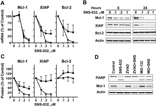
The most sensitive targets of transcription inhibitors are probably transcripts and proteins that intrinsically turn over rapidly. SNS-032 induced a rapid concentration-dependent decrease in the mRNA levels of Mcl-1 (Figure 4A). After 6 hours with 0.3 μM SNS-032, Mcl-1 mRNA was reduced to 27% of controls and to 10% by 1 μM SNS-032 (Figure 4A). There was no apparent difference between 6 and 24 hours, probably because of the intrinsically short half-life of Mcl-1 mRNA. In contrast, there was a time- and concentration-dependent decrease of XIAP and Bcl-2 mRNA. This was associated with decreases in the protein levels of Mcl-1 and XIAP (Figure 4B), although there was no apparent decrease in Bcl-2 protein levels. Similar analyses from all 6 patient samples were quantitated and were summarized in Figure 4C. The rate of decrease in the protein levels of Mcl-1 and XIAP was proportional to their intrinsic half-lives, with Mcl-1 being the most labile. There was no significant change in the Bcl-2 protein, consistent with a much longer protein half-life.37
SNS-032 reduced the expression of antiapoptotic proteins. The total RNA and protein of CLL cells from the same set of 6 patients in Figure 1A were isolated after 6 hours (■) and 24 hours (●) of incubation with 0.1, 0.3, and 1 μM SNS-032. (A) The mRNA levels of Mcl-1, XIAP, and Bcl-2 were measured by real-time reverse-transcribed polymerase chain reaction, each performed in duplicate, and compared with time-matched controls. (B) A representative immunoblot from patient 4. (C) Quantitations of immunoblots of Mcl-1, XIAP, and Bcl-2 from the samples used in panel A. Levels of Mcl-1, XIAP, and Bcl-2 were normalized to actin and expressed as percentage of time-matched controls. (D) SNS-032–induced Mcl-1 reduction is independent of caspase activity. CLL cells were preincubated with pan-caspase inhibitor ZVAD-FMK (100 μM) or proteosome inhibitor MG-132 (10 μM) for 1 hour before incubating with 0.3 μM SNS-032 for 8 hours. PARP cleavage and Mcl-1 protein levels were visualized by immunoblotting.
SNS-032 reduced the expression of antiapoptotic proteins. The total RNA and protein of CLL cells from the same set of 6 patients in Figure 1A were isolated after 6 hours (■) and 24 hours (●) of incubation with 0.1, 0.3, and 1 μM SNS-032. (A) The mRNA levels of Mcl-1, XIAP, and Bcl-2 were measured by real-time reverse-transcribed polymerase chain reaction, each performed in duplicate, and compared with time-matched controls. (B) A representative immunoblot from patient 4. (C) Quantitations of immunoblots of Mcl-1, XIAP, and Bcl-2 from the samples used in panel A. Levels of Mcl-1, XIAP, and Bcl-2 were normalized to actin and expressed as percentage of time-matched controls. (D) SNS-032–induced Mcl-1 reduction is independent of caspase activity. CLL cells were preincubated with pan-caspase inhibitor ZVAD-FMK (100 μM) or proteosome inhibitor MG-132 (10 μM) for 1 hour before incubating with 0.3 μM SNS-032 for 8 hours. PARP cleavage and Mcl-1 protein levels were visualized by immunoblotting.
When the supply of mRNA for translation is shut down by SNS-032, the decrease in total Mcl-1 protein reflects the degradation of existing Mcl-1 protein by proteases, such as through the proteosome degradation pathway.38 Alternatively, Mcl-1 protein can also be degraded by activated caspase-3,39 which may cleave Mcl-1 into small fragments. To determine the extent to which each of these mechanisms accounts for Mcl-1 protein turnover, CLL cells were preincubated with the caspase-3 inhibitor, ZVAD, for 1 hour before exposure to 0.3 μM SNS-032 for 8 hours. ZVAD inhibited the activity of caspase-3, shown by the blocking of PARP cleavage (Figure 4D). However, ZVAD did not block the degradation of Mcl-1 protein, indicating that this was independent of the caspase activity. In contrast, the proteosome inhibitor, MG-132, stabilized Mcl-1 protein in the cells and partially restored the Mcl-1 protein level. That Mcl-1 protein was only partially restored by MG-132 is probable because caspase-3 is already activated in the cells, diminishing the protein. Therefore, this experiment indicated that the SNS-032–induced decrease of Mcl-1 was the result of intrinsic turnover through the proteosome pathway after transcription inhibition. The more limited proteolysis by caspase-3 probably occurred secondary to activation of the apoptotic process.
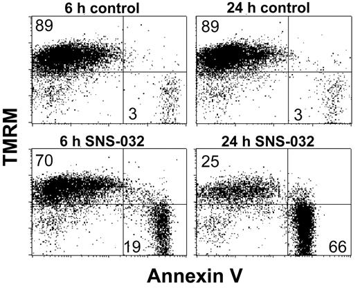
On removal of Mcl-1 protein, the freed proapoptotic binding partners are able to disrupt the mitochondrial membrane. Depolarization of mitochondrial membranes was quantitated by the loss of binding to the cationic dye TMRM. As shown in Figure 5, SNS-032 induced a time-dependent shifting of cells from the TMRM+/annexin− to the TMRM−/annexin+ population, indicating that loss of mitochondrial membrane potential was associated with the onset of cell death, probably initiated by the reduced Mcl-1 protein.
SNS-032 induced loss of mitochondrial membrane potential. CLL cells were incubated with 0.3 μM SNS-032 for 6 and 24 hours. Mitochondrial membrane potential and viability were measured by TMRM and annexin V–FITC double staining.
SNS-032 induced loss of mitochondrial membrane potential. CLL cells were incubated with 0.3 μM SNS-032 for 6 and 24 hours. Mitochondrial membrane potential and viability were measured by TMRM and annexin V–FITC double staining.
Removal of SNS-032 reactivated transcription and restored Mcl-1 protein levels
To relate the decrease in antiapoptotic protein levels with the induction of cell death, it was important to determine the duration of exposure to SNS-032 that was optimal for CLL cell killing. Cells from 4 samples were incubated with 0.3 μM SNS-032 for as long as 24 hours; apoptosis was determined by annexin V/PI staining at 2-hour intervals. Cell killing by SNS-032 in vitro was evident at 6 hours but was maximized between 10 and 12 hours (Figure 6A). In contrast, the viability of mock-treated control samples remained stable for 24 hours. To determine whether additional cell killing occurred after removal of SNS-032, we measured cell death at 24 hours after a 6-hour incubation with SNS-032 followed by washing cells into drug-free medium. Cells incubated continuously with SNS-032 for 24 hours were used as the comparator for maximum cell kill. After washing cells into fresh medium, there was no evidence of additional cell death beyond that observed after the 6-hour incubation (Figure 6B). Although there was a modest concentration-dependent cell killing after 6-hour incubation, death did not increase during the 18 hours after removal of SNS-032, even at the greatest concentration, 1 μM. Continuous incubation with these SNS-032 concentrations for 24 hours generated a prominent concentration-dependent killing, as was also seen in Figure 1A. To investigate the reason for this lack of continued killing, CLL cells were incubated with 0.3 μM SNS-032 for 6 hours, and one portion of the culture was washed into drug-free medium, whereas the other portion was maintained in the presence of drug. Immunoblotting analysis over time demonstrated that there was a rapid rephosphorylation of Ser2 and Ser5 of the RNA Pol II CTD that was clearly evident 3 hours after washing cells of the drug (Figure 6C). This was accompanied by the recovery of RNA synthesis (Figure 6D), again suggesting the linkage between these parameters. The kinetics of decrease in Mcl-1 and XIAP proteins was halted on washing, and some recovery occurred (Figure 6C). Thus, on SNS-032 removal, new proteins were synthesized to overcome their rapid degradation and restore the protein levels.
Recovery of RNA synthesis and resynthesis of antiapoptotic proteins when the CLL cells were washed into fresh media without SNS-032. (A) Time dependence of SNS-032–induced cell death. CLL cells were incubated with 0.3 μM SNS-032, and cell death was measured every 2 hours by annexin V/PI staining (●) compared with time-matched controls (○). Data represent mean plus or minus SD of 4 CLL patient samples. (B) CLL cells were incubated with SNS-032 for 6 hours, and apoptosis was measured by annexin V/PI staining ( ). One portion of the cells was washed and then incubated in fresh medium without SNS-032 until 24 hours (
 ), and cell death was compared with cells that were continuously exposed to SNS-032 for 24 hours (
 ). Data represent mean plus or minus SD of 5 CLL patient samples. (C) Recovery of RNA Pol II phosphorylation and antiapoptotic proteins when SNS-032 was washed out. After incubating with 0.3 μM SNS-032 for 6 hours, the cells were washed into fresh media and collected for analysis every 3 hours. The phosphorylation status of RNA Pol II at Ser2 and Ser5 and protein levels of Mcl-1 and XIAP were measured by immunoblotting and compared between time-matched controls (ctrl), cells exposed to SNS-032 continuously (SNS), and cells washed at 6 hours and placed in drug-free media (wash). *Nonspecific band. (D) Recovery of RNA synthesis after washing out SNS-032. [3H]Uridine incorporation was presented as percentage of time-matched controls (■). Cells were washed at 6 hours into drug-free media (●); cells were incubated with SNS-032 (▲) continuously. Data represent mean ± SD of 3 experiments, each done in triplicate.
Recovery of RNA synthesis and resynthesis of antiapoptotic proteins when the CLL cells were washed into fresh media without SNS-032. (A) Time dependence of SNS-032–induced cell death. CLL cells were incubated with 0.3 μM SNS-032, and cell death was measured every 2 hours by annexin V/PI staining (●) compared with time-matched controls (○). Data represent mean plus or minus SD of 4 CLL patient samples. (B) CLL cells were incubated with SNS-032 for 6 hours, and apoptosis was measured by annexin V/PI staining ( ). One portion of the cells was washed and then incubated in fresh medium without SNS-032 until 24 hours (
 ), and cell death was compared with cells that were continuously exposed to SNS-032 for 24 hours (
 ). Data represent mean plus or minus SD of 5 CLL patient samples. (C) Recovery of RNA Pol II phosphorylation and antiapoptotic proteins when SNS-032 was washed out. After incubating with 0.3 μM SNS-032 for 6 hours, the cells were washed into fresh media and collected for analysis every 3 hours. The phosphorylation status of RNA Pol II at Ser2 and Ser5 and protein levels of Mcl-1 and XIAP were measured by immunoblotting and compared between time-matched controls (ctrl), cells exposed to SNS-032 continuously (SNS), and cells washed at 6 hours and placed in drug-free media (wash). *Nonspecific band. (D) Recovery of RNA synthesis after washing out SNS-032. [3H]Uridine incorporation was presented as percentage of time-matched controls (■). Cells were washed at 6 hours into drug-free media (●); cells were incubated with SNS-032 (▲) continuously. Data represent mean ± SD of 3 experiments, each done in triplicate.
Discussion
This study investigated the hypothesis that depriving cells of antiapoptotic proteins by inhibition of transcription would initiate the irreversible process of apoptosis. CLL was used as a model because of the dependence of this disease on the continued expression of antiapoptotic proteins for survival. Further, 2 such survival factors, Mcl-1 and XIAP, have intrinsically rapid turnover rates in both transcripts and proteins. SNS-032, a potent inhibitor of both Cdk9 and Cdk7, blocked the transcription-enabling phosphorylations on RNA Pol II by these kinases. This was closely associated with decreases in Mcl-1 and XIAP transcripts and, subsequently, of their proteins. Consistent with the working hypothesis, CLL cells initiated apoptosis within a few hours of Mcl-1 depletion. However, the action of SNS-032 was readily reversible, as its removal was followed by reactivation RNA Pol II, resumption of Mcl-1 expression, and halting further of cell death, emphasizing the importance of sustained suppression of these oncoproteins for cell killing.
Because of the heterogeneous clinical responses of CLL patients, cellular and molecular markers have been identified to predict the disease tendency or outcome of therapy. Rai40 and Binet41 staging systems have been widely used to assess disease status and treatment options, with Rai stages III and IV considered high-risk and aggressive disease. Using interphase fluorescence in situ hybridization, cytogenetic lesions were identified in CLL patient cells, including the deletion of the short arm of chromosome 17 where the TP53 gene is located, or the long arm of chromosome 11 associated with the ATM gene. Evidence showed that these chromosomal aberrations can be an unfavorable prognosis to standard chemotherapy containing alkylating agents and purine nucleoside analogs.42 The absence of somatic mutation IgVH gene, or high expression of ZAP-7033 or CD3843 in the leukemia cells, is also associated with aggressive disease. The combination of fludarabine, cyclophosphamide, and rituximab is the most active regimen in the current treatment of CLL.35 Still, patients with high β2-microglobulin have significantly inferior responses than those who express lesser amounts of this protein. When responses to SNS-032 in vitro were compared in patient samples with either favorable or poor prognostic characteristics, there were no significant differences found among samples in each group (Figure 1C). These results indicate that SNS-032 induces apoptosis by a mechanism that is independent of these variables. The fact that CLL samples from 16 patients who failed other therapies were responsive to SNS-032 in vitro suggests that SNS-032 may have the potential to overcome drug resistance in CLL therapy.
The activity of SNS-032 takes advantage of the dependence of CLL cells on antiapoptotic proteins for survival. The phenomenon that some tumors are dependent on the continuous activity of specific oncogenes for sustaining their malignant phenotype was characterized as being “addicted” to the activity of the oncogene.44 This concept has provided a rationale for the development of targeted therapeutics specifically directed at inhibiting the activity of the particular oncogene product.45 In such an approach, the biologic context of the dependency of the tumor on oncogene function provides a basis for the therapeutic index. Recently, this rationale for cancer therapeutics has proved to be effective in the clinic.45 CLL cells are characterized by their addiction to the continuous presence of antiapoptotic proteins to maintain their survival.8 Therapeutic strategies that remove these blocks will release the proapoptotic signal and initiate cell death. In addition, antiapoptotic proteins, such as Mcl-1 and XIAP, which sustain the CLL cell viability, are intrinsically short lived; thus, their mRNA and protein levels will decrease rapidly by degradation when synthesis is stopped after inhibition of transcription. This biologic context is probably critical for the clinical success of flavopiridol in CLL compared with its lack of activity in other diseases.23
There is controversy of how important Mcl-1 is for CLL cell survival relative to Bcl-2. When measured quantitatively, CLL cells express a 4- to 14-fold less Mcl-1 compared with Bcl-2.3 In addition, in BH3 profiling assay, peptides derived from Noxa, which binds and inhibits Mcl-1, failed to cause cytochrome c release from mitochondria isolated from CLL cells.3 These data support the argument that Bcl-2 may be the dominant antiapoptotic protein sustaining the CLL cells. However, other evidence suggests that Mcl-1 plays an important role in CLL survival as well. Decreasing Mcl-1 alone is sufficient to induce a substantial amount of apoptosis in CLL cells in vitro: for example, when Mcl-1 expression was specifically knocked down by siRNA,46 there was a significant reduction in cell survival and enhancement of chemosensitivity. In addition, our results with CLL cells treated with either SNS-032 or flavopiridol15 showed that inhibition of transcription substantially reduced Mcl-1 and XIAP expression and induced cell death, whereas the Bcl-2 protein level remained stable, indicating that Mcl-1 may also be critical in sustaining the CLL cells. Thus, it is possible that it is the balance of the pools of the antiapoptotic and proapoptotic proteins, rather than expression of an individual protein, that controls the fate of the cells. For example, cellular resistance to Bcl-2 antagonists was associated with an overexpression of Mcl-1.47 Strategies that reduced Mcl-1 expression have shown synergy with the BH3 mimetic, ABT-737,47,48 which itself has little effect on Mcl-1 binding to proapoptotic proteins. Furthermore, it was reported that the proapoptotic proteins released after reduction of Mcl-1 may again be sequestered by Bcl-2, thereby delaying the onset of apoptosis.49 These observations indicate that either diminishing Mcl-1 expression or antagonizing Bcl-2 binding will lower the buffering capacity of the pool of antiapoptotic proteins. When this capacity is reduced below a critical point, the excess proapoptotic proteins will disrupt the mitochondrial membrane and induce cell death.
The observation that the inhibition of transcription by SNS-032 appeared to be reversible has important implications for therapeutic strategies. On washing out SNS-032 after 6 hours of incubation with CLL cells, RNA Pol II phosphorylation and uridine incorporation recovered within 3 hours, followed by repletion of short-lived proteins, such as Mcl-1 and XIAP (Figure 6C). This indicated that synthesis of these proteins had resumed and viability of cells that had not initiated apoptosis was maintained. As maximal killing in vitro occurred after 10 to 12 hours of exposure to SNS-032, these results suggest that it may be necessary to expose the CLL cells to SNS-032 for such a duration in the clinic. In addition, these experiments indicate the importance of sustained suppression of Mcl-1 and XIAP to maximize induction of cell death, and emphasize the importance of these proteins for CLL survival. Finally, these experiments raise the possibility that protein synthesis inhibitors that block the resynthesis of Mcl-1 may extend the cytotoxicity of SNS-032. One such inhibitor, homoharringtonine, has activity to reduce the Bcr-Abl oncoprotein in a chronic myelogenous leukemia cell line.50 It is currently being evaluated in a phase 2 clinical trial in imatinib-resistant chronic myelogenous leukemia patients with the T315I mutation. The combination effect of SNS-032 with homoharringtonine is under investigation.
SNS-032 was selected for development based on its favorable characteristics, such as low protein binding, potency, and selective inhibition on a small subset of Cdks (Cdk2, 7, and 9). In comparison with other compounds with similar activities, flavopiridol is known for its high plasma protein binding, whereas only approximately 63% of SNS-032 was bound in human serum.24 Its potency is evident in assays of both inhibition of RNA synthesis and induction of apoptosis compared with flavopiridol and roscovitine. In addition, although both flavopiridol and SNS-032 have similar IC50 values for Cdk9/cyclin T, SNS-032 is approximately 5 times more potent against Cdk7/cyclin H than is flavopiridol.36 In contrast, although roscovitine has inhibitory activity against each of these targets, concentrations required are 10 to 100 times greater. Further, flavopiridol and roscovitine inhibit Cdk1/cyclin B, Cdk4/cyclin D, Cdk6/cyclin D, and Cdk5/p35 in similar concentration ranges as against Cdk7 and Cdk9, whereas SNS-032 is considerably less effective against these proliferation-related Cdks.28 Moreover, SNS-032 had little activity (IC50 > 1000 nM) against 190 other kinases.28 Therefore, SNS-032 has biochemical and pharmacologic properties that differ from flavopiridol and roscovitine, which probably predict differing activities in the clinic.
In conclusion, this study demonstrated in vitro the action of a novel Cdk inhibitor, SNS-032, in CLL cells. In a recent report of an ongoing trial, SNS-032 concentrations above in vitro IC90 (115 ng/mL or 0.3 μM)30 were maintained for more than 6 hours in patients who received total doses of 75 mg/m2.31 Target modulation, such as reduced Pol II phosphorylation and decrease of Mcl-1 and XIAP levels, was demonstrated in CLL cells isolated from patients in this cohort. Apoptosis was detected by PARP cleavage at the completion of infusion.31 Thus, pharmacodynamic actions observed in vivo are consistent with the predictions of the present investigations in CLL cells in vitro. Additional investigations are required to provide rigorous proof-of-principle evidence for the actions of SNS-032 in the clinical setting.
The publication costs of this article were defrayed in part by page charge payment. Therefore, and solely to indicate this fact, this article is hereby marked “advertisement” in accordance with 18 USC section 1734.
Acknowledgments
The authors thank Susan C. Smith and Susan Lerner of the Department of Leukemia, University of Texas M. D. Anderson Cancer Center, Houston, TX, for providing the patient information.
This study was supported in part by research funding from Sunesis Pharmaceuticals (W.P.), CLL Global Research Foundation (Houston, TX), the National Cancer Institute, Department of Health and Human Services (Washington, DC; grants CA81534 and CA100632), and Cancer Center Support (Houston, TX; grant CA16672).
National Institutes of Health
Authorship
Contribution: R.C. conceptualized the project, designed and performed research, analyzed the data, and wrote the paper; W.G.W. and M.J.K. identified CLL patients for inclusion in the study; S.C. performed research and analyzed the data; R.E.H. contributed to experimental design and analysis; J.A.F. contributed to experimental design and analysis and provided SNS-032; V.G. contributed to experimental design and analysis; and W.P. conceptualized the project, directed experiment design and data analysis, and wrote the paper.
Conflict-of-interest disclosure: R.E.H. and J.A.F. are employees of Sunesis Pharmaceuticals. W.P. and W.G.W. received research funding from Sunesis Pharmaceuticals. All the authors declare no other competing financial interests.
Correspondence: William Plunkett, Department of Experimental Therapeutics, Unit 71, University of Texas M. D. Anderson Cancer Center, Houston, TX 77030; e-mail: wplunket@mdanderson.org.


![Figure 2. SNS-032 inhibited RNA synthesis in the CLL cells. CLL cells were incubated with 0.1, 0.3, and 1 μM SNS-032 for 6 and 24 hours, the phosphorylation of RNA Pol II and Cdk7, Cdk9 expression levels were analyzed by immunoblotting, using antibodies toward the phosphorylated Ser2 or Ser5 sites of the CTD, as well as total RNA Pol II, total Cdk7, and Cdk9. (A) A representative immunoblot from patient 4. (B) Inhibition of phosphorylation of RNA Pol II at Ser2 (■) and Ser5 (●) sites and protein levels of Cdk7 (▲) and Cdk9 (▼) after 6-hour (left) and 24-hour (right) incubation with SNS-032 from the 6 patient samples in Figure 1A. Levels of phosphorylation were quantified from the blots, normalized to total Pol II, and then expressed as a percentage of time-matched controls (mean ± SD). Levels of Cdk7 and Cdk9 were normalized to actin and expressed as percentage of time-matched controls (mean ± SD). (C) Inhibition of [3H]uridine incorporation by SNS-032 in CLL cells. The inhibition of [3H]uridine incorporation was measured in the same set of CLL samples at 6 hours (■) and 24 hours (●) in SNS-032. Data are percentage of time-matched controls (mean ± SD; n = 6 samples, each performed in triplicate). The average dpm value for untreated samples was 109 740 plus or minus 59 359 (mean ± SD).](https://ash.silverchair-cdn.com/ash/content_public/journal/blood/113/19/10.1182_blood-2008-12-190256/7/m_zh80200934860002.jpeg?Expires=1770401681&Signature=X31621ZkOxcv5rXovBPu0PNIS0YpaVURdmt0Z3t8NOBNJ6SwlzCz4FGuEEQEwjOKeUQLWQeD0w3AEEKUAUouQ3aaIW3Za-UvHu5aJAhPQ~reJi-cfUYNR5dVQhC~DhZYRxmneLsftyHXoLBnMPzS9VhcL0rwuuks1VC9FgMA2NhX3VQ5x7FUwedDy3XVBkee-~onKgtu-fIpH6tVoSndniTSaDhEzoLuxVE9tt3eh-PkhmAsWC3ccTVJkHQn43NPdXKNvBdw2CruJcHruFS6Oz5IrZcMcwg1BasMz5W1t31LrpmtWuJMGKpUJkM-bmFLJr9ie9hCxqy8-ZDkkp9-CA__&Key-Pair-Id=APKAIE5G5CRDK6RD3PGA)


![Figure 6. Recovery of RNA synthesis and resynthesis of antiapoptotic proteins when the CLL cells were washed into fresh media without SNS-032. (A) Time dependence of SNS-032–induced cell death. CLL cells were incubated with 0.3 μM SNS-032, and cell death was measured every 2 hours by annexin V/PI staining (●) compared with time-matched controls (○). Data represent mean plus or minus SD of 4 CLL patient samples. (B) CLL cells were incubated with SNS-032 for 6 hours, and apoptosis was measured by annexin V/PI staining (). One portion of the cells was washed and then incubated in fresh medium without SNS-032 until 24 hours (), and cell death was compared with cells that were continuously exposed to SNS-032 for 24 hours (). Data represent mean plus or minus SD of 5 CLL patient samples. (C) Recovery of RNA Pol II phosphorylation and antiapoptotic proteins when SNS-032 was washed out. After incubating with 0.3 μM SNS-032 for 6 hours, the cells were washed into fresh media and collected for analysis every 3 hours. The phosphorylation status of RNA Pol II at Ser2 and Ser5 and protein levels of Mcl-1 and XIAP were measured by immunoblotting and compared between time-matched controls (ctrl), cells exposed to SNS-032 continuously (SNS), and cells washed at 6 hours and placed in drug-free media (wash). *Nonspecific band. (D) Recovery of RNA synthesis after washing out SNS-032. [3H]Uridine incorporation was presented as percentage of time-matched controls (■). Cells were washed at 6 hours into drug-free media (●); cells were incubated with SNS-032 (▲) continuously. Data represent mean ± SD of 3 experiments, each done in triplicate.](https://ash.silverchair-cdn.com/ash/content_public/journal/blood/113/19/10.1182_blood-2008-12-190256/7/m_zh80200934860006.jpeg?Expires=1770401681&Signature=KMY09IGRqbecbMGOl3EhmC88duhIpRHCY0nzQiJaJSl2N3jmk0dHjB96cVqhW2Jhpq3ddeV58AOR~SQQ2SEhoFgyEguvkvO9vx6aleVreeCcJS7p0KZIgCWWPYDKov7KJAWLHNdSOEU71EyDO9PGdrhtj0cHfOKmK6rRiRJc3cRorby-ZuxfP5YbcBz70JIdScZcQ~e~goNePN84StwePuD2Q7Um4tmOlMfRqLTNwsk7UKvq8KCaPqd6pSgbEj4vnFVl899rK7REKDF1IlZtdh~W2wQdQ-LKgcmP1lM1453MVcnMAmSR0wY3jg8Aydf8QYZE35cK0uqExwlYYyfLog__&Key-Pair-Id=APKAIE5G5CRDK6RD3PGA)