Abstract
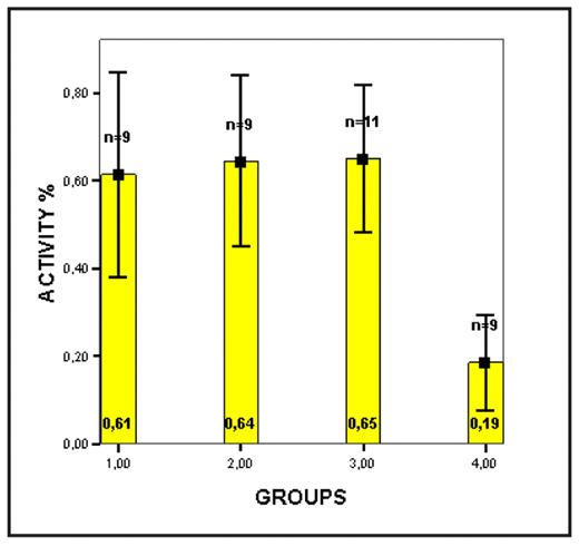
Imatinib is currently the most chosen agent in the treatment of CML patients. There are published articles reporting that imatinib has suppressive effects on T lymphocytes. However there is little information avaliable about the effects of imatinib on the immune regulation after allogeneic stem cell transplantation and effect of graft versus leukemia. In light of these observations, in our study, primary objective was in vivo analysis of T lymphocyte functions by flow cytometry and the secondary objective was to evaluate the possible functional changes that might occur under imatinib therapy in CML patients. A total of 29 patients and 9 healthy control subjects were enrolled in this cross sectional, clinical-laboratory study. CML patients were divided into three groups as newly diagnosed patients having no treatment (group 1), patients receiving imatinib for 1 year (group 2) and patients receiving imatinib more than 1 year (group 3), respectively. Healthy control subjects were regarded as group 4. To evaluate T lymphocyte functions, cells were induced by phorbol myristate acetate (PMA) and ionomisin then CD4+ T-cells were selected and IL-4 and IFN-γ expression on these cells; how much percentage of CD3+ T cells were activated (CD3+CD69+); CD8+ T lymphocytes and the ratio and grade of expression of HLA-ABC and HLA-DR on those cells were evaluated, respectively. In our study, there was no significant difference in terms of mean number of CD4+ cells between the groups (p=0.125). However, there was a tendency toward higher CD4+ cells in group 4. Cytokine expression analyses (IL-4 and IFNgamma) were found not to be statistically significant between the groups. (p values 0.112 and 0.165 respectively). It was striking that group 4 has lower IL-4 and IFNgamma expression values but just failed to reach a statistical significant level. On the other hand, mean number of CD4+ cells, which did not express IL-4 and IFNgamma, were statistically higher in group 4 when compared to other groups. There were no significant differences in terms of CD3+ cells and CD69+ expression, which was an early activation marker of T cells. However, it was found differences in % activation values (p=0.002) and % activation of subjects in group 4 was found to be decreased when compared to that of other groups (Figure 1). CD8+ cell ratio was found to be statistically lower in all CML patient groups when compared to that of healthy control subjects (p=0.001). The expression of HLA-ABC and HLA-DR on CD8+ cells was similar between the groups. We could not show any inhibitory effect of imatinib on T cell functions that could support clinical observations. It will be a research area how this interaction will be in new and more potent 2nd and 3rd generation tyrosine kinase inhibitors.
Graph showing distribution of mean percent activity between the groups. Note that group 4 shows significant lower values of mean percent activity.
Graph showing distribution of mean percent activity between the groups. Note that group 4 shows significant lower values of mean percent activity.
Disclosures: No relevant conflicts of interest to declare.
Author notes
Corresponding author


This feature is available to Subscribers Only
Sign In or Create an Account Close Modal