Selective targeting of endothelial cells in tumor vessels requires delineation of key molecular events in formation and survival of blood vessels within the tumor microenvironment. To this end, proteins transiently up-regulated during vessel morphogenesis were screened for their potential as targets in antiangiogenic tumor therapy. The molecular chaperone αB-crystallin was identified as specifically induced with regard to expression level, modification by serine phosphorylation, and subcellular localization during tubular morphogenesis of endothelial cells. Small interfering RNA–mediated knockdown of αB-crystallin expression did not affect endothelial proliferation but led to attenuated tubular morphogenesis, early activation of proapoptotic caspase-3, and increased apoptosis. αB-crystallin was expressed in a subset of human tumor vessels but not in normal capillaries. Tumors grown in αB-crystallin−/− mice were significantly less vascularized than wild-type tumors and displayed increased areas of apoptosis/necrosis. Importantly, tumor vessels in αB-crystallin−/− mice were leaky and showed signs of caspase-3 activation and extensive apoptosis. Ultrastructural analyses showed defective vessels partially devoid of endothelial lining. These data strongly implicate αB-crystallin as an important regulator of tubular morphogenesis and survival of endothelial cell during tumor angiogenesis. Hereby we identify the small heat shock protein family as a novel class of angiogenic modulators.
Introduction
Genetic changes and low oxygen pressure in proliferating tumor tissue promote production of several proangiogenic factors such as vascular endothelial growth factor (VEGF), which constitute interesting targets for angiogenesis inhibition in the treatment of cancer. In accordance, neutralization of VEGF in combination with chemotherapy prolongs the life expectancy of patients with metastatic colon, breast, and lung cancer.1 However, the plasticity of the tumor cell compartment is likely to offer means of escape from long-term suppression of VEGF function. An alternative approach to optimizing antiangiogenic treatment is to identify and manipulate key proteins regulating endothelial cell apoptosis. This requires a detailed understanding of the molecular mechanisms that regulate angiogenesis and survival of endothelial cells within the tumor environment
Tumor vascularization may occur through sprouting angiogenesis from preexisting vessels or through recruitment of circulating endothelial cells or progenitors, which may contribute to different extents depending on the molecular context.2,–4 Both processes involve migration of endothelial cells and/or progenitors into the surrounding tissue, followed by proliferation and differentiation of cells to create new lumen-containing vessels. The stability of the new vessel is dependent on production of a vascular basement membrane and recruitment of supporting perivascular cells, pericytes.5 VEGF promotes survival of endothelial cells, at least in part, through activation of the phosphoinositide 3′ kinase and Akt/protein kinase B pathways.6 Less is known about regulation of endothelial cell apoptosis and how this process could be exploited therapeutically.
To identify intrinsic regulators of endothelial apoptosis/survival during angiogenesis, we used a proteomic screen based on tubular morphogenesis of capillary endothelial cells, which recapitulates the different steps in sprouting angiogenesis. We found that αB-crystallin/HSPB5, a member of the small heat shock protein (sHsp) family, was specifically up-regulated during tubular morphogenesis of endothelial cells in a 3-D collagen matrix. Targeted deletion of αB-crystallin and HspB2 (heat shock 27-kDa protein 2), the genetic loci of which overlap as a result of an ancient duplication of an ancestral gene, is compatible with vascular development but leads to progressive myopathy.7 In accordance, αB-crystallin repression of caspase-3 activity is important for regulation of survival during myogenic differentiation.8 Moreover, together with its partner αA-crystallin, αB-crystallin is required for maintaining lens fiber integrity during lens terminal cell differentiation.9 A recently described function of αB-crystallin is in F-box protein-mediated recognition and subsequent ubiquitination of cyclin D1, suppressing proliferation through inhibition of S-phase entry.10
Heat shock proteins were first identified as molecular chaperones that ensure proper folding of proteins and inhibit protein aggregation during stress. Recent evidence has demonstrated that sHsp family proteins may directly interact with the apoptotic machinery of the cell and act as prosurvival factors. αB-crystallin is expressed at low levels in most cells, and its expression is induced in response to various types of cellular stress.11 Specifically, αB-crystallin enhances cell survival by inhibiting the autoproteolytic maturation of caspase-3.12 Caspase-3 is an effector caspase activated downstream of both death receptor– and mitochondrial-induced apoptosis, leading to cleavage of various cellular proteins and activation of nucleases.13 αB-crystallin has also been shown to directly bind proapoptotic Bcl-2 family members Bax and Bcl-Xs, leading to restricted release of cytochrome c from the mitochondria.14 Moyano et al recently identified αB-crystallin expression as a predictor of poor survival in breast cancer, indicating that the antiapoptotic property of αB-crystallin plays a role in tumor progression.15 In this report, we provide the first evidence that αB-crystallin is an important survival factor for endothelial cells during tumor angiogenesis in vivo. Our data suggest that αB-crystallin promotes tumor progression by enhancing survival of endothelial cells, allowing efficient vascularization of the expanding tumor mass.
Methods
Cell culture and in vitro angiogenesis assays
Bovine capillary endothelial (BCE) cells from calf adrenal cortex were maintained in Dulbecco modified Eagle medium (DMEM; Invitrogen, Carlsbad, CA) supplemented with 10% newborn calf serum (NCS), 2 ng/mL fibroblast growth factor-2 (FGF-2; Boehringer Ingelheim, Ingelheim, Germany) on gelatinized dishes. Cells were starved in DMEM with 1% NCS overnight, then tube formation was induced on a collagen matrix in the presence of DMEM with 10% NCS, 10 ng/mL FGF-2, as described previously.16 Telomerase-immortalized human microvascular endothelial (TIME) cells17 were cultured on gelatinized dishes in endothelial basal medium (EBM) MV2 growth medium with supplements (5 ng/mL endothelial growth factor, 0.2 μg/mL hydrocortisone, 0.5 ng/mL VEGF-A, 10 ng/mL FGF-2, and 20 ng/mL insulin-like growth factor-1; PromoCell, Heidelberg, Germany). Cells were starved overnight in EBM MV2 medium with 1% fetal bovine serum before tube formation in the presence of 50 ng/mL VEGF-A (PeproTech, Rocky Hill, NJ) in EBM MV2 medium with supplements as described.18 Mouse embryonic stem cell line R119 was maintained on growth-arrested mouse embryonic fibroblasts in DMEM-Glutamax medium supplemented with 15% fetal bovine serum, 25 mM HEPES, pH 7.4, 1.2 mM sodium pyruvate, 0.12% monothiolglycerol, and 1000 U/mL leukemia inhibitory factor. Embryoid bodies were induced as described previously in the presence of VEGF-A (30 ng/mL; PeproTech), seeded in a collagen matrix to induce formation of endothelial sprouts, and analyzed at day 8.20
Cell lysates and Western blot analysis
Cells were washed in phosphate-buffered saline (PBS) and lysed in 70°C lithium dodecyl sulfate–sample buffer (Invitrogen). Samples were homogenized using a syringe and centrifuged for 5 minutes at 14 000 rpm at 4°C in a chilled micro centrifuge. Extracts were fractionated on Novex NuPAGE (4%-12%) precast gels using the Novex electrophoresis and blotting system (Invitrogen). The membrane (Hybond-C extra; GE Healthcare, Chalfont St Giles, United Kingdom) was blocked in 5% dry milk in 0.1%Tween, Tris-buffered saline (TTBS) for 1 hour at room temperature (rt). Incubation with primary antibody was done at 4°C overnight, the membrane was then washed 3 times in TTBS and incubated with horseradish peroxidase–linked secondary antibodies (GE Healthcare) for 1 hour at rt. After washing the membranes extensively in TTBS, antibody binding was detected using enhanced chemiluminescence-plus (GE Healthcare). Primary antibodies used were: α-αB-crystallin (SPA-222), α-αB-crystallin pSer59 (SPA227; Assay Designs, Ann Arbor, MI), α-β-catenin (Transduction Laboratories, Lexington, KY), α-actin (Santa Cruz Biotechnology, Santa Cruz, CA), and α-cleaved caspase-3 (Cell Signaling Technology, Danvers, MA).
Immunofluorescence and immunohistochemistry
Collagen gels with tube-forming BCE cells or embryoid bodies, grown in 8-well tissue culture slides, were washed for 30 minutes in TBS, fixed for 30 minutes at rt or at 4°C overnight in zinc fix (0.1 M Tris-HCl, pH 7.5, 3 mM calcium acetate, 23 mM zinc acetate, and 37 mM zinc chloride) with 0.2% Triton X-100, blocked in 3% bovine serum albumin (BSA) in TBS for 1 hour at rt, and incubated with primary antibodies in blocking solution for 2 hours at rt. Samples were washed in TBS and incubated with secondary antibodies in blocking solution for 1 hour at rt, followed by extensive washing steps. Hoechst 33 342 was added to one of the washing steps to visualize the nuclei. Collagen gels were transferred to microscope slides and mounted with Fluoromount-G (Southern Biotechnology) before analysis using an LSM 510 confocal microscope (Carl Zeiss International, Oberkochen, Germany).
Fully anonymized human tissue samples were used in accordance with the Swedish biobank legislation. The use of human tissue was approved by the ethical review board in Uppsala (no. Ups 03-412/2003). Tape-transfer frozen sections (6 μm) were obtained from the Fresh Tissue Biobank, Clinical Pathology, Uppsala University Hospital: renal carcinoma (n = 5), non–small cell lung carcinoma (n = 5), normal kidney (n = 4), and normal lung (n = 1). Human and mouse frozen tumor sections (6 μm) were fixed in ice-cold methanol for 15 minutes and washed in PBS. Sections were blocked in 3% BSA for 1 hour followed by incubation with primary antibodies in blocking solution for 2 hours at rt. The sections were washed in PBS and incubated with Alexa-conjugated secondary antibodies (Invitrogen) and/or Fluorescein Ulex Europeus Agglutinin-I (FITC-UEA-1; Vector Laboratories, Burlingame, CA) in blocking buffer for 1 hour, followed by nuclear staining with Hoechst 33 342. Sections were mounted using Fluoromount-G (Southern Biotechnology Associates, Birmingham, AL) and analyzed by confocal microscopy (LSM 510 META; Carl Zeiss).
For immunohistochemistry analysis of mouse tumor sections, methanol-fixed sections were incubated with primary antifibrinogen antibody overnight, washed, and incubated with biotinylated anti–rabbit immunoglobulin (Dako Denmark, Glostrup, Denmark). Antibody binding was detected using the Strept ABComplex/HRP method (Dako Denmark) and visualized by the diaminobenzidine reaction. Sections were counterstained with hematoxylin. Primary antibodies and reagents used were: α-vimentin (Dako Denmark), α-β-tubulin (Invitrogen), α-αB-crystallin SPA-222 (Assay Designs), α-αB-crystallin SPA-223 (Assay Designs), α-cleaved caspase-3 (Cell Signaling Technology), α-fibrinogen (Dako Denmark), α-NG-2 (Chemicon International, Temecula, CA), α-Flk1 (R&D Systems, Minneapolis, MN), α-CD31 (BD PharMingen, San Diego, CA), α-α-smooth muscle actin (ASMA; Sigma-Aldrich, St Louis, MO) and Texas Red-Phalloidin (Invitrogen).
αB-crystallin small interfering RNA transfection
TIME cells (2.1 × 10⁵ cells/well) were seeded in 6-well tissue culture dishes 36 hours before transfection with small interfering RNA (siRNA) targeting αB-crystallin (CRYAB-1, CRYAB-2) or control siRNA (QIAGEN, Valencia, CA) using RNAi-Fect (QIAGEN) according to the manufacturer's protocol. The medium was changed after 6 hours, and the cells were transferred to 10-cm culture dishes 24 hours after transfection. At 48 hours after transfection, the cells were seeded on collagen matrices in 12-well plates for tube formation as described above. Tubular structures in collagen gels were fixed after 24 hours with zink-fix and incubated with Texas Red-phalloidin (Invitrogen) and examined using a Nikon Eclipse E1000 microscope (Nikon, Tokyo, Japan). Images spanning the entire wells were analyzed using Easy Image Analysis 2000 software (Rainfall, Stockholm, Sweden). Total area and length of tubular structures were calculated, and the values of CRYAB-1– and CRYAB-2–treated cells were compared with nonsilencing control siRNA-treated cells.
siRNA duplexes used were: CRYAB-1: Sense: r(GGC CCA AAU UAU CAA GCU A)dTdT; antisense: r(UAG CUU GAU AAU UUG GGC C)dTdG; CRYAB-2: Sense: r(CCA GGG AGU UCC ACA GGA A)dTdT; antisense: r(UUC CUG UGG AAC UCC CUG G)dAdG; nonsilencing control siRNA (Alexa 488–conjugated and unconjugated): sense: r(UUC UCC GAA CGU GUC ACG U)dTdT; and antisense: r(ACG UGA CAC GUU CGG AGA A)dTdT.
Annexin-V staining
Collagen gels with tube-forming TIME cells transfected with CRYAB-1, CRYAB-2, or unconjugated nonsilencing siRNA were washed for 30 minutes in PBS at 4°C and then stained with annexin-V staining solution (annexin-V-Fluos Kit; Roche Diagnostics, Indianapolis, IN) for 3 hours at 4°C. Gels were washed 4 times in PBS at 4°C and fixed in 1% paraformaldehyde (Sigma-Aldrich) in PBS for 15 minutes at rt. For counterstaining, Hoechst 33 342 (2 μg/mL) was added to the first washing step. Seven images (10× magnification) were captured from the central area of each gel using a TE300 Eclipse inverted fluorescence microscope (Nikon, Tokyo, Japan) equipped with a Spot camera (Diagnostic Instruments, Sterling Heights, MI). The numbers of total, annexin-V–positive, propidium iodide (PI)–positive, and double-positive cells were determined using the Easy Image Analysis software (Rainfall). The proportion of annexin-V–positive/PI-negative cells were normalized to the 24-hour control siRNA-treated well in each experiment.
Animal studies
Animal work was approved by the Uppsala University board of animal experimentation and thus performed according to the United Kingdom Coordinating Committee on Cancer Research guidelines for the welfare of animals in experimental neoplasia.21 The mice were anesthetized with isoflurane (Forene; Abbott Laboratories, Abbott, IL) during all manipulations. Six-week-old female αB-crystallin/HSPB2−/− mice (Cryab−/−)7 in the 129S4/SvJae X 129S6/SvEvTac background and the corresponding wild-type controls (129Sv-TacJae, Mollegard/Bomhultgard, Denmark) were injected subcutaneously with 106 syngeneic F9 teratocarcinoma cells (American Type Culture Collection, Manassas, VA) on their left flank (n = 15 animals/group). Palpable tumors were established within 1 week, and animals were killed at day 13 after injection of tumor cells, following ethical regulations regarding tumor size. The tumors were measured with a caliper, in a blind procedure, and volumes were calculated by the formula π/6×width2×length. Tumor material was snap-frozen in dry ice/isopentane and sectioned (3-μm sections). The sections were stained by immunofluoresence as described, and images covering the entire area of one middle section in each tumor were analyzed using Easy Image Analysis software (Rainfall). Vessels were counted and results are presented as the total number of CD31- and ASMA-positive vessels normalized to area. The incidence of apoptosis in the sections was determined as cleaved caspase-3–positive area normalized to total area.
Transmission electron microscopy
A separate tumor study using 8-week-old male αB-crystallin/HSPB2−/− mice (Cryab−/−),7 and the corresponding wild-type controls (129Sv-TacJae, Mollegard/Bomhultgard; n = 4 tumors/group) was performed as described above to provide material for transmission electron microscopy analysis. At dissection, small (1 × 1×1 mm) samples of the tumor were immediately transferred into 2% glutaraldehyde (Agar Scientific, Stansted, United Kingdom) in 0.1 M sodium cacodylate buffer, pH 7.2, supplemented with 0.1 M sucrose for 6 hours. The primary fixation was followed by 1.5-hour postfixation in 1% osmium tetroxide (Agar Scientific) in the cacodylate buffer, dehydration in graded series of ethanol, and embedding in epoxy plastic Agar 100 (Agar Scientific). Ultrathin sections were cut and placed on polyvinyl formal plastic (Formvar; Agar Scientific)–coated copper grids and contrasted with uranyl acetate and lead citrate before analysis using a Hitachi H-1700 electron microscope (Hitachi, Tokyo, Japan).
Immunohistochemical and immunofluorescence microscopy
Mounted samples were analyzed by the use of a Nikon Eclipse E100 microscope (Nikon; see Figures 4A and 5A), a Nikon TE300 Eclipse inverted fluorescence microscope (Nikon; Figures 1A and 2C,G) or an LSM 510 META confocal laser-scanning inverted microscope (Carl Zeiss; Figures 1D, 3A-F, and 4D). The following objectives were used: Nikon Plan Apochromat 4×/0.2, 10×/0.45, 20×/0.75; Nikon TE300 Eclipse Plan Fluor 10×/0.3; Zeiss confocal Plan Neofluar 20×/0.75 UV Plan Apoochromat 63×/1.4 oil immersion 100×/1.45 oil immersion. Microphotographs were captured using a Nikon Eclipse DXM 1200 camera or a Spot camera (Diagnostic Instruments). The following software programs were used: Laser Scanning Microscope LSM 510 version 3.2 (Carl Zeiss), Spot Advanced version 3.5.9 for MacOS, and Easy Image Analysis version 2.7.4.03 (Rainfall). Processing of microphotographs was done using Adobe Photoshop version CS2 (Adobe Systems, San Jose, CA).
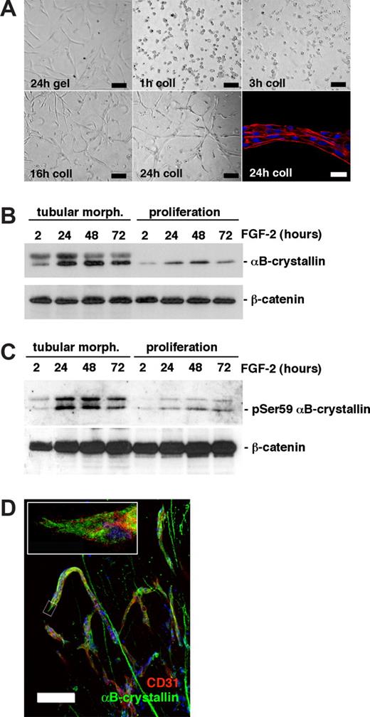
αB-crystallin is up-regulated and phosphorylated during in vitro angiogenesis. (A) Tubular morphogenesis (morph.) of BCE cells cultured in a 3-D collagen (coll) matrix in the presence of 10 ng/mL FGF-2. Black bar = 100 μm. Bottom right panel shows a magnified confocal image of a tubular structure with actin filaments (red) connecting several cells, visualized by nuclear staining (Hoechst 33 342; blue). White bar=20 μm. (B,C) Western blot analyses of αB-crystallin expression (B) and the level of Ser59-phosphorylated αB-crystallin (C) in whole cell lysates of BCE cell monolayers on gelatin (proliferating cells) or 3-D collagen cultures forming tubular structures in the presence of 10 ng/mL FGF-2 (tubular morph.). β-catenin expression was used as a loading control. (D) Immunofluorescence analysis of embryoid bodies cultured in a 3-D collagen matrix in the presence of 30 ng/mL VEGF-A. αB-crystallin (green) was expressed in endothelial sprouts (visualized by α-CD31 staining, red). Bar = 100 μM. The boxed area is shown at higher magnification in the top left corner.
αB-crystallin is up-regulated and phosphorylated during in vitro angiogenesis. (A) Tubular morphogenesis (morph.) of BCE cells cultured in a 3-D collagen (coll) matrix in the presence of 10 ng/mL FGF-2. Black bar = 100 μm. Bottom right panel shows a magnified confocal image of a tubular structure with actin filaments (red) connecting several cells, visualized by nuclear staining (Hoechst 33 342; blue). White bar=20 μm. (B,C) Western blot analyses of αB-crystallin expression (B) and the level of Ser59-phosphorylated αB-crystallin (C) in whole cell lysates of BCE cell monolayers on gelatin (proliferating cells) or 3-D collagen cultures forming tubular structures in the presence of 10 ng/mL FGF-2 (tubular morph.). β-catenin expression was used as a loading control. (D) Immunofluorescence analysis of embryoid bodies cultured in a 3-D collagen matrix in the presence of 30 ng/mL VEGF-A. αB-crystallin (green) was expressed in endothelial sprouts (visualized by α-CD31 staining, red). Bar = 100 μM. The boxed area is shown at higher magnification in the top left corner.
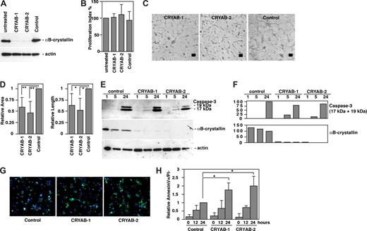
Knockdown of αB-crystallin in endothelial cells inhibits tubular morphogenesis. (A) TIME cells were transfected with siRNA oligos as described in experimental procedures. Whole cell lysates were prepared 3 hours after induction of tubular morphogenesis, and the knockdown of αB-crystallin expression by siRNA was analyzed by Western blot. Actin expression was used as a loading control. (B) SiRNA-transfected cells were counted 48 hours and 96 hours after transfection. Proliferation index was calculated as the increase in total cell number compared with untransfected control (n = 3; mean ± SD). (C) Tubular morphogenesis of endothelial cells transfected with αB-crystallin–specific siRNA (CRYAB-1 and CRYAB-2) or Alexa 488–conjugated control siRNA (bar = 100 μm). (D) Quantification of tube formation (area and length) compared with control siRNA-transfected cells analyzed by the Easy Image Analysis software (n = 3; mean ± SD; **P < .05; *P < .01). (E) Whole cell extracts from siRNA-transfected cells were prepared 1, 5, or 24 hours after induction of tubular morphogenesis, and the expression of αB-crystallin and cleaved caspase-3 (17-kDa and 19-kDa fragments) were analyzed by immunoblotting. (F) αB-crystallin and cleaved caspase-3 expression was quantified and normalized to actin expression using Image Gauge software. Values are given as percentage of expression compared with the 24-hour time point of control siRNA–transfected cells (set to 100%). (G) Annexin-V (green), PI (red), and Hoechst 33 342 (blue) staining of endothelial cells transfected with αB-crystallin–specific siRNA (CRYAB-1, CRYAB-2) and unconjugated control siRNA after 24 hours of tube formation in collagen gel. (H) Quantification of apoptosis in siRNA-transfected endothelial cells after 0 hours, 12 hours, and 24 hours of tube formation. Bars represent mean values and SD of the relative apoptosis rate (fraction of annexin-V–positive, PI-negative cells normalized to control siRNA at 24 hours) of 4 independent experiments (n = 4; mean ± SD; *P < 0,05).
Knockdown of αB-crystallin in endothelial cells inhibits tubular morphogenesis. (A) TIME cells were transfected with siRNA oligos as described in experimental procedures. Whole cell lysates were prepared 3 hours after induction of tubular morphogenesis, and the knockdown of αB-crystallin expression by siRNA was analyzed by Western blot. Actin expression was used as a loading control. (B) SiRNA-transfected cells were counted 48 hours and 96 hours after transfection. Proliferation index was calculated as the increase in total cell number compared with untransfected control (n = 3; mean ± SD). (C) Tubular morphogenesis of endothelial cells transfected with αB-crystallin–specific siRNA (CRYAB-1 and CRYAB-2) or Alexa 488–conjugated control siRNA (bar = 100 μm). (D) Quantification of tube formation (area and length) compared with control siRNA-transfected cells analyzed by the Easy Image Analysis software (n = 3; mean ± SD; **P < .05; *P < .01). (E) Whole cell extracts from siRNA-transfected cells were prepared 1, 5, or 24 hours after induction of tubular morphogenesis, and the expression of αB-crystallin and cleaved caspase-3 (17-kDa and 19-kDa fragments) were analyzed by immunoblotting. (F) αB-crystallin and cleaved caspase-3 expression was quantified and normalized to actin expression using Image Gauge software. Values are given as percentage of expression compared with the 24-hour time point of control siRNA–transfected cells (set to 100%). (G) Annexin-V (green), PI (red), and Hoechst 33 342 (blue) staining of endothelial cells transfected with αB-crystallin–specific siRNA (CRYAB-1, CRYAB-2) and unconjugated control siRNA after 24 hours of tube formation in collagen gel. (H) Quantification of apoptosis in siRNA-transfected endothelial cells after 0 hours, 12 hours, and 24 hours of tube formation. Bars represent mean values and SD of the relative apoptosis rate (fraction of annexin-V–positive, PI-negative cells normalized to control siRNA at 24 hours) of 4 independent experiments (n = 4; mean ± SD; *P < 0,05).
αB-crystallin is expressed in a subset of tumor vessels. (A) Immunofluorescence analysis of αB-crystallin expression (red) and endothelial cells (visualized by UEA-1 binding [green]) in normal human kidney. (B) Expression of αB-crystallin in endothelial cells (indicated by arrows) in human renal cancer. Arrowheads indicate vessels lacking αB-crystallin expression. (C) Magnification of tumor vessel in human renal cancer showing expression of αB-crystallin in endothelial cells. (D) Expression of αB-crystallin in smooth muscle cells and endothelial cells (arrows) in human renal cancer. (E) Normal human lung vessels lacking expression of αB-crystallin in endothelial cells. (F) αB-crystallin expression in endothelial cells in human lung cancer. Bars equal 100 μm.
αB-crystallin is expressed in a subset of tumor vessels. (A) Immunofluorescence analysis of αB-crystallin expression (red) and endothelial cells (visualized by UEA-1 binding [green]) in normal human kidney. (B) Expression of αB-crystallin in endothelial cells (indicated by arrows) in human renal cancer. Arrowheads indicate vessels lacking αB-crystallin expression. (C) Magnification of tumor vessel in human renal cancer showing expression of αB-crystallin in endothelial cells. (D) Expression of αB-crystallin in smooth muscle cells and endothelial cells (arrows) in human renal cancer. (E) Normal human lung vessels lacking expression of αB-crystallin in endothelial cells. (F) αB-crystallin expression in endothelial cells in human lung cancer. Bars equal 100 μm.
Statistical examination
Statistical examination was performed on all data using analysis of variance or unpaired Student t test using the Statview software. We considered a P value less than .05 to be significant.
Results
αB-crystallin is up-regulated and activated during in vitro angiogenesis
To define the proteome of endothelial cells engaged in tubular morphogenesis, BCE cells, seeded in a 3-D collagen matrix, were harvested after different time periods of exposure to FGF-2 (Figure 1A). We have shown previously that lumenized tubules are formed under these conditions.16 The pattern of protein expression was compared with that of proliferating, monolayer BCE cells cultured on gelatin by 2-D gel electrophoresis followed by mass spectrometry. In such a screen, we identified the small heat shock protein αB-crystallin as specifically up-regulated during tubular morphogenesis in collagen; this pattern of expression was confirmed by immunoblotting (Figure 1B). αB-crystallin function is regulated, at least in part, by serine phosphorylation, and Ser59 has been suggested to regulate its prosurvival function.22 We found that αB-crystallin was phosphorylated on Ser59 after FGF-2 treatment, and that this phosphorylation was considerably enhanced during tubular morphogenesis in collagen (Figure 1C). Phosphorylation and up-regulation of αB-crystallin was also noted in BCE cells forming tubular structures in response to 10% NCS only, in the absence of FGF-2 (Figure S1, available on the Blood website; see the Supplemental Materials link at the top of the online article), and in human microcapillary endothelial cells undergoing VEGF-induced tubular morphogenesis (data not shown). Differentiating mouse embryonic stem cell cultures (embryoid bodies) represent a suitable model to study expression and function of endothelial cell proteins during blood vessel formation. Embryoid bodies placed in 3-D collagen form angiogenic sprouts, invading the collagen gel, in response to VEGF-treatment. Figure 1D shows that αB-crystallin was expressed in CD31-positive endothelial cells in angiogenic sprouts. We conclude that αB-crystallin is expressed in several different kinds of endothelial cells analyzed in 2 different models of in vitro angiogenesis.
Knockdown of αB-crystallin inhibits tubular morphogenesis in vitro
We next examined the role of αB-crystallin in endothelial cell tubular morphogenesis by introduction of αB-crystallin–specific siRNA into human microcapillary endothelial cells, with close to 100% efficiency (as judged from the efficiency of introduction of Alexa 488–conjugated control siRNA; data not shown). This resulted in an efficient knockdown of αB-crystallin protein expression as determined by immunoblotting (Figure 2A). SiRNA knockdown of αB-crystallin using either of 2 different αB-crystallin–specific siRNA (CRYAB-1 and CRYAB-2) did not affect endothelial cell proliferation (Figure 2B), nor endothelial cell migration (data not shown). However, αB-crystallin–deficient endothelial cells failed to efficiently form tubular structures in 3-D collagen in response to VEGF compared with control siRNA-treated cells (Figure 2C; quantification is shown in Figure 2D). Failure to form tubular structures was accompanied by an early cleavage of pro–caspase-3 into active 17- and 19-kDa isoforms, evident after 5 hours of tubular formation in CRYAB-1 and CRYAB-2 siRNA-treated cells (Figure 2E, quantified in Figure 2F). This was followed by increased apoptosis in αB-crystallin siRNA-treated cells after 24 hours of tubular morphogenesis, as judged by the presence of annexin V–positive/PI-negative cells (Figure 2G, quantified in Figure 2H). These data indicate that αB-crystallin expression is required for certain endothelial cell functions, such as regulation of apoptosis and tubular morphogenesis, but not for others, such as endothelial cell migration and proliferation.
Serine phosphorylation of αB-crystallin may regulate interactions with different cytoskeletal components.23,24 We examined the subcellular localization of αB-crystallin in relation to that of actin and the intermediate filament protein vimentin, which form separate cytoskeletal structures in endothelial cells (Figure S2A top panel). In tubular structures formed in 3-D collagen, actin filaments lined up during the cell fusion process (Figures 1A and S2B). αB-crystallin colocalized with vimentin in the monolayer cultures (Figure S2A bottom panel) but not to the same extent in tube forming cells after 24 hours of induction. Instead αB-crystallin was found in small cytoplasmic granules (Figure S2B bottom panel). Immunoprecipitation of αB-crystallin, followed by Western blot analysis, confirmed that the association between vimentin and αB-crystallin was regulated during tubular formation, with the highest association noted after 12 hours (Figure S2C, supported by immunofluorescence analysis in Figure S2D). The dramatic fluctuations in subcellular localization of αB-crystallin and its presence in cytoplasmic granules may be regulated by serine phosphorylation. However, available reagents did not allow immunostaining to detect localization of phosphorylated αB-crystallin in tube-forming endothelial cells.
αB-crystallin is expressed in a subset of renal and lung tumor vessels
αB-crystallin is expressed in the wall of some large vessels, such as the aorta, and may be released into the circulation upon vessel injury.25 Expression in intermediate and small vessels and capillaries has not been analyzed previously. Immunostaining showed expression of αB-crystallin in a subset of small vessels in human kidney tumors (Figure 3B,C) as well as abundant expression in the tumor cells. Quantification of tumor vessels in sections from 5 separate renal cancer samples showed colocalization of αB-crystallin and UEA-1 in endothelial cells in between 0.7% and 15% of the vessels. αB-crystallin expression was occasionally found in pericytes as well as in vascular smooth muscle cells surrounding larger vessels in the tumor (Figure 3D). Similar to expression in renal tumor vessels, αB-crystallin was often expressed in small vessels in human lung cancer samples (Figure 3F). Coexpression of αB-crystallin and UEA-1 was found in between 16% and 40% of the vessels when 4 separate lung tumors were analyzed. In contrast, expression was never detected in endothelial cells in normal human lung tissue or healthy human or mouse kidney tissue (Figure 3A,E; data not shown). These data are in agreement with a transient expression of αB-crystallin during 3-D morphogenesis of endothelial cells to form the vascular tube (Figure 1B).
Reduced tumor angiogenesis in the absence of αB-crystallin
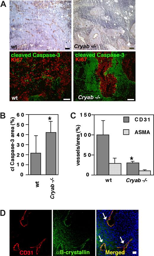
Mice lacking αB-crystallin and HspB2 (Cryab−/−) develop normally but are slightly smaller than wild-type mice and acquire a muscular dysfunction later in life.7 HspB2 expression is largely restricted to heart and muscle and is not induced by heat shock.26 In accordance, we did not detect expression of HspB2 in endothelial cells, as determined by real-time polymerase chain reaction (data not shown). To determine whether αB-crystallin is important in tumor angiogenesis, Cryab−/− mice and 129Sv wild-type mice were inoculated with syngenic F9 teratocarcinoma cells. F9 cells grow aggressively as a subcutaneous tumor and reached a volume of 2 cm3 by 2 weeks. We found no significant difference in tumor size between Cryab−/− and wild-type mice. However, hematoxylin staining of tumor sections from Cryab−/− animals showed large areas of apoptosis and necrosis (Figure 4A top). The total apoptotic area, as determined by expression of cleaved caspase-3, was on average 40% of the tumor area in Cryab−/− animals and 20% in wild-type mice (Figure 4A bottom, quantified in 4B). Quantification of CD31-positive vessels in tumor sections revealed a dramatic decrease in blood vessel count/area in tumors from the Cryab−/− animals, to 30% of that in tumors from wild-type animals (Figure 4C). Vessels in the Cryab−/− tumors were surrounded by α-smooth muscle actin–positive cells to approximately the same extent as in the wild-type tumors (Figure 4C). Immunofluorescent analysis of F9 tumors showed that a subset of tumor vessels from wild-type mice expressed αB-crystallin, whereas, as expected, no expression was noted in tumor vessels in Cryab−/− mice (Figure 4D; data not shown).
Tumors grown in Cryab−/− mice are less vascularized and show larger areas of apoptosis than tumors from wild-type (wt) mice. (A) Top panels, Immunohistochemistry of representative tumors grown in wild-type (left) or Cryab−/− mice, showing CD31-positive vessels (brown) in viable areas of the tumors and large areas of necrosis/apoptosis (bar = 100 μM). Bottom panels, Immunofluorescent staining of representative tumors from each group showing apoptotic cells expressing cleaved caspase-3 (green) and proliferating cells expressing Ki-67 (red) in viable tumor areas (bar = 100 μm). (B) Quantification of mean cleaved caspase-3–positive area was done by analyzing 4× optical fields spanning all across a middle section of 4 different tumors per group using Easy Image Analysis. Bars represent mean values and SD of cleaved caspase-3−positive area (n = 4; mean ± SD; *P < .05). (C) The number of vessels/area was determined by counting the total number of CD31-positive and ASMA-positive vessels in each tumor by analyzing 10× optical fields spanning all across a middle section of 5 different tumors per group and normalizing relative to tumor area using Easy Image Analysis software. Results are given as percentage of vessels/area compared with wild-type mice, and bars represent mean values and SD (n = 5; mean ± SD; *P < .01). (D) Expression of αB-crystallin (red) was detected in a subset of CD31-positive vessels (green) in tumors grown in wild-type animals. Bar equals 20 μM.
Tumors grown in Cryab−/− mice are less vascularized and show larger areas of apoptosis than tumors from wild-type (wt) mice. (A) Top panels, Immunohistochemistry of representative tumors grown in wild-type (left) or Cryab−/− mice, showing CD31-positive vessels (brown) in viable areas of the tumors and large areas of necrosis/apoptosis (bar = 100 μM). Bottom panels, Immunofluorescent staining of representative tumors from each group showing apoptotic cells expressing cleaved caspase-3 (green) and proliferating cells expressing Ki-67 (red) in viable tumor areas (bar = 100 μm). (B) Quantification of mean cleaved caspase-3–positive area was done by analyzing 4× optical fields spanning all across a middle section of 4 different tumors per group using Easy Image Analysis. Bars represent mean values and SD of cleaved caspase-3−positive area (n = 4; mean ± SD; *P < .05). (C) The number of vessels/area was determined by counting the total number of CD31-positive and ASMA-positive vessels in each tumor by analyzing 10× optical fields spanning all across a middle section of 5 different tumors per group and normalizing relative to tumor area using Easy Image Analysis software. Results are given as percentage of vessels/area compared with wild-type mice, and bars represent mean values and SD (n = 5; mean ± SD; *P < .01). (D) Expression of αB-crystallin (red) was detected in a subset of CD31-positive vessels (green) in tumors grown in wild-type animals. Bar equals 20 μM.
Impaired endothelial function in αB-crystallin −/− tumors
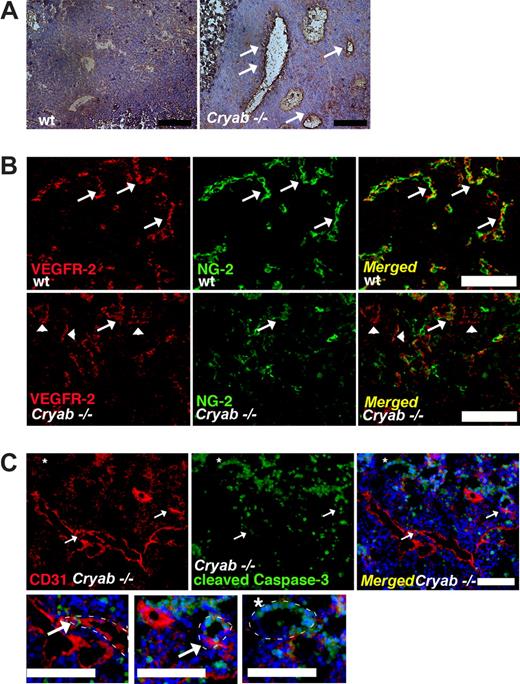
Tumor vessels in Cryab−/− mice were morphologically distinct from those of wild-type mice, with thinner, often partially disrupted vessel walls. Immunohistochemical staining to detect plasma-derived fibrin showed distinct areas of fibrin deposits around tumor vessels in Cryab−/− mice (Figure 5A), indicating increased vessel leakiness under αB-crystallin–deficient conditions. Further, the majority of vessels in tumors from wild-type animals were covered by NG-2–positive pericytes. In contrast, vessels in tumors from the Cryab−/− animals were often partially devoid of a pericyte coat, and NG-2–positive cells were more loosely arranged around the endothelial cells and occasionally localized at a distance from the vessel (Figure 5B). This was not seen in the wild-type tumors. In addition, vessels with incomplete endothelial lining were often observed in tumors from Cryab−/− mice (Figure 5B). Coimmunostaining for cleaved caspase-3 and CD31 revealed that certain vessels in tumors grown in Cryab−/− mice were undergoing apoptosis (Figure 5C). Such vessels were not found in tumors from wild-type animals, apart from occasional vessels in nonviable areas of the tumor. Coexpression of CD31 and cleaved caspase-3 was rarely detected in individual endothelial cells, most likely because CD31 is shed during endothelial cell apoptosis.27 However, cells expressing either CD31 or cleaved caspase-3 were frequently detected together in vessel structures characterized by partially or extensively disrupted vessel walls (Figure 5C bottom row of magnifications).
Tumor vessels in Cryab−/− mice are leaky, partially lack NG-2–positive pericyte coverage, and show signs of apoptosis. (A) Fibrin deposits (brown, indicated by arrows) around tumor vessels in wild-type (wt; left) and Cryab−/− (right) mice (bar = 100 μM). (B) NG-2–positive pericytes (green) in VEGFR-2 positive vessels (red) in tumors grown in wild-type (top) and Cryab−/− (bottom) mice (bar = 100 μm). Arrows show vessels covered by NG-2–positive pericytes, and arrowheads show vessels lacking pericyte coverage. (C) CD31-positive tumor vessels (red) in Cryab−/− mice with partial expression of proapoptotic cleaved caspase-3 (green, arrow) and (*) apoptotic vessel expressing cleaved caspase-3, magnified below (bar = 100 μM). White dotted lines in magnifications indicate the vessel borders.
Tumor vessels in Cryab−/− mice are leaky, partially lack NG-2–positive pericyte coverage, and show signs of apoptosis. (A) Fibrin deposits (brown, indicated by arrows) around tumor vessels in wild-type (wt; left) and Cryab−/− (right) mice (bar = 100 μM). (B) NG-2–positive pericytes (green) in VEGFR-2 positive vessels (red) in tumors grown in wild-type (top) and Cryab−/− (bottom) mice (bar = 100 μm). Arrows show vessels covered by NG-2–positive pericytes, and arrowheads show vessels lacking pericyte coverage. (C) CD31-positive tumor vessels (red) in Cryab−/− mice with partial expression of proapoptotic cleaved caspase-3 (green, arrow) and (*) apoptotic vessel expressing cleaved caspase-3, magnified below (bar = 100 μM). White dotted lines in magnifications indicate the vessel borders.
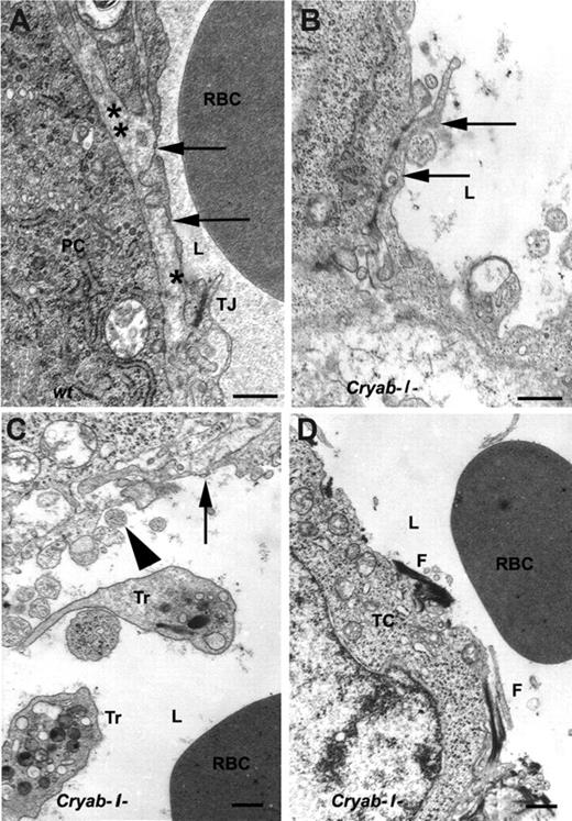
Tumors grown in Cryab−/− and wild-type animals were examined by high-resolution transmission electron microscopy to further investigate vessel morphology. Vessels in tumors from wild-type mice were lined with fenestrated endothelium resting on a basal lamina and sealed to neighboring cells with tight junctions (Figure 6A). In contrast, tumor vessels from Cryab−/− mice were often only partially lined with fenestrated endothelium (Figure 6B) or showed distorted endothelial lining with only remnants of normal endothelium present (Figure 6C). Some vessels completely lacked endothelial lining, although red blood cells were present in the lumen, and fibrous deposits were present on the surface of surrounding tumor cells (Figure 6D). Taken together, our results suggest that αB-crystallin–deficient vessels present several hallmarks of increased apoptosis such as the presence of cleaved caspase-3–positive cells in the vessel walls, partially or completely detached endothelial lining, increased leakiness, and impaired pericyte coverage.
Transmission electron microscopy analysis reveals distorted and defective endothelium in tumor vessels in Cryab−/− mice. (A) A representative micrograph of a small tumor vessel in a wild-type (wt) mouse shows a fenestrated endothelial cell (arrow) resting on a basal lamina (*) and sealed to the neighboring endothelial cell with tight junctions (TJ). A pericyte (PC) with its own basal lamina (**) is located in close contact with the endothelium. A red blood cell (RBC) is seen in the lumen (L). (B-D) Representative micrographs of tumor vessels in Cryab−/− mice. (B) A blood vessel partially lined with fenestrated endothelium (arrow). (C) Defective endothelial lining (arrowheads) and remnants of an endothelial cell with fenestration (arrow). A red blood cell and 2 thrombocytes (Tr) are present in the lumen. (D) A vessel lacking endothelial lining with fibrous deposits (F) located directly on the surface of one of the surrounding tumor cells (TC). Original magnification for A,B, ×25 000; C,D, ×15 000. Bar equals 500 nm.
Transmission electron microscopy analysis reveals distorted and defective endothelium in tumor vessels in Cryab−/− mice. (A) A representative micrograph of a small tumor vessel in a wild-type (wt) mouse shows a fenestrated endothelial cell (arrow) resting on a basal lamina (*) and sealed to the neighboring endothelial cell with tight junctions (TJ). A pericyte (PC) with its own basal lamina (**) is located in close contact with the endothelium. A red blood cell (RBC) is seen in the lumen (L). (B-D) Representative micrographs of tumor vessels in Cryab−/− mice. (B) A blood vessel partially lined with fenestrated endothelium (arrow). (C) Defective endothelial lining (arrowheads) and remnants of an endothelial cell with fenestration (arrow). A red blood cell and 2 thrombocytes (Tr) are present in the lumen. (D) A vessel lacking endothelial lining with fibrous deposits (F) located directly on the surface of one of the surrounding tumor cells (TC). Original magnification for A,B, ×25 000; C,D, ×15 000. Bar equals 500 nm.
Discussion
This study identifies the small heat shock protein αB-crystallin as a novel regulator of tumor angiogenesis. We identified αB-crystallin as up-regulated in endothelial cells using a tubular morphogenesis assay in a 3-D collagen matrix, first described by Montesano et al.28 The relevance of this assay has been validated by the observation that tubular structures in a 3-D collagen slab can connect to the host vasculature when implanted in mice and form long-lasting, functional vessels in vivo.29 In addition, time-lapse imaging shows striking similarities between vessel formation in zebra fish and tubular morphogenesis in a 3-D collagen matrix.30 αB-crystallin has not been implicated previously in blood vessel formation, but the highly homologous family member Hsp27 appears, at least in vitro, to regulate migration of endothelial cells and smooth muscle cells downstream of p38 mitogen-activated protein kinase (p38MAPK) activation, through a phosphorylation-dependent association with the actin cytoskeleton.31,32 In addition, treatment with several different angiogenesis inhibitors, such as thrombospondin, has been suggested to target the endothelial cytoskeleton through regulation of Hsp27 function.33 Several members of the small heat shock protein family influence cytoskeletal events in a phosphorylation-dependent manner, but there are conflicting data regarding the role of phosphorylation depending on the context.24 We show that αB-crystallin is phosphorylated on Ser-59 during tubular morphogenesis, and that this coincides with translocation from a pool colocalized with the intermediate filament vimentin into granular structures (see Figure S2B). These αB-crystallin–positive structures are reminiscent of “heat shock granules,” composed of small heat shock proteins, which have been identified in response to heat shock in several systems and which have high chaperone activity.34,,–37 Serine phosphorylation of αB-crystallin during tubular morphogenesis occurred downstream of p38MAPK activation (data not shown), which is in accordance with previous reports.38 The role of αB-crystallin serine phosphorylation in its chaperone function is not clear. Phosphorylation of αB-crystallin Ser-59 in response to stress has been associated with decreased oligomerization and chaperone activity.39,40 However, Ser-59 phosphorylation has also been described as both necessary and sufficient for protection of cardiac myocytes from apoptosis, an effect that was coupled to a decreased activation of caspase-3.22
Our results show that αB-crystallin plays an important role in regulation of endothelial cell survival by inhibition of caspase 3-activation, in conjunction with vessel morphogenesis and the accompanying dramatic cytoskeletal reorganization required for creation of the 3-D vascular tube. This is inferred from the fact that siRNA knockdown of αB-crystallin in endothelial cells attenuated tubular morphogenesis in vitro, an effect that was accompanied by enhanced kinetics of caspase-3 activation and increased apoptosis. Moreover, immunofluorescence and ultrastructural analyses of tumor sections showed distorted vessels and increased caspase-3 activation in tumors in Cryab−/− mice in vivo. It should be noted that αB-crystallin and HspB2 may be expressed in pericytes and smooth muscle cells, and that the lack of αB-crystallin and HspB2 in these cell types in Cryab−/− animals may affect angiogenesis and vessel stability. However, we did not detect a difference in the proportion of ASMA-positive vessels between wild-type and Cryab−/− mice (Figure 4C). In addition, the presence of defective endothelial cells in tumors in Cryab−/− mice indicates that an endothelial dysfunction is the primary cause of reduced vessel formation in these mice. αB-crystallin was up-regulated in endothelial cells during vessel formation in vitro, and in endothelial sprouts formed by embryoid bodies, but we did not detect expression in apparently quiescent vessels in healthy tissues. In contrast, we identified a subset of smaller vessels in both murine and human tumors that expressed αB-crystallin. Our data suggest that αB-crystallin is transiently up-regulated during vessel formation, enhancing survival of endothelial cells before stabilization. The preserved developmental angiogenesis, in combination with inhibited tumor angiogenesis, suggests that αB-crystallin may be required specifically when endothelial cells are exposed to certain stress stimuli during pathologic vessel formation. In accordance, αB-crystallin is up-regulated in response to osmotic or sodium arsenite stress in cultured endothelial cells in vitro and in cerebral arteriovenous malformations in vivo.41,42 In many cell types, αB-crystallin is up-regulated in response to different kinds of cellular stress, increasing cell survival during potentially harmful conditions.11 Hitherto, αB-crystallin has been shown to inhibit apoptosis induced by etopside, tumor necrosis factor-α (TNF-α), staurosporine, and TNF-related apoptosis-inducing ligand (TRAIL).12,14,43 A differential requirement for αB-crystallin, depending on the microenvironment, was also supported by a partial inhibition of blood vessel formation in matrigel plugs in Cryab−/− mice (Figure S3). We found that growth factor–induced angiogenesis was reduced by roughly 35% in Cryab−/− implanted plugs. There was no difference in the distance the vessels invaded into the plugs; instead, there were fewer and smaller hot spots of vessel formation in mice lacking αB-crystallin. These data support the concept that αB-crystallin is important for vessel formation and/or maintenance.
αB-crystallin is expressed in many tumors and has recently been suggested to be a novel oncogene. Stable expression of αB-crystallin in a mouse xenograft model led to enhanced tumor growth and inhibition of TRAIL-induced apoptosis.43 Importantly, αB-crystallin expression independently predicted poor survival in breast cancer, and over-expression of αB-crystallin resulted in transformation of immortalized human mammary epithelial cells.15 Therefore, treatment targeting αB-crystallin could be predicted to affect both the tumor compartment as well as the pathologic vasculature. The emerging role of chaperones such as Hsp27 and αB-crystallin in various diseases is likely to promote the development of new drugs specifically targeting this protein family.44 Small molecular weight inhibitors or peptides inhibiting αB-crystallin binding to target proteins could be administered using novel technologies for specific retention at the tumor bed and ultimately affect both vessels and tumor cells. Importantly, αB-crystallin may be secreted into the blood stream in response to vessel injury and regulate platelet activation,25 thus influencing secretion of proangiogenic factors and inflammatory response. In this regard, αB-crystallin, injected intravenously into mice, has been shown to modulate inflammation in experimental autoimmune encephalitis.45 An important future goal is to determine whether secreted αB-crystallin affects the tumor microenvironment, further influencing tumor progression and blood vessel formation.
An Inside Blood analysis of this article appears at the front of this issue.
The online version of this article contains a data supplement.
The publication costs of this article were defrayed in part by page charge payment. Therefore, and solely to indicate this fact, this article is hereby marked “advertisement” in accordance with 18 USC section 1734.
Acknowledgments
This study was supported by the Association for International Cancer Research (AICR Grant 04-0069), the Astrid Karlsson foundation, the Gustaf Adolf Johansson foundation, Svenska Sällskapet för Medicinsk Forskning, the Magnus Bergvall foundation, Swedish Research Council (project no. K2005-32X-12 552-08A), and the Swedish Cancer Society (project 4828-B03-01PAA and 3820-B05-10XBC). The Fresh Tissue Biobank at the Unit of Clinical Pathology, Uppsala University Hospital, was supported by the National Biobank Platform funded by Wallenberg Consortium North and Swegene.
Authorship
Contribution: A.D. designed experiments, performed research, analyzed and interpreted data, and wrote the paper. L.C.D., S.R., A.-K.O., P.R., C.W., and S.B. performed research and analyzed data; J.B. contributed vital new reagents and collected data; A.L. performed electron microscopy analysis; E.F.W. contributed vital new reagents; and L.C.-W. designed experiments, interpreted data, and wrote the paper.
Conflict-of-interest disclosure: The authors declare no competing financial interests.
Correspondence: Anna Dimberg, Department of Genetics and Pathology, The Rudbeck Laboratory, Uppsala University, SE-751 85 Uppsala, Sweden; e-mail: Anna.Dimberg@genpat.uu.se.
References
Author notes
S.R. and L.C.D. contributed equally to this study.

![Figure 3. αB-crystallin is expressed in a subset of tumor vessels. (A) Immunofluorescence analysis of αB-crystallin expression (red) and endothelial cells (visualized by UEA-1 binding [green]) in normal human kidney. (B) Expression of αB-crystallin in endothelial cells (indicated by arrows) in human renal cancer. Arrowheads indicate vessels lacking αB-crystallin expression. (C) Magnification of tumor vessel in human renal cancer showing expression of αB-crystallin in endothelial cells. (D) Expression of αB-crystallin in smooth muscle cells and endothelial cells (arrows) in human renal cancer. (E) Normal human lung vessels lacking expression of αB-crystallin in endothelial cells. (F) αB-crystallin expression in endothelial cells in human lung cancer. Bars equal 100 μm.](https://ash.silverchair-cdn.com/ash/content_public/journal/blood/111/4/10.1182_blood-2007-04-087841/3/m_zh80040815260003.jpeg?Expires=1769083453&Signature=uppur5XiSC-VbIj7z25B2p2lTPJk~DoYPNtRzzys8mHqgze6ti07QVP1HOEmYKFRZUdw8T-qC8sSeMqoPew19W69JsslvlFtnp3ps2NSs2gCU7VjXl9vIkH9Xj3Kn9~ok4qraa9YpvxqS3nSpjsOVLgb32bDTMTKTw8qkpPluDqXoGDbIs~urbQyKcKKlhiWI5so~2OKkWQ-JxUpcuJfNgxWrkPZ4ljvxVUAN54Vu3B9AdcrWDAqGUCWOPue7lzlM00NAGEdkERuYQo5uA1UalCkmwuvmzEm5YxjnFPEmbrhfBinsd2gZ8RXwVStiBx7Yx~RwN6xjds10Rq83-~czA__&Key-Pair-Id=APKAIE5G5CRDK6RD3PGA)
This feature is available to Subscribers Only
Sign In or Create an Account Close Modal