Abstract
Vorinostat (suberoylanilide hydroxamic acid, SAHA) is a histone deacetylase inhibitor active clinically in cutaneous T-cell lymphoma and preclinically in leukemia. A phase 1 study was conducted to evaluate the safety and activity of oral vorinostat 100 to 300 mg twice or thrice daily for 14 days followed by 1-week rest. Patients with relapsed or refractory leukemias or myelodysplastic syndromes (MDS) and untreated patients who were not candidates for chemotherapy were eligible. Of 41 patients, 31 had acute myeloid leukemia (AML), 4 chronic lymphocytic leukemia, 3 MDS, 2 acute lymphoblastic leukemia, and 1 chronic myelocytic leukemia. The maximum tolerated dose (MTD) was 200 mg twice daily or 250 mg thrice daily. Dose-limiting toxicities were fatigue, nausea, vomiting, and diarrhea. Common drug-related adverse experiences were diarrhea, nausea, fatigue, and anorexia and were mild/moderate in severity. Grade 3/4 drug–related adverse experiences included fatigue (27%), thrombocytopenia (12%), and diarrhea (10%). There were no drug-related deaths; 7 patients had hematologic improvement response, including 2 complete responses and 2 complete responses with incomplete blood count recovery (all with AML treated at/below MTD). Increased histone acetylation was observed at all doses. Antioxidant gene expression may confer vorinostat resistance. Further evaluation of vorinostat in AML/MDS is warranted.
Introduction
Histone acetylation facilitates active gene transcription and is highly regulated by histone deacetylases (HDACs) and histone acetyltransferases (HATs).1 Numerous HDAC or HAT defects leading to reduced or abnormal acetylation have been identified in leukemia, lymphoma, and solid tumor cell lines, including mutation, translocation, or overexpression of p300, CBP, TIF-2, RARα, BCL6, AML1, STAT5, or HDAC1.1,2 HDAC inhibition has therefore been postulated to restore normal acetylation of histone proteins and transcription factors, and to be of benefit in the treatment of cancer
Vorinostat (Zolinza, Merck, Whitehouse Station, NJ), suberoylanilide hydroxamic acid (SAHA), is a small molecule inhibitor of class I and II HDAC enzymes3 that has been shown to promote cell-cycle arrest and apoptosis of cancer cells through regulation of gene expression.4-6 Vorinostat has demonstrated activity against leukemia and other hematologic malignancies in vitro7-14 and also improved survival and/or produced antitumor effects in rodent models of leukemia.8,15 Oral vorinostat has been reported to be tolerable at 400 mg daily or 200 mg twice daily for continuous daily dosing and 300 mg twice daily for 3 days per week dosing in phase 1 trials (Table 1).16-19 In these trials, vorinostat was shown to inhibit HDAC activity in tumors or peripheral blood. Dose-limiting toxicities (DLTs) included anorexia, dehydration, diarrhea, and fatigue. Based on the safety and efficacy demonstrated in phase 1/2 trials,16-21 vorinostat was approved in October 2006 by the US Food and Drug Administration (FDA) for the treatment of cutaneous manifestations in patients with cutaneous T-cell lymphoma who have progressive, persistent or recurrent disease on or after 2 systemic therapies.
Summary of Phase I vorinostat trials
| Trial . | N . | Population . | Maximum tolerated dose . | Activity . |
|---|---|---|---|---|
| Kelly et al16 | 73 | Advanced solid and hematologic malignancies | 400 mg orally 4 times a day or 200 mg orally twice a day for continuous daily dosing; 300 mg orally twice a day for 3 consecutive days per week dosing | CR: DLBCL; PR: CTCL,* mesothelioma, laryngeal and thyroid cancer; prolonged SD: renal cell and thyroid cancer |
| Richardson et al18 | 13 | Advanced multiple myeloma | Not determined† | Of 10 evaluable patients: 1 MR, 9 SD |
| Rubin et al19 | 23 | Advanced solid tumors | Not determined‡ | PR: ovarian cancer, prolonged; SD: breast and non–small cell lung cancer |
| Kelly et al37 | 37 | Advanced solid and hematologic malignancies | 300 mg/m2 per day intravenously by 5 days for 3 weeks followed by a 1-week rest§ | PR: Hodgkin disease*; MR: bladder cancer; SD: Hodgkin disease |
CR indicates complete response; CTCL, cutaneous T-cell lymphoma; DLBCL, diffuse large B-cell lymphoma; MR, minor response; PR, partial response; SD, stable disease.
*O'Connor et al.17
†The maximum administered doses were 250 mg orally twice a day for 5 days/week (4-week cycle) and 200 mg orally twice a day for 14 days (3-week cycle).
‡400 mg orally on days 1, 5, and 7 to 28 was evaluated.
§For patients with hematologic malignancies; not determined for patients with solid tumors.
Other HDAC inhibitors have been evaluated in early clinical trials in patients with leukemias and/or myelodysplastic syndromes (MDSs).22-30 The primary objective of this trial was to determine the maximum tolerated dose (MTD) of oral vorinostat administered 3 times daily for 14 days followed by 7 days of rest in patients with advanced leukemias or MDSs. Once the MTD on this schedule was established, the MTD on the twice daily schedule was studied. The more intensive schedule and re-covery period were proposed to optimize the therapeutic index. Secondary objectives were to assess the biologic effects of vorinostat in peripheral mononuclear cells and bone marrow blasts and to evaluate antitumor activity in patients with advanced leukemias or MDSs.
Methods
This open-label, nonrandomized phase 1 study (Protocol 003) was approved by the Institutional Review Board of the MD Anderson Cancer Center, and all patients provided written informed consent in accordance with the Declaration of Helsinki before enrollment following institutional guidelines.
Eligibility criteria
Patients with relapsed or refractory acute myeloid leukemia (AML), acute lymphoblastic leukemia (ALL), chronic lymphocytic leukemia (CLL), or MDS, or chronic myelocytic leukemia (CML) in accelerated phase or blast crisis that had progressed after treatment with imatinib mesylate were eligible for enrollment. Patients with AML, ALL, CLL, CML (accelerated phase or blast crisis), or MDS who refused or were not candidates for conventional chemotherapy were also eligible. Other eligibility criteria included age of at least 18 years, Eastern Cooperative Oncology Group (ECOG) Performance Status of 2 or less, adequate hepatic (bilirubin no more than 25.7 μmol/L [1.5 mg/dL]; aspartate aminotransferase/alanine aminotransferase no more than 2.5 times the upper limit of normal), and renal (creatinine no more than 176.8 μmol/L [2.0 mg/dL] or creatinine clearance more than mL/s [60 mL/min]) function, and a life expectancy greater than 3 months. Patients must have discontinued prior therapy for at least 2 weeks and recovered from prior toxicities.
Patients with central nervous system involvement by leukemia, HIV infection, or clinically significant illness (including active infection, uncontrolled hypertension, symptomatic congestive heart failure, unstable angina pectoris, myocardial infarction within the past 6 months, or uncontrolled cardiac arrhythmia) were excluded. Patients who planned to undergo allogeneic bone marrow transplantation within 4 weeks, or who were pregnant, lactating, or receiving concurrent complementary or alternative medicines were also excluded.
Treatment plan
Vorinostat was provided by Merck in dosage strengths of 50, 100, or 200 mg. The starting dose of oral vorinostat was 100 mg 3 times daily for 14 consecutive days, followed by 7 days of rest. This initial total daily dose was below the MTD in a previous phase 1 trial.16 The rest period was instated to reduce the likelihood of DLTs and allow patients time for recovery from any adverse events.
The study used a “3 + 3” dose escalation design. Three patients were to be enrolled at a dose level. If at least 2 of 3 patients experienced DLT in the first cycle, the dose was not escalated. If 1 of 3 patients experienced DLT in the first cycle, 3 additional patients were enrolled into the cohort. The dose was not escalated if at least 2 of 6 patients in a cohort experienced DLT during the first treatment cycle. If none of the first 3 patients or no more than 1 of 6 patients completed the first treatment cycle without DLT, 3 patients were entered into the next cohort. Additional patients could be enrolled at any given dose level for further evaluation of toxicity. To potentially prolong the duration of HDAC enzyme inhibition, twice and 3 times daily dosing regimens were used. Dose escalation continued until the MTD was reached. The dose regimens evaluated were 100, 150, 200, 250, and 300 mg 3 times daily, as well as 200 and 300 mg twice daily. During the course of the study, the 50 mg capsule was no longer available, and a 250 mg twice- daily regimen was not evaluated.
Any patient who experienced DLT was to have the drug held until the toxicity resolved to grade 1 or less, and daily dosing was reduced by one level in that patient. Up to 12 patients were to be enrolled at the MTD. Continuation of treatment up to 12 months was permitted at the discretion of the physician if the patient had no excessive toxicity or evidence of disease progression.
Evaluation
Pretreatment evaluation included a complete history and physical examination, ECOG Performance Status, complete blood count, electrolytes, prothrombin time, partial thromboplastin time, hepatic, renal, and thyroid function tests, urinalysis, pregnancy test (if appropriate), chest x-ray, electrocardiogram, bone marrow biopsy and aspiration, and computerized tomography scan for patients with measurable disease. Patients were reevaluated weekly and within 8 weeks after the last dose of study medication by physical examination, ECOG Performance Status, complete blood count, electrolytes, and hepatic and renal function tests. Patients were reevaluated each cycle by prothrombin time, partial thromboplastin time, urinalysis, and electrocardiogram.
Safety and response criteria
The National Cancer Institute Common Terminology Criteria for Adverse Events, version 3.0, was used to grade adverse events.31 All clinical adverse events and laboratory abnormalities were evaluated by the investigators for potential relationship to vorinostat. Those considered possibly, probably, or definitely related to vorinostat were classified as drug-related. DLT was defined as: pancytopenia with no more than 5% bone marrow cellularity and no evidence of leukemia, lasting greater than 42 days; grade 3 or greater nausea, vomiting, or diarrhea despite maximum supportive care; or other grade 3 or greater nonhematologic toxicity related to the study drug. The MTD was the maximum dose level at which no more than 1 of 6 patients experienced DLT during the first treatment cycle.
Responses were evaluated using the revised guidelines by the International Working Group32 for AML, ALL, and MDS, and the updated National Cancer Institute-Sponsored Working Group Guidelines33 for CLL as shown in Table S1 (available on the Blood website; see the Supplemental Materials link at the top of the online article). Per protocol, all responses, except complete response with incomplete blood count recovery (CRi), must have lasted at least 4 weeks for AML, ALL, and CML, and at least 8 weeks for CLL. The duration of response was measured from the time when all response criteria were first met until recurrent or progressive disease was documented.
Histone acetylation
Collection of blood and bone marrow samples for correlative studies was optional for enrolled patients. Unsorted peripheral blood samples (10-30 mL) and bone marrow biopsies obtained before vorinostat administration on day 1, 2 hours after vorinostat on day 14, and before vorinostat on day 22 of cycle 1 were obtained from participating patients. Histone extracts were isolated from these samples and analyzed by enzyme-linked immunosorbent assay for induction of histone H3 acetylation as previously described.16 Samples from 18 patients were analyzed (3 patients each receiving 100 and 150 mg, and 6 patients each receiving 200 and 250 mg 3 times daily). Histone acetylation was also assessed in bone marrow biopsies by immunohistochemical staining. Formalin-fixed, decalcified biopsies were embedded in paraffin, and 5-μm sections were incubated with polyclonal antibodies directed against acetylated histone H3, lysine 9 (1:25 dilution, Cell Signaling Technology, Danvers, MA) or acetylated histone H4, lysine 8 (1:25 dilution, Cell Signaling Technology), followed by a biotinylated secondary antibody (0.5 μg/mL, Jackson ImmunoResearch, West Grove, PA) and ABC/DAB (Vector Laboratories, Burlingame, CA), then hematoxylin counterstaining. Brightfield micrographs were acquired with an AxioImager Z1 microscope (Carl Zeiss Microimaging, Thornwood, NY) fitted with a 63×/1.4 NA oil objective, an Axiocam HRc camera (Carl Zeiss Microimaging), and AxioVision image acquisition software (v. 4.5, Carl Zeiss Microimaging). Flat-field and color temperature corrections were applied during image acquisition, and no further processing of captured images was performed.
Gene-expression profiling
Total RNA was extracted from the unsorted peripheral blood samples and amplified using standard protocols.34,35 Because the initial samples were not evaluable, the protocol was modified to include stabilization of the RNA with TRIZOL Reagent (Invitrogen, Carlsbad, CA) before storage at −80°C. Expression of approximately 25 000 human genes was determined by hybridization to 60-mer oligonucleotide arrays (Agilent Technologies, Santa Clara, CA), using Stratagene Universal Human Reference RNA. Gene expression was measured and normalized using standard methods as previously described.34,35 Gene-expression data from peripheral blood RNA samples of 20 AML patients (5 with hematologic improvement, complete response (CR) or CRi, and 15 with no improvement) were analyzed. At baseline, 8 samples from the 5 patients with hematologic improvement (> 50% decrease in blast counts with incomplete blood count recovery) or response (CR or CRi), and 21 samples from the 15 patients with no improvement were evaluable. Gene-expression data are shown with genes agglomeratively clustered and samples split into response/improvement and no improvement groups. The samples within groups were sorted using the average expression of the genes shown in the heat map. The average values of the gene set or signature in the samples were correlated with clinical outcome.
Results
Patient characteristics and treatment administration
A total of 41 patients were enrolled in the study. The baseline demographics, disease characteristics, and baseline laboratory values are shown in Table 2. The most common diagnosis was AML, and the majority of patients had received 2 or more prior therapies. Forty-one patients received a total of 111 treatment cycles. The mean number of cycles completed was 2 (range, 1-25).
Baseline demographics, disease characteristics, and laboratory values (N = 41)
| Characteristic . | Value . |
|---|---|
| Median age, y (range) | 68 (18-90) |
| Male sex, no. (%) | 27 (66) |
| AML, no. (%) | 31 (76) |
| CLL, no. (%) | 4 (10) |
| MDS, no. (%) | 3 (7) |
| ALL, no. (%) | 2 (5) |
| CML, no. (%) | 1 (2) |
| Median no. of prior treatments (range) | 2 (0-7) |
| Median white blood cell count, × 109/L (range) | 2.1 (0.2-87.7) |
| Median hemoglobin, g/L (range) | 94 (74-126) |
| Median absolute neutrophil count, × 109/L (range) | 0.5 (0-10.1) |
| Median platelet count, × 109/L (range) | 36 (5-232) |
| Median bilirubin, μmol/L (range) | 5.8 (4.6-7.8) |
| Median aspartate aminotransferase, units/L (range) | 25.5 (0-85) |
| Median alanine aminotransferase, units/L (range) | 22.5 (12-148) |
| Median creatinine, μmol/L (range) | 8.6 (3.4-15.4) |
| Cytogenetics, no. (%) | |
| Diploid | 11 (27) |
| Core binding factor | 1 (2) |
| Other | 29 (71) |
AML indicates acute myeloid leukemia; CLL, chronic lymphocytic leukemia; MDS, myelodysplastic syndromes; ALL, acute lymphoblastic leukemia; and CML, chronic myelocytic leukemia.
Safety and tolerability
The number of patients enrolled and the DLTs in each cohort are shown in Table 3. In total, DLTs were observed in 7 patients. These were fatigue, nausea, vomiting, and diarrhea. The DLTs occurred in 1 of 12 patients in cohort 3 (200 mg 3 times daily), 2 of 3 patients in cohort 5 (300 mg 3 times daily), 3 of 7 patients in cohort 6 (300 mg twice daily), and 1 of 6 patients in cohort 6a (200 mg twice daily). The MTD of oral vorinostat was 200 mg administered twice daily for 14 days every 21 days or 250 mg administered 3 times daily for 14 days every 21 days.
Dose-limiting toxicities and response or hematologic improvement by cohort
| Cohort . | Dose, mg . | N . | N with DLT . | DLT . | Response or improvement . | Cytogenetics . | Cycle of response or improvement . | Duration of response or improvement, wk . |
|---|---|---|---|---|---|---|---|---|
| 1 | 100 (3×/d) | 3 | 0 | NA | 1 CRi | 47 (+8) | 4 | 0.1 |
| 2 | 150 (3×/d) | 3 | 0 | NA | — | — | — | — |
| 3 | 200 (3×/d) | 12 | 1 | Grade 3 fatigue | 1 CR | Diploid | 2 | 9 |
| 3 | — | — | — | Grade 3 nausea | 1 CRi | Diploid | 1 | 2 |
| 3 | — | — | — | — | 2 HI | Hyper + other, pseudodiploid | 3, 1 | 3, 9* |
| 4 | 250 (3×/d) | 7 | 0 | NA | — | — | — | — |
| 5 | 300 (3×/d) | 3 | 2 | Grade 2 vomiting | — | — | — | — |
| 5 | — | — | — | Grade 3 diarrhea | — | — | — | — |
| 5 | — | — | — | Grade 3 vomiting | — | — | — | — |
| 6 | 300 (2×/d) | 7 | 3 | Grade 3 fatigue (2) | — | — | — | — |
| 6 | — | — | — | Grade 3 nausea | — | — | — | — |
| 6 | — | — | — | Grade 3 vomiting | — | — | — | — |
| 6a | 200 (2×/d) | 6 | 1 | Grade 3 fatigue | 1 CR | Diploid | 8 | 53* |
| 6a | — | — | — | — | 1 HI | 47 (+8) | 2 | 6* |
CR indicates complete response (neutrophils of at least 104/L, platelets more than 106/L, and marrow blasts less than 5% lasting at least 4 weeks); CRi, CR with incomplete blood count recovery (marrow blasts <5%, with peripheral neutrophils of at least 104/L or platelets greater than 106/L); Grade, grade based on National Cancer Institute Common Terminology Criteria for Adverse Events, version 3.0.31 ; HI, hematologic improvement (>50% decrease in blast counts with incomplete blood count recovery); NA, not applicable; and —, no data.
*Patient went on to bone marrow transplantation.
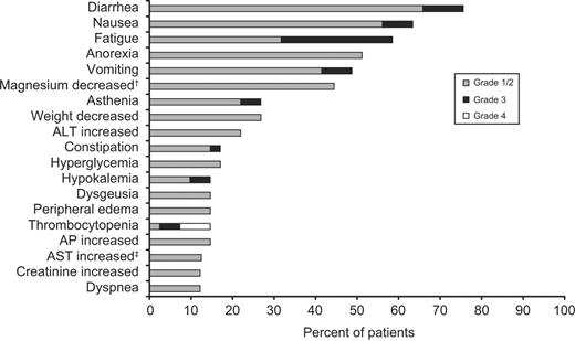
The most common drug-related adverse experiences and laboratory abnormalities observed were diarrhea (76%), nausea (63%), fatigue (59%), anorexia (51%), and vomiting (49%) (Figure 1). These events were mostly mild to moderate in severity. Grade 3/4 drug-related adverse experiences or laboratory abnormalities included fatigue (27%), thrombocytopenia (12%), diarrhea (10%), nausea (7%), vomiting (7%), asthenia (5%), and hypokalemia (5%). The median time to resolution was 12 days (range, 2-73+ days). It should be noted that the overall rates of grade 3/4 thrombocytopenia and febrile neutropenia were 51% and 32%, respectively; however, most of these events were not considered to be related to vorinostat. The overall and drug-related incidences of all other adverse experiences were similar or identical, with the exception of hyperglycemia. The overall rate (regardless of causality) of hyperglycemia was 63% and grade 3 in 2%. Serious adverse experiences occurred in 61% of patients and included febrile neutropenia (29%), AML (10%), gastrointestinal hemorrhage (5%), pneumonia (5%), and subdural hematoma (5%). A serious adverse experience of death occurred in one patient (cause unknown, day 40). However, drug-related serious adverse experiences were observed in only one patient (grade 3 diarrhea, vomiting, and hypertension). This patient was in the 300 mg 3 times daily cohort and recovered from these events after 8 days without dose modification or interruption.
Most common (10% or greater) drug-related adverse experiences and laboratory abnormalities (N = 41). Graded based on National Cancer Institute Common Terminology Criteria for Adverse Events, v3.0. ALT indicates alanine transaminase; AP, alkaline phosphatase; AST, aspartate aminotransferase. † Four of 9 patients tested. ‡Five of 40 patients tested.
Most common (10% or greater) drug-related adverse experiences and laboratory abnormalities (N = 41). Graded based on National Cancer Institute Common Terminology Criteria for Adverse Events, v3.0. ALT indicates alanine transaminase; AP, alkaline phosphatase; AST, aspartate aminotransferase. † Four of 9 patients tested. ‡Five of 40 patients tested.
Five of the 41 patients (12%) had adverse experiences that required dose modification. These included 2 patients who were reduced from 300 mg 3 times daily to 250 mg 3 times daily for grade 3 diarrhea or grade 3 nausea and vomiting, 2 patients who were reduced from 300 mg twice daily to 200 mg twice daily for grade 3 fatigue, and 1 patient who was reduced from 200 mg 3 times daily to 100 mg 3 times daily for grade 3 fatigue and nausea. The median time to dose modification was 10 days (range, 2-28 days). The 2 patients who were reduced from 300 mg twice daily to 200 mg twice daily were subsequently discontinued from the study because of grade 3 asthenia or fatigue. In addition, 4 patients had dose interruptions because of adverse experiences. After dose modification, discontinuation, or dose interruption, all but 2 of the patients recovered from these events within 30 days of the last visit. There were no other discontinuations because of adverse experiences. Twenty-nine patients discontinued because of progressive disease, 7 withdrew consent, 2 discontinued for adverse experiences, 2 went on to bone marrow transplantation, and 1 patient died during the second treatment cycle (cause unknown, day 40). Four patients ultimately died of progressive disease and 1 died because of complications of pulmonary mycosis. The deaths were determined by the investigator to be unrelated to vorinostat.
Response
The numbers of patients enrolled and achieving clinical benefit in each cohort are shown in Table 3. Overall, 7 of 41 patients (17%) had hematologic improvement (> 50% decrease in blast counts with incomplete blood count recovery) or response, including 2 CR and 2 CRi. All patients with hematologic improvement or response had AML and were treated at or below the MTD (in the 100 mg 3 times daily, 200 mg 3 times daily, and 200 mg twice daily cohorts). The median cycle of response or improvement was 2 (range, 1-8), and the median duration of response or improvement was 6 weeks (range, 0.1-53 weeks). Responses occurred in patients with normal and abnormal (trisomy 8) karyotypes. After vorinostat therapy, 3 of the patients, including 1 with a CR on study for 18 months and 2 with hematologic improvement each on study for 10.9 weeks (1 had discontinued because of progressive disease), underwent bone marrow transplantation with successful stem-cell mobilization and engraftment.
Histone acetylation
Evaluation of histone acetylation in patient peripheral blood cells and bone marrow samples was performed. By enzyme-linked immunosorbent assay, the extent of histone acetylation 2 hours after vorinostat dosing on day 14 and before vorinostat on day 22 relative to that before vorinostat on day 1 are shown in Figure 2A. Acetylation of histone H3 was rapidly induced 2- to 3-fold in all patients evaluated regardless of dose level or response. Acetylation generally returned to baseline levels during the week of rest. Similar results were observed in patient bone marrow cells (Figure 2B).
Evaluation of histone acetylation in patient peripheral blood samples and bone marrow biopsies. Induction of histone H3 acetylation was rapidly induced in (A) patient peripheral blood (100 mg, n = 3; 150 mg, n = 3; 200 mg, n = 6; 250 mg, n = 6) and (B) bone marrow cells, and generally returned to baseline levels by the end of cycle 1. Some of the hyperacetylated cells appear to be leukemic blasts based on morphology. Error bars represent SD.
Evaluation of histone acetylation in patient peripheral blood samples and bone marrow biopsies. Induction of histone H3 acetylation was rapidly induced in (A) patient peripheral blood (100 mg, n = 3; 150 mg, n = 3; 200 mg, n = 6; 250 mg, n = 6) and (B) bone marrow cells, and generally returned to baseline levels by the end of cycle 1. Some of the hyperacetylated cells appear to be leukemic blasts based on morphology. Error bars represent SD.
Gene-expression analysis
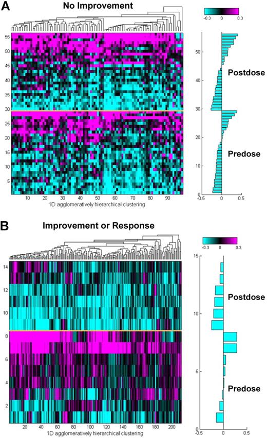
An analysis of gene-expression changes in response to vorinostat therapy was conducted. mRNA levels of proliferation-associated genes were down-regulated after therapy in peripheral blood samples of AML patients who had hematologic improvement or response (Figure 3A; Table S2). In contrast, expression of the same proliferation-associated genes in peripheral blood samples was generally unchanged after vorinostat treatment of AML patients who did not show improvement (Figure 3B; Table S2).
Analysis of proliferation-associated gene expression in peripheral blood samples of AML patients. The expression levels of proliferation associated genes were (A) similar after vorinostat treatment in blood samples of AML patients who did not show clinical improvement, but (B) down-regulated after dose in those who had hematologic improvement or response. The average resistance signature values for each of the samples are shown in the horizontal bar graphs.
Analysis of proliferation-associated gene expression in peripheral blood samples of AML patients. The expression levels of proliferation associated genes were (A) similar after vorinostat treatment in blood samples of AML patients who did not show clinical improvement, but (B) down-regulated after dose in those who had hematologic improvement or response. The average resistance signature values for each of the samples are shown in the horizontal bar graphs.
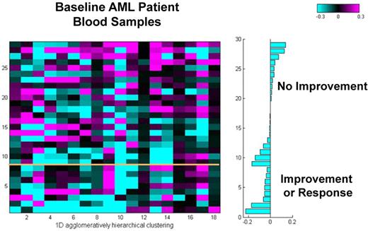
In preclinical studies, vorinostat treatment resulted in reactive oxygen species generation in vorinostat-sensitive, but not vorinostat-resistant cell lines (data not shown). Vorinostat-induced cytotoxicity has furthermore been shown to be blocked by preexposure to antioxidants.6,36 Because up-regulation of reactive oxygen species scavengers appears to be a mechanism of vorinostat resistance, expression of a panel of 17 antioxidant genes was analyzed as a potential vorinostat resistance biomarker (Table S3). Increased expression of the antioxidant signature in lymphoma cell lines (data not shown) and baseline peripheral blood samples of AML patients (Figure 4) as well as all patients (data not shown) correlated with vorinostat resistance. Seventy-six percent (16 of 21) of the samples from AML patients who did not show improvement had higher average baseline expression levels of the 17 antioxidant genes than of those from patients with hematologic improvement or response.
A baseline gene-expression resistance signature in AML patient peripheral blood cells was predictive of clinical outcome. Expression levels of the 17 antioxidant genes in baseline AML patient samples as well as all patients (data not shown) correlated with clinical outcome.
A baseline gene-expression resistance signature in AML patient peripheral blood cells was predictive of clinical outcome. Expression levels of the 17 antioxidant genes in baseline AML patient samples as well as all patients (data not shown) correlated with clinical outcome.
Discussion
In this open-label, nonrandomized phase 1 trial, the primary objective was to determine the MTD of oral vorinostat administered twice or 3 times daily for 14 consecutive days in a 21-day cycle to patients with advanced leukemias or MDS. DLTs included fatigue, nausea, vomiting, and diarrhea. Five of the 7 DLTs occurred in patients treated at dose levels of vorinostat above the MTD. On this schedule, the MTD of oral vorinostat was 200 mg twice daily or 250 mg 3 times daily.
The most common toxicities were gastrointestinal (diarrhea, nausea, anorexia, and vomiting) and fatigue. The events were mostly mild to moderate in severity, and these findings are similar to those observed in previous phase 1 trials with vorinostat.16,19,37 The 7-day rest included in each treatment cycle may therefore have prevented any increased toxicity because of more frequent daily dosing used in this study. The most common drug-related grade 3/4 toxicities were gastrointestinal (nausea, vomiting, and diarrhea), fatigue, and thrombocytopenia. The patients rapidly recovered from the majority of these events within 2 weeks on dose modification, interruption, discontinuation, or no action. Two of the 41 patients (5%) discontinued because of adverse events. The only patient to develop any drug-related serious adverse events was treated at a dose level above the MTD (300 mg 3 times daily). One patient developed neutropenic colitis (300 mg twice daily) and another acquired Clostridium difficile colitis (300 mg 3 times daily). These patients were not rechallenged with study drug and were discontinued because of lack of response and progressive disease, respectively. These experiences were not considered to be drug-related by the investigator and, thus, not DLTs. Further assessment of vorinostat safety may best be accomplished through future randomized trials.
A secondary objective of this trial was to evaluate the antitumor activity of vorinostat in patients with advanced leukemias or MDS. Overall, 7 of 41 patients (17%) achieved CR, CRi, or hematologic improvement; most occurring by cycle 2 and lasting at least 6 weeks. Patients with normal and abnormal karyotypes responded. It is important to note that all of the patients who achieved hematologic improvement or response had AML and were treated at or below the MTD. Bona fide responses were not observed in the other 10 patients with CLL, ALL, CML, or MDS.
Other HDAC inhibitors have been evaluated in early clinical trials in patients with AML or MDS.22-30 These compounds have been associated with distinct safety profiles and various degrees of activity. Combination regimens of HDAC inhibitors with DNA hypomethylating agents such as azacitidine or decitabine have also shown promising antileukemia/MDS activity.38-41
As observed in other phase 1 trials of vorinostat,16,17,37 histone acetylation was rapidly induced by vorinostat in this study in patient peripheral blood and bone marrow cells. Histone acetylation returned to baseline levels during the week of rest before the next cycle, and the level of histone acetylation was not associated with response or dose level.
Vorinostat treatment also resulted in decreased expression levels of proliferation-associated genes in patients who later achieved hematologic improvement or response. These findings are consistent with the decreased peripheral blood blast counts observed in these patients. Proliferation signatures are differentially expressed across many datasets, reflecting the relative rates of proliferation of cells in compared samples.42,43 Expression of the panel of proliferation-associated genes may potentially serve as a postdose vorinostat efficacy biomarker.
Preclinical experiments have also led to the development of an additional gene panel that is being evaluated as a predictive biomarker of vorinostat resistance. Increased expression of this panel of 17 antioxidant genes correlated with vorinostat resistance in unsorted peripheral blood samples of AML patients as well as all patients. As with other agents,44 induction of oxidative stress through generation of reactive oxygen species appears to be a mechanism of vorinostat action, whereas increased antioxidant levels may be a mechanism of vorinostat resistance.6,36,45-47 Indeed, increased expression of the panel of antioxidant genes was observed after dose and also correlated with vorinostat resistance in preclinical studies.
In a retrospective analysis of this clinical study, measurement of the mRNA levels of the antioxidant gene panel before therapy (a preliminary predictive biomarker) would have allowed for the exclusion of more than 50% of patients who did not benefit from vorinostat treatment. Although this preliminary predictive biomarker requires further study and validation, such gene-expression patterns may allow for future enrichment of patients who are more likely to benefit from treatment with vorinostat. Eliminating unnecessary treatments and toxicities without denying treatment to patients who are likely to respond are objectives for which to strive.48
In conclusion, the MTD of oral vorinostat in this phase 1 trial was determined to be 250 mg administered 3 times daily for 14 consecutive days in a 21-day treatment cycle. The MTD on the twice-daily schedule for 14 consecutive days in a 21-day treatment cycle was 200 mg. Significant antileukemia activity was observed at or below the MTD in patients with AML, and vorinostat effectively inhibited HDAC activity in peripheral blood and bone marrow blasts. Based on these results, further evaluation of vorinostat in patients with leukemias and MDS, including analysis of potentially predictive gene expression signatures for efficacy or resistance, is warranted. Phase 1 and 2 trials of vorinostat as monotherapy or in combination regimens in AML/MDS are ongoing.
Presented in part at the 40th Annual Meeting of the American Society of Clinical Oncology, New Orleans, LA, June 7, 2004; at the 47th Annual Meeting and Exposition of the American Society of Hematology, Atlanta, GA, December 12, 2005; and at the 48th Annual Meeting and Exposition of the American Society of Hematology, Orlando, FL, December 10, 2006.
The online version of this article contains a data supplement.
The publication costs of this article were defrayed in part by page charge payment. Therefore, and solely to indicate this fact, this article is hereby marked “advertisement“ in accordance with 18 USC section 1734.
Acknowledgments
This study was supported by research funding from Merck and the University of Texas M. D. Anderson Cancer Center's Physician-Scientist Program Award funded by the Commonwealth Cancer Foundation for Research (G.G.M.).
Authorship
Contribution: G.G.-M. designed and directed the study, performed research, analyzed data, and wrote the manuscript; C.B.-R. performed research, including histopathologic analysis and built the tissue array platform; H.Y., A.F., J.C., W.W., S.F., C.K., G.M., A.L., V.R.F., J.S.H., J.F.R., J.P.S., and H.K. performed research, collected data, and analyzed and interpreted data; G.R. analyzed and interpreted data; S.S.R., V.M.R. S.R.F. designed research and analyzed and interpreted data; C.C. analyzed and interpreted data and performed statistical analysis; and J.L.R. analyzed and interpreted data and drafted the manuscript.
Conflict-of-interest disclosure: A.L., V.R.F., S.S.R., J.S.H., J.F.R., C.C., J.L.R., J.P.S., V.M.R., and S.R.F. are employed by Merck. All other authors declare no competing financial interests.
Correspondence: Guillermo Garcia-Manero, Leukemia Department, University of Texas MD Anderson Cancer Center, 1515 Holcombe Blvd, Unit 428, Houston, TX 77030; ggarciam@mdanderson.org.


