Stat5 proteins are critical signaling molecules activated by many cytokines. Within the immune system, Stat5 plays important roles related to the development of thymocytes and proliferation of T cells. Stat5 has been implicated in malignant transformation, and moreover, the activated tyrosine phosphorylated form of Stat5 is frequently observed in human lymphomas. We previously demonstrated the oncogenic potential of Stat5, with thymic lymphoblastic lymphomas developing in a significant proportion of transgenic (TG) mice overexpressing Stat5a or Stat5b in lymphocytes. In addition, immunization or expression of a T-cell receptor (TCR) transgene augmented the rate of tumor formation. Here, we investigate the mechanism of Stat5-mediated lymphomagenesis by exploring the contributions of major histocompatibility complex (MHC)/TCR and pre-TCR signals. We present data demonstrating that Stat5b TG mice unexpectedly develop CD8+ lymphoma even in the absence of either pre-TCR signaling or normal thymic selection. Indeed, acceleration of Stat5b transgene-mediated lymphoma occurred on TCRα−/− and pre-TCRα−/− backgrounds. In light of these data, we propose a model in which alterations in T-cell development at the double-negative/double-positive (DN/DP) stages cooperate with cytokine-mediated pathways in immature thymocytes to give rise to lymphoblastic T-cell lymphomas in Stat5b TG mice.
Introduction
The JAK-STAT pathway is a major signaling pathway used by numerous cytokines and growth factors.1 Consistent with its role in cell proliferation and survival, Stat5 has been implicated as an oncogene, and the activated (tyrosine-phosphorylated) form has been observed in many human lymphomas and leukemias.2 T-cell acute lymphoblastic leukemias (T-ALLs) and lymphomas comprise a heterogeneous group of malignancies in which cells are arrested at various stages of T-cell development that largely reflect progressive stages in normal thymic development.3,4 CD4 and CD8 are often expressed on T lymphocytes in T-ALL, with single and dual expression described.5,6 We recently demonstrated a role for Stat5 in lymphomagenesis; T-ALL developed in a significant proportion of mice overexpressing either Stat5a or Stat5b as a transgene.7
The complex multistep process of TCRαβ+ T-cell development is dependent on signals transduced by both the T-cell receptor (TCR) and cytokine receptors.8,–10 These signals orchestrate the survival and development of thymocytes as they progress from the most immature CD8−CD4− (double-negative [DN]) cells, through the immature single-positive (ISP) CD4−CD8+11 and subsequent CD8+CD4+ (double-positive [DP]) stages, to become CD4+ or CD8+ (SP) T cells.12 Stat5 is activated by many cytokine receptors, but its role in response to TCR signaling is controversial.13,,–16 Importantly, dysregulation of these upstream signals has been implicated in lymphomagenesis, as has dysregulation of their downstream mediators, including Stat5. For example, overexpression of either IL-717,,–20 or IL-1521,22 can result in CD8+ T-cell expansion and hematopoietic malignancies. Likewise, pre-TCR signaling cooperates with TEL-JAK2 to transform immature thymocytes,23 and Stat5 is implicated in this process.24,25 Furthermore, lymphomagenesis is accelerated in Stat5 transgenic (TG) mice by immunization or by coexpression of a rearranged αβTCR.7 Thus, there are several stages where Stat5 signaling might influence developing thymocytes and where Stat5 overexpression could promote CD8+ T-cell expansion and the development of lymphoma.
Here, we use a genetic approach to distinguish the contribution of upstream signals to lymphomagenesis by mating Stat5b TG mice onto genetic backgrounds deficient in either normal TCR signaling/thymic selection or pre-TCR signaling. We show that Stat5b TG mice can develop lymphoma in the absence of normal TCR/major histocompatibility complex (MHC) interactions. Furthermore, pre-TCRα expression was not required for Stat5b-mediated lymphomagenesis. Indeed, acceleration of Stat5b transgene-mediated lymphoma occurred on TCRα−/− and pre-TCRα−/− backgrounds. In light of these data, we propose a model in which alterations in T-cell development at the DN/DP stages cooperates with cytokine-mediated pathways in immature thymocytes to give rise to lymphoblastic T-cell lymphomas.
Methods
Generation of mice
The Stat5b transgene construct was under the control of the H-2Kb promoter and heavy-chain enhancer, as previously described.26 Stat5 TG mice were mated with mice that express the OT-I TCR transgene specific for an ovalbumin peptide (amino acids 257-264) presented by the MHC class I molecule H2Kb, and onto various genetic backgrounds, including TCRα−/− (Jackson Laboratory, Bar Harbor, ME), β2m−/− (Taconic, Germantown, NY) and pre-TCRα−/− mice (graciously provided by Harold von Boehmer, Dana-Farber Cancer Institute, Boston, MA). Because of the complex nature of multiple crossings, for many experiments, homozygous with heterozygous matings were performed. In these instances, heterozygous littermates were used as phenotypically normal controls.
We have previously noted thymic expansion of CD8+ T cells in Stat5b TG mice prior to obvious lymphoma development. Because we analyzed mice at various time points prior to cervical adenopathy and massive thymic enlargement, we used the following criteria to define tumors: (1) total thymocyte number greater than 200 million; (2) CD8+ greater than 15%; and (3) abnormal CD3/forward scatter (FSC) profile.27,28 If mice were positive for 2 of 3 criteria, they were deemed to have lymphoma, whereas if they were negative for 2 of 3 criteria, they were classified as not having lymphoma. If data were not available (eg, CD3 staining was not available for all mice) and the 2 remaining criteria were not conclusive, the mice were excluded from analysis.
Experiments were performed under an Animal Component of Research Protocol (ACORP) approved by the Institutional Animals Care and Use Committees (IACUCs) of the White River Junction Veteran's Association (WRJ VA) and Dartmouth Medical School.
Flow cytometric analysis
Single-cell suspensions from the thymus and spleen were stained and analyzed using a FACScan with CELLQuest software (Becton Dickinson, San Jose, CA). The following antibodies, all from BD PharMingen (San Diego, CA), were used: anti-CD4–FITC, anti-CD4–PE, and anti-CD4–Cy-Chrome; anti-CD8–FITC, anti-CD8–PE, and anti-CD8–Cy-Chrome; anti–TCR-γ/δ–FITC; anti–IL-2Rα (CD25)–FITC and anti–IL-2Rα–PE; anti-CD44–Cy-Chrome; anti–IL-2Rβ–FITC and anti–IL-2Rβ–PE; anti–pan–natural killer (NK) cells (DX5)–FITC; and anti-CD3–Cy-Chrome, anti–heat-stable antigen (HSA; CD24)–FITC, and anti-CD5–FITC. TCRβ variable (Vβ) staining was performed using a screening panel (Vβ 2,3,4,5,6,7,8,9,10,11,12,13,14,17; BD PharMingen). NKT cells were evaluated with CD1d tetramer loaded with PBS-57, an analog of α-GalCer (provided by the National Institute of Allergy and Infectious Diseases [NIAID], National Institutes of Health [NIH] tetramer facility).
For intracellular staining, cells were fixed and permeabilized using Cytofix/Cytoperm solution (BD PharMingen), followed by anti-TCRα/β (H57–597 to TCRβ)–FITC, anti-Vβ–FITC, or rabbit anti-TdT (Supertechs, Bethesda, MD), or polyclonal serum control with subsequent treatment with anti-rabbit IgG-PE (Jackson ImmunoResearch Laboratories, West Grove, PA).
Statistical analysis
The results were expressed as means plus or minus standard error of the mean (SEM). The Student t test was applied to compare the statistical difference between 2 groups. The Fisher exact test was used for analysis of categorical variables. A probability (P) value of less than .05 was considered statistically significant.
Results
Stat5b TG mice develop CD8+ T-cell lymphoma
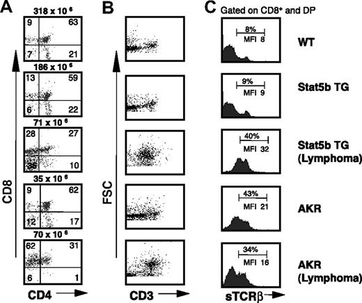
Stat5b TG mice develop lymphoma7 with characteristics of T-ALL.29 Representative Stat5b TG mice with and without lymphoma are shown in Figure 1 along with AKR mice with and without lymphoma. The latter are shown in comparison because AKR mice have a virus in their genome that leads to a high incidence of thymic lymphoblastic lymphoma within 1 year.30 The phenotypic pattern, including surface CD3 (sCD3) expression versus FSC, can help discriminate immature thymocytes from T-ALL.27,28 The Stat5b TG and AKR mice with lymphoma have abnormal CD4/CD8 profiles (Figure 1A) and also demonstrate characteristic FSC/CD3 profiles, with a well-defined population of large cells with less variability in sCD3 expression compared with controls with small cells (FSC) and variable expression of sCD3 (Figure 1B). The CD8+ cells (gated on SP and DP) express surface (s) TCRβ (Figure 1C).
Lymphoblastic lymphoma in Stat5b TG mice. (A) CD4/CD8 profiles in Stat5b TG mice with and without lymphoma are compared with AKR mice with and without lymphoma and a WT control. Numbers indicate the percentage of cells in each quadrant, and total cellularity is indicated above each dot plot. (B) sCD3 expression versus FSC profiles reveals a characteristic pattern in the mice with lymphoma. (C) sTCRβ staining gated on CD8+ cells (CD8 SP and CD4/CD8 DP) is consistent with a T-cell origin of these lymphomas. Numbers indicate the mean fluorescence intensity (MFI) for the CD8+ (SP + DP) populations as well as the percentage within the gated region.
Lymphoblastic lymphoma in Stat5b TG mice. (A) CD4/CD8 profiles in Stat5b TG mice with and without lymphoma are compared with AKR mice with and without lymphoma and a WT control. Numbers indicate the percentage of cells in each quadrant, and total cellularity is indicated above each dot plot. (B) sCD3 expression versus FSC profiles reveals a characteristic pattern in the mice with lymphoma. (C) sTCRβ staining gated on CD8+ cells (CD8 SP and CD4/CD8 DP) is consistent with a T-cell origin of these lymphomas. Numbers indicate the mean fluorescence intensity (MFI) for the CD8+ (SP + DP) populations as well as the percentage within the gated region.
CD8+ T-cell lymphoma does not require MHC I/TCR interaction in Stat5b TG mice
Immunization or coexpression of a TCR transgene greatly augmented thymic CD8+ T-cell expansion in Stat5b TG mice.7 Class I– and class II–restricted transgenes normally direct development of CD8+ or CD4+ TCR TG T cells, respectively. However, Stat5b-mediated CD8+ T-cell expansion occurred not only with the MHC class I–restricted HY TCR transgene, but also remarkably with the MHC class II–restricted 5C.C7 TCR transgene.7 The expansion of CD8+ TCR TG T cells with coexpression of a class II TCR transgene suggests a unique and powerful effect of Stat5 overexpression within the thymus. Indeed, we previously noted that introduction of a TCR transgene enhanced the incidence and reduced the latency for tumor formation in Stat5b TG mice (25% vs 100% and 8 months vs 3 months, respectively, for Stat5b transgene vs Stat5b/TCR double transgene).7 Thus, we first investigated the requirement for MHC/TCR interaction in this process.
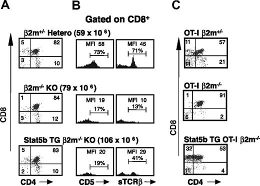
The typical developmental pathway of a CD8+ T cell requires MHC class I/self-antigen interaction with an αβTCR.31,32 This interaction contributes to T-cell development and CD8+ lineage commitment. To investigate the contribution of MHC I to the expansion of CD8+ T cells in Stat5b TG mice, we crossed our Stat5b TG mice onto a genetic background deficient in MHC I expression due to a null mutation in the β2m gene (β2m−/− mice).33 β2m−/− mice have reduced CD8+ SP cells (Figure 2A middle panel). Interestingly, Stat5b overexpression results in expansion of CD8+ T cells on the β2m−/− background (Figure 2A; Table 1). The CD8+ T cells in these mice had an immature phenotype, as evidenced by low expression of both CD5 and sTCRβ (Figure 2B) when compared with wild-type (WT) heterozygous controls, as well as heightened expression of HSA (CD24; data not shown). Furthermore, as Stat5b TG/β2m−/− mice age, they developed features of lymphoblastic lymphoma (Table 2).
Stat5b-mediated thymocyte expansion in MHC class I–deficient (β2m−/−) mice. (A) Thymic CD4/CD8 profiles of 8-week-old β2m−/− mice with and without the Stat5b transgene. β2m+/− littermates served as controls. Numbers indicate the percentage of cells in each quadrant, and total cellularity is indicated above in parentheses. (B) CD5 and sTCRβ expression on CD8+ SP cells. Numbers indicate the MFI for the CD8+ SP population as well as the percentage within the gated region. The expanded CD8+ SP population is immature, as evidenced by fewer sTCRβhi and CD5hi cells when compared with WT heterozygous controls. (C) Thymic CD4/CD8 profiles of 10-week-old β2m−/− mice expressing the ova-specific MHC class I–restricted OT-I TCR transgene. The Stat5b transgene-mediated expansion of CD8+ cells in MHC class I (β2m−/−)–deficient mice is enhanced by coexpression of the TCR transgene. OT-I TG/ β2m+/− littermates served as controls. Numbers indicate the percentage of cells in each quadrant. Representative data are shown from 1 of 5 similar experiments involving a total of 27 Stat5b TG/β2m−/− and 20 β2m−/− mice.
Stat5b-mediated thymocyte expansion in MHC class I–deficient (β2m−/−) mice. (A) Thymic CD4/CD8 profiles of 8-week-old β2m−/− mice with and without the Stat5b transgene. β2m+/− littermates served as controls. Numbers indicate the percentage of cells in each quadrant, and total cellularity is indicated above in parentheses. (B) CD5 and sTCRβ expression on CD8+ SP cells. Numbers indicate the MFI for the CD8+ SP population as well as the percentage within the gated region. The expanded CD8+ SP population is immature, as evidenced by fewer sTCRβhi and CD5hi cells when compared with WT heterozygous controls. (C) Thymic CD4/CD8 profiles of 10-week-old β2m−/− mice expressing the ova-specific MHC class I–restricted OT-I TCR transgene. The Stat5b transgene-mediated expansion of CD8+ cells in MHC class I (β2m−/−)–deficient mice is enhanced by coexpression of the TCR transgene. OT-I TG/ β2m+/− littermates served as controls. Numbers indicate the percentage of cells in each quadrant. Representative data are shown from 1 of 5 similar experiments involving a total of 27 Stat5b TG/β2m−/− and 20 β2m−/− mice.
Thymic cellularity and CD8+ expansion in Stat5b TG mice at 2 time intervals
| . | 0 to 10 wk . | 10 to 20 wk . | ||||
|---|---|---|---|---|---|---|
| Thymic cellularity, 10−6 (SEM) . | CD8, % (SEM) . | n . | Thymic cellularity, 10−6(SEM) . | CD8, % (SEM) . | n . | |
| WT | 106 (13) | 6 (0.6) | 28 | 74 (13) | 9 (2.3) | 10 |
| 5bTG WT | 128 (14) | 7 (1.2) | 16 | 55 (6) | 17* (3.7) | 8 |
| β2m−/− | 88 (9) | 3 (0.7 | 5 | 76* (13) | 4 (0.9) | 8 |
| 5bTG β2m−/− | 106 | 5 | 1 | 41 (8) | 13* (4) | 9 |
| TCRα−/− | 107 (26) | 3 (0.7) | 4 | 85 | 5 | 1 |
| 5bTG TCRα−/− | 165 | 5 | 1 | 198 (83) | 10* (0.3) | 2 |
| preTCRα−/− | 6 (2) | 3 (1.4) | 4 | 5 (0.3) | 6 (1.3) | 4 |
| 5bTG preTCRα−/− | 30* (10) | 9* (0.4) | 2 | 10 (5.5) | 7 (3.2) | 2 |
Mean thymic cellularity and percentage of CD8+ cells (SEM) in WT, β2m−/−, TCRα−/−, and pre-TCRα−/−mice with and without the Stat5b transgene are shown. Only mice without lymphoma as defined by criteria outlined in ″Generation of mice″ were included in the analysis. Because of tumor development in older mice, we do not have an expansion group beyond 20 weeks. Also shown are total numbers (n) in each group that met criteria for analysis; for example, there was only a single Stat5b TG/TCRα−/− mouse that met the criteria for not having lymphoma in the 0- to 10-week group.
Significant differences (P < .05) between the mice without and with the transgene.
Frequency and latency of lymphoma in Stat5b TG mice
| . | Mean age of onset, wk (SEM) . | n1 . | Tumor at 20 wk, % . | n2 . |
|---|---|---|---|---|
| 5bTG WT | 26 (2.4) | 16 | 21 | 28 |
| 5bTG β2m−/− | 21 (1.8) | 9 | 39 | 13 |
| 5bTG TCRα−/− | 11* (1.9) | 5 | 100* | 5 |
| 5bTG preTCRα−/− | 17* (1.0) | 7 | 86* | 7 |
| . | Mean age of onset, wk (SEM) . | n1 . | Tumor at 20 wk, % . | n2 . |
|---|---|---|---|---|
| 5bTG WT | 26 (2.4) | 16 | 21 | 28 |
| 5bTG β2m−/− | 21 (1.8) | 9 | 39 | 13 |
| 5bTG TCRα−/− | 11* (1.9) | 5 | 100* | 5 |
| 5bTG preTCRα−/− | 17* (1.0) | 7 | 86* | 7 |
Mean age of detection and percentage of mice with tumors at 20 weeks are shown for β2m−/−, TCRα−/−, and pre-TCRα−/−mice with the Stat5b transgene. Stat5b transgene on WT background (C57BL/6) are also shown for comparison. Also shown are total numbers of tumors in each group (n1) as well as total number of mice (n2) that met criteria for analysis as outlined in “Generation of mice.” Please note that some mice developed tumors after the 20-week time point.
Significant differences (P < .05) between the mice on WT background and those on knock-out backgrounds.
Stat5b TG/β2m−/− mice were then crossed with OT-1 TCR TG mice to generate Stat5b/TCR double TG mice on a β2m−/− background. Whereas the OT-1 TCR TG expressed on an MHC class I–deficient background failed to cause expansion of CD8+ T cells, coexpression of this TCR transgene and Stat5b augmented CD8+ expansion as well as lymphoma formation, evidenced by grossly enlarged thymi and the typical CD4/CD8 profiles characteristic of T-ALL. Moreover, these double TG mice exhibited accelerated CD8+ tumor formation on an MHC class I–deficient background (Figure 2C). The expanded population of CD8+ SP thymocytes in Stat5b TG/β2m−/− mice express sTCRβ and stain with an antibody to terminal deoxynucleotidyl transferase (TdT; Figure S1, available on the Blood website; see the Supplemental Materials link at the top of the online article). This antibody labels normal cortical thymocytes and primitive lymphocytes and represents a useful marker for differentiating acute lymphoblastic lymphoma/leukemia from other lymphomas.34
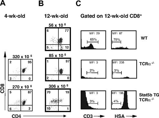
To confirm that the expanded thymic CD8+ T-cell population in Stat5b TG/β2m−/− mice represents an immature T-cell population as opposed to mature MHC class II–restricted CD8+ T cells,35 we crossed our Stat5b TG mice onto the TCRα−/− background. TCRα-deficient mice have a block in T-cell development at the DP stage.36 Consistent with an effect of Stat5 on immature thymocyte expansion, 4-week-old Stat5b TG/TCRα−/− mice exhibited expansion of DN cells (16 million vs 10 million) and CD8+ SP thymocytes (14 million vs 6 million) when compared with TCRα−/− mice (Figure 3A; Table 1). In addition, as Stat5b/TCRα−/− mice aged, they developed thymic lymphomas as evidenced by grossly enlarged thymi and CD4/CD8 profiles characteristic of Stat5b-mediated T-ALL (Figure 3B). Furthermore, T-ALL was accelerated as it occurred in Stat5b TG/TCRα−/− mice at a mean age of 11 weeks compared with a mean age of 26 weeks on a WT background (P < .05; Table 2). The expanded population of CD8+ T cells was CD3lo and HSAhi, strongly suggesting that it was comprised of immature cells (Figure 3C). The expanded population did not express sTCRβ, but did exhibit cytoplasmic (c) TCRβ staining, and stained with anti-TdT (Figure S2). We examined cTCR Vβ staining in a 15-week-old Stat5b TG/TCRα−/− mouse with grossly enlarged thymus and spleen and confirmed an expanded population of oligoclonal cells (Figure S3). Taken together, our results indicate that Stat5b TG mice can develop T-cell lymphomas in experimental situations where normal TCR/MHC interactions do not occur. Furthermore, Stat5b TG on a TCRα−/− background mice have acceleration of lymphoma formation, similar to that seen previously with coexpression of a TCR transgene.7
Stat5b-mediated CD8+ thymic T-cell expansion and lymphoma on the TCRα−/− background. (A) Thymic CD4/CD8 profiles of 4-week-old TCRα−/− mice with and without the Stat5b transgene. Numbers indicate the percentage of cells in each quadrant, and total cellularity is indicated above each dot plot. Note the increase in the percentage of CD8+ SP cells (with comparable total cellularity) in Stat5b TG/TCRα−/− mice. (B) CD4/CD8 profiles of 12-week-old TCRα−/− mice with and without the Stat5b transgene. The Stat5b TG/TCRα−/− mice had grossly enlarged thymi and spleens consistent with T-cell lymphoblastic lymphoma. (C) CD3 and HSA surface expression on CD8+ SP thymocytes in TCRα−/− mice. Numbers indicate the MFI for the CD8+ SP population as well as the percentage of mature cells (gated on CD3hi/HSAlo). Representative data are shown from 1 of 4 similar experiments involving a total of 12 Stat5b TG/TCRα−/− and 10 TCRα−/− mice.
Stat5b-mediated CD8+ thymic T-cell expansion and lymphoma on the TCRα−/− background. (A) Thymic CD4/CD8 profiles of 4-week-old TCRα−/− mice with and without the Stat5b transgene. Numbers indicate the percentage of cells in each quadrant, and total cellularity is indicated above each dot plot. Note the increase in the percentage of CD8+ SP cells (with comparable total cellularity) in Stat5b TG/TCRα−/− mice. (B) CD4/CD8 profiles of 12-week-old TCRα−/− mice with and without the Stat5b transgene. The Stat5b TG/TCRα−/− mice had grossly enlarged thymi and spleens consistent with T-cell lymphoblastic lymphoma. (C) CD3 and HSA surface expression on CD8+ SP thymocytes in TCRα−/− mice. Numbers indicate the MFI for the CD8+ SP population as well as the percentage of mature cells (gated on CD3hi/HSAlo). Representative data are shown from 1 of 4 similar experiments involving a total of 12 Stat5b TG/TCRα−/− and 10 TCRα−/− mice.
Stat5b-mediated CD8+ T-cell expansion and lymphoma does not require pre-TCR signaling
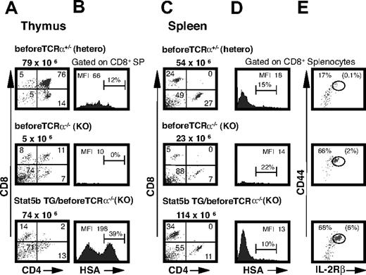
The effect of Stat5 on immature T-cell expansion could result from altered pre-TCR–mediated signaling23,37,–39 or from pre-TCR–independent mechanisms40 such as amplified cytokine signals. To distinguish these possibilities, we first investigated the contribution of the pre-TCR to Stat5-mediated lymphomagenesis by mating Stat5b TG mice with mice lacking the expression of pre-TCRα.41 Stat5b TG/pre-TCRα−/− mice exhibit increased thymic cellularity when compared with pre-TCRα−/− mice lacking the Stat5b transgene (Figure 4A; Table 1). The expanded thymic CD8+ SP cells were immature as evidenced by high HSA staining (Figure 4B). In addition, these cells had lower sTCRβ staining than the preTCR+/− heterozygous controls, but did exhibit cTCRβ staining (Figure S4). The spleens were also enlarged with an expanded CD8+ T-cell population (Figure 4C). These CD8+ splenocytes in younger Stat5b TG/pre-TCRα−/− mice were mature as evidenced by low HSA expression (Figure 4D). Furthermore, a significant proportion of these CD8+ splenocytes were CD44hi IL-2Rβhi memory phenotype cells (Figure 4E), consistent with a Stat5/cytokine-mediated effect on peripheral T-cell homeostasis.26 As they age, Stat5b TG/pre-TCRα−/− mice develop features consistent with T-ALL at a mean age of 17 weeks compared with a mean age of 26 weeks on a WT background (P < .05; Table 2). In these Stat5b TG/pre-TCRα−/− mice with lymphoma, the splenic CD8+ population was immature (Figure S5). This is consistent with our previous findings of splenic spread of lymphomas.7 Thus, not only is pre-TCRα signaling not required for abnormal CD8+ T-cell expansion and lymphomagenesis in Stat5b TG mice, absence of pre-TCRα, like TCRα, resulted in accelerated lymphoma formation.
Stat5b-mediated thymocyte and splenic expansion in pre-TCRα−/− mice. (A) Thymic CD4/CD8 profiles of 10-week-old pre-TCRα−/− mice with and without the Stat5b transgene, with pre-TCRα+/− serving as controls. Numbers indicate the percentage of cells in each quadrant, and total cellularity is indicated above each dot plot. (B) HSA surface expression on CD8+ SP thymocytes in pre-TCRα−/− mice. Numbers indicate the MFI for the CD8+ SP population as well as the percentage within the gated region. (C) Splenic CD4/CD8 profiles from the same mice. Numbers indicate the percentage of cells in each quadrant, and total cellularity is indicated above each dot plot. (D) HSA staining on CD8+ splenocytes. Numbers indicate the MFI for the CD8+ SP population as well as the percentage within the gated region. (E) CD44/IL-2Rβ profile gated on CD8+ T cells. The CD44hiIL-2Rβhi memory population are expanded in Stat5b TG/pre-TCRα−/− mice. Numbers indicate percentage of CD44hiIL-2Rβhi memory phenotype CD8+ cells within the gated region, and numbers in parentheses indicate percentage of total splenocytes within gated region. Representative data are shown from 1 of 4 similar experiments involving a total of 8 Stat5b TG/pre-TCRα−/− and 8 pre-TCRα−/− mice.
Stat5b-mediated thymocyte and splenic expansion in pre-TCRα−/− mice. (A) Thymic CD4/CD8 profiles of 10-week-old pre-TCRα−/− mice with and without the Stat5b transgene, with pre-TCRα+/− serving as controls. Numbers indicate the percentage of cells in each quadrant, and total cellularity is indicated above each dot plot. (B) HSA surface expression on CD8+ SP thymocytes in pre-TCRα−/− mice. Numbers indicate the MFI for the CD8+ SP population as well as the percentage within the gated region. (C) Splenic CD4/CD8 profiles from the same mice. Numbers indicate the percentage of cells in each quadrant, and total cellularity is indicated above each dot plot. (D) HSA staining on CD8+ splenocytes. Numbers indicate the MFI for the CD8+ SP population as well as the percentage within the gated region. (E) CD44/IL-2Rβ profile gated on CD8+ T cells. The CD44hiIL-2Rβhi memory population are expanded in Stat5b TG/pre-TCRα−/− mice. Numbers indicate percentage of CD44hiIL-2Rβhi memory phenotype CD8+ cells within the gated region, and numbers in parentheses indicate percentage of total splenocytes within gated region. Representative data are shown from 1 of 4 similar experiments involving a total of 8 Stat5b TG/pre-TCRα−/− and 8 pre-TCRα−/− mice.
Neither NKT cells nor γ/δ T cells are expanded in Stat5b TG mice
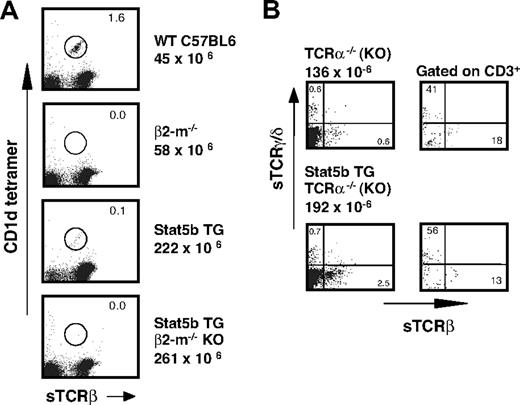
To investigate whether the expanded CD8+ T-cell population in Stat5b TG mice represented a cell subset other than TCRα/β+ cells, we examined Sta5b TG thymi for the presence of NKT cells and γ/δ T cells (Figure 5A,B, respectively). The term “NKT cell” was originally used to describe T cells coexpressing markers of the NK lineage, but NKT cells are now largely defined by their recognition of CD1d for their development.42,–44 CD1d is a nonclassical MHC class I molecule comprised of a glycoprotein associated with β2m.42 However, CD1d can be expressed on the cell surface independently of β2m,43 and CD1d-independent NKT cells have been described in β2m−/− mice.44 Furthermore, a proportion of these CD1d-independent NKT cells are CD8+.44 Most CD1d-restricted NKT cells can be identified by the ability to bind CD1d tetramers loaded with the glycosphingolipid antigen alpha-galactosyl ceramide (α-GalCer).45 We therefore incubated thymocytes from 18-week-old β2m−/− mice with and without the Stat5b transgene (Figure 5A) with CD1d tetramer loaded with PBS-57, an analog of α-GalCer (provided by NIAID, NIH tetramer facility). As expected, this CD1d-dependent population was not expanded in Stat5b TG/β2m−/− mice with evidence of lymphoma. Interestingly, this NKT population was not expanded in Stat5b TG mice with lymphoma on the WT (C57BL/6) background but was, indeed, reduced (2.2 × 10⁵ vs 7.2 × 10⁵ in TG and WT mice, respectively). This phenotype might reflect the effect of lymphomas on NKT cells. Because Stat5 is critical for γ/δ T-cell development,46 we next evaluated the presence of this population in Stat5b TG lymphomas. γ/δ are present in some human T-ALL,47,48 and this population is expanded in mice that express a constitutively active form of Stat5.49 However, we did not see Stat5b-mediated expansion of γ/δ T cells on either the TCRα−/− (Figure 5B) or WT backgrounds (data not shown). These data, coupled with lymphoma cell TCRβ expression and response to CD3 stimulation in vitro7 (data not shown), are consistant with an αβTCR lineage origin of Stat5b transgene-mediated lymphomas.
CD1d-dependent NKT cells in β2m−/− mice with and without the Stat5b transgene. (A) Thymic sTCRβ/CD1d tetramer profiles of 18-week-old β2m−/− mice with and without the Stat5b transgene. Studies of age-matched C57BL/6 mice, with and without the Stat5b transgene, are shown for comparison. Both the Stat5b TG mice on the WT background and Stat5b TG β2m−/− mice had evidence of lymphoma (enlarged thymus, increased cellularity, and characteristic CD3/FSC and CD4/CD8 profiles; data not shown). Percentage of gated cells is indicated with each profile, and the total cellularity of the thymus indicated on the right. (B) γδ T cells in TCRα−/− mice. The left panels show 2-color profiles for sTCRγδ and sTCRαβ in TCRα−/− mice with and without the Stat5b transgene. Numbers indicate the percentage of cells in each quadrant, and total cellularity is indicated above the dot plots. The right panels show the same comparison gated on CD3+ cells. Representative data are shown from 1 of 2 similar experiments.
CD1d-dependent NKT cells in β2m−/− mice with and without the Stat5b transgene. (A) Thymic sTCRβ/CD1d tetramer profiles of 18-week-old β2m−/− mice with and without the Stat5b transgene. Studies of age-matched C57BL/6 mice, with and without the Stat5b transgene, are shown for comparison. Both the Stat5b TG mice on the WT background and Stat5b TG β2m−/− mice had evidence of lymphoma (enlarged thymus, increased cellularity, and characteristic CD3/FSC and CD4/CD8 profiles; data not shown). Percentage of gated cells is indicated with each profile, and the total cellularity of the thymus indicated on the right. (B) γδ T cells in TCRα−/− mice. The left panels show 2-color profiles for sTCRγδ and sTCRαβ in TCRα−/− mice with and without the Stat5b transgene. Numbers indicate the percentage of cells in each quadrant, and total cellularity is indicated above the dot plots. The right panels show the same comparison gated on CD3+ cells. Representative data are shown from 1 of 2 similar experiments.
Discussion
The critical role that Stat5 plays in signaling in hematologic malignancies has generated increased interest in the mechanisms whereby Stat5 functions as an oncogene.50 Previous studies using mice overexpressing WT Stat5a or Stat5b26 or the constitutively-activated form of Stat5a (H299R; S711F)49 have suggested important roles for Stat5 in T- and B-cell development. Importantly, dysregulation of Stat5 activity in these mice was associated with T-cell7 and B-cell51 lymphoid malignancies. Stat5 is critical not only for early T-cell development but also for the proper homeostasis of mature T cells, especially CD8+ T cells.46 Thus, there are multiple stages in TCRαβ+ T-cell development where Stat5 overexpression could determine the developmental fate of the emerging cell population.
We have previously shown that coexpression of a TCR transgene greatly augments thymic CD8+ T-cell expansion in the Stat5b TG mice and predisposes these Stat5b/TCR double TG mice to develop lymphomas at a much younger age than Stat5b TG mice without the TCR transgene.7 Stat5 can cooperate with pathways downstream of TCRβ/CD3. Lck overexpressing cells display activated Stat5,52 and elevated WT Stat5b may cooperate with active Lck kinase in promoting cell proliferation.53 Moreover, it has been recently shown that calcineurin activation is critical for the maintenance of the leukemic phenotype.54 Stat5b TG thymocytes, particularly from mice with lymphoma, exhibit a synergistic response to the combination of IL-7 and TCR stimulation,7 and cyclosporin A inhibits proliferation of Stat5b TG lymphocytes in vitro (data not shown). The importance of TCR-mediated signaling in lymphomagenesis is supported by spontaneous thymic lymphoma development in mice expressing TCR transgenes.55 In this regard, TCR/ligand interactions may influence selection events in developing lymphomas.56,57 To investigate the role of typical TCR/MHC in the development of Stat5-mediated T-ALL, we looked at the effect of Stat5b overexpression on β2m−/− or TCRα−/− backgrounds, settings in which normal TCR/MHC interactions do not occur. Stat5b TG β2m−/− and Stat5b TG TCRα−/− mice developed lymphomas, strongly suggesting that at least one of the primary events contributing to lymphoma formation in Stat5b TG mice must occur in the DN/DP population, prior to the point at which TCR/MHC interactions play a role in developmental decisions. In the RasGRP1 model of thymic lymphomagenesis, wherein the development of lymphomas is also accelerated by coexpression of a TCR transgene, lymphomas originate in thymocytes that are not undergoing positive selection.40
The most likely mechanisms through which Stat5b could exert effects on expansion of immature T-lineage cells is through cooperation with pre-TCR signals,23,–25 or through its actions in transmitting signals from γc cytokines.46 TEL/JAK2 TG mice develop α/β lineage lymphomas within the thymus.23 Similar to our system, absence of TCR did not lengthen the latency of lymphoma in TEL-JAK2 TG mice; however, in contrast to our system, the absence of pre-TCR signaling results in delayed TEL-JAK2–mediated T-cell transformation,23 whereas the absence of pre-TCR signaling actually accelerated lymphoma development in Stat5b TG mice. Accelerated lymphoma development can occur in settings that interfere with thymocyte maturation and promote the DN-to-DP transition in the absence of pre-TCR expression.58 Acceleration of Stat5b TG-mediated lymphoma occurred in various settings, including coexpression of a TCR transgene7 and on TCRα−/− and pre-TCRα−/− backgrounds (Table 2). In most TCR TG model systems, both TCRα and TCRβ chains are expressed early in T-cell development.59 The presence of an αβTCR is associated with impaired transition from DN to DP thymocytes.60,61 Thus, the alterations in T-cell development at the DN/DP stages common to all these backgrounds may account for the acceleration of Stat5b-mediated lymphomagenesis in these mice.
The activated Stat5 pathway is important for lymphocyte homeostasis,26 and can supersede the need for both TCR engagement and cytokine stimulation.62 Moreover, Stat5 activation acts as a stabilizer of gene regulation initiated by TCR signals, contributing to the development of a complete CD8 T-cell effector program.63 Indeed, we see splenic CD8+ T-cell expansion (relative to control) in Stat5b TG mice,26 including TG mice on β2m−/−, TCRα−/−, and pre-TCRα−/− backgrounds (Figure 4; data not shown). A high proportion of the expanded cells have a memory phenotype. This CD44hi IL-2Rβhi population is also present in β2m−/−,64 TCRα−/−, and pre-TCRα−/− mice without the Stat5b transgene (Figures 4, S5; data not shown). Thus, this splenic expansion is likely an effect of Stat5 on cytokine-mediated peripheral T-cell homeostasis.26 CD8+ T-cell expansion is seen in Stat5b TG mice without evidence of lymphoma, and we believe the effect in the spleen is separate from the immature expansion and lymphomas that arise in the thymus. In mice with lymphoma that has spread to the spleen, the splenocytes have phenotypic characteristics similar to the primary lymphoma in thymus7 (Figures S3, S5; data not shown). Furthermore, the gene expression pattern in Stat5b TG lymphomatous spleens is similar to that of thymi.7 To investigate the lineage of the Stat5b-mediated lymphoma cells in the thymus, we evaluated the expression of genes previously shown to correlate with CD4/CD8 lineage commitment.65 OX40 is an example of a CD4 lineage gene, while RUNX3 is a CD8 lineage-specific gene.66 When compared with WT total thymus, Stat5b TG lymphomas have decreased expression of CD8 and CD4, with no increase in either OX40 or RUNX3, suggesting that these cells are immature cells (data not shown).
Of the γc-dependent cytokines that signal through Stat5, IL-7 is most crucial for thymic T-cell precursor development,67,68 survival of naive CD8+ T-cells, and homeostatic proliferation in lymphopenic states.69 IL-7 is produced by thymic epithelial cells (TECs), and the interaction between IL-7 and its receptor has a major role in modulating T-ALL survival within the microenvironment generated by the T-ALL/TEC interaction.70 Analysis of the signaling requirements involved in IL-7 transgene-induced lymphomas demonstrated that mice bearing one targeted allele each for Stat5a and Stat5b exhibited a greatly prolonged latency and significantly reduced incidence of lymphomas, supporting a role for Stat5 in IL-7–dependent oncogenesis.20
In summary, whereas we previously demonstrated that TG expression of Stat5a or Stat5b induced lymphomas in a manner that was augmented by expression of a TCR transgene, we now demonstrate that neither pre-TCR expression nor MHC/TCR interaction is critical for oncogenesis in these animals. Furthermore, conditions interfering with normal T-cell development at the DN/DP stages resulted in acceleration of Stat5b-mediated lymphomagenesis. We suggest a mechanism whereby alterations in thymocyte development at the DN/DP stage, resulting from TCR transgene expression or TCRα or pre-TCRα deficiency, could cooperate with cytokine-mediated effects of Stat5 overexpression in immature thymocytes to induce lymphomagenesis.
The online version of this article contains a data supplement.
The publication costs of this article were defrayed in part by page charge payment. Therefore, and solely to indicate this fact, this article is hereby marked “advertisement” in accordance with 18 USC section 1734.
Acknowledgments
We thank Dr Harald von Boehmer for graciously providing pre-TCR alpha KO mice. We thank Drs Christopher Cook, DO, Peter Morganelli, PhD, William Green, PhD, and William Rigby, MD, for valuable discussions and/or critical comments. Dawn Carbonneau provided technical assistance.
This work was supported by National Center for Research Resources (NCRR) Centers of Biomedical Research Excellence (COBRE) grant no. 2P20RR016437-06, and VA Merit Review and Hitchcock Foundation grants. This work was also supported in part by the Intramural Research Program of the National Institutes of Health; National Heart, Lung and Blood Institute; and the National Institute of Allergy and Infectious Diseases.
National Institutes of Health
Authorship
Contribution: K.B., M.L., and J.H. performed research and analyzed data. R.F. contributed to writing the paper. M.G. and L.H. performed research. R.S. designed research. A.A.-S. and H.M. contributed to writing the paper. W.L. designed research and contributed to writing the paper. J.K. designed research, performed research, and wrote the paper.
Conflict-of-interest disclosure: The authors declare no competing financial interests.
Correspondence: John A. Kelly, 2-123 Building 44, 215 N Main Street, White River Junction, VT 05009; e-mail: john.a.kelly@dartmouth.edu.

This feature is available to Subscribers Only
Sign In or Create an Account Close Modal