Abstract
Cell surface major histocompatibility complex (MHC) I molecules are associated with self peptides that are collectively referred to as the self MHC I immunopeptidome (sMII). Despite the tremendous importance of the sMII, very little is known on its genesis and molecular composition. On the other hand, it is well established that the signalling pathway involving mammalian target of rapamycin (mTOR) plays an essential role in the regulation of processes such as ribosome biogenesis and protein translation which are critical for cell growth, proliferation and differentiation. In this work, we studied the influence of mTOR on the sMII for two major reasons:
the tremendous importance of this pathway in oncogenic processes
its role in the control of protein synthesis, which is at the origin of the generation of the sMII. To achieve this goal, we developed a novel high-throughput mass spectrometry approach that yields an accurate definition of the nature and relative abundance of unlabeled peptides presented by MHC I molecules.
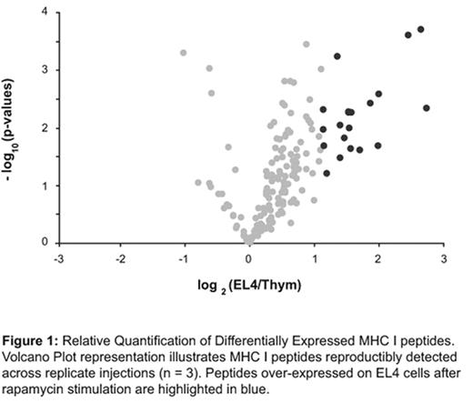
Starting from EL4 thymoma peptide extracts, more than 200 MHC I-associated peptides were sequenced with high confidence level across more than 5500 peptide clusters reproductibly identified through replicate injections (n = 3). Comparison of the sMII of EL4 thymomas before and after rapamycin stimulation revealed that 13% of the MHC I-associated peptides were significant overexpressed (fold change ≥ 2, p-value ≤ 0.05) after mTOR inhibition. Out of the 27 MHC I peptide candidates showing differential expression, 60% of peptide source proteins were linked to cell development and/or proliferation including ITFIFKSL and NAIKNHWNSTM assigned to FK506 binding protein 12 (mTOR) and myeloblastosis oncogene (c-myb) proteins, respectively. These results indicate that cell signalling events have a major impact on the composition of the sMII that can be monitored by analyses of high-throughput MS-based quantitative profiles.
Relative Quantification of Differentially Expressed MHC I peptides. Volcano Plot representation illustrates MHC I peptides reproductibly detected across replicate injections (n=3). Peptides over-expressed on EL4 cells after rapamycin stimulation are highlighted in blue.
Relative Quantification of Differentially Expressed MHC I peptides. Volcano Plot representation illustrates MHC I peptides reproductibly detected across replicate injections (n=3). Peptides over-expressed on EL4 cells after rapamycin stimulation are highlighted in blue.
Author notes
Disclosure: No relevant conflicts of interest to declare.


This feature is available to Subscribers Only
Sign In or Create an Account Close Modal