Abstract
Human T-cell leukemia virus type-1 (HTLV-1) is associated with adult T-cell leukemia (ATL) and neurological syndromes. HTLV-1 encodes the oncoprotein Tax-1, which modulates viral and cellular gene expression leading to T-cell transformation. Guanine nucleotide–binding proteins (G proteins) and G protein–coupled receptors (GPCRs) constitute the largest family of membrane proteins known and are involved in the regulation of most biological functions. Here, we report an interaction between HTLV-1 Tax oncoprotein and the G-protein β subunit. Interestingly, though the G-protein β subunit inhibits Tax-mediated viral transcription, Tax-1 perturbs G-protein β subcellular localization. Functional evidence for these observations was obtained using conditional Tax-1–expressing transformed T-lymphocytes, where Tax expression correlated with activation of the SDF-1/CXCR4 axis. Our data indicated that HTLV-1 developed a strategy based on the activation of the SDF-1/CXCR4 axis in the infected cell; this could have tremendous implications for new therapeutic strategies.
Introduction
Human T-cell leukemia virus type-1 (HTLV-1), the first pathogenic retrovirus discovered in humans 26 years ago,1 is the causative agent of 2 major diseases: a rapidly fatal leukemia designated adult T-cell leukemia (ATL)2 and a neurological degenerative disease known as tropical spastic paraparesis (TSP) or HTLV-1–associated myelopathy (HAM).3 Malignancy develops in approximately 1 in 20 HTLV-1–infected persons after 40 to 50 years of latency.4 The viral transcriptional activator and oncoprotein Tax-1 has been the major focus of scientific investigation because of its numerous and crucial roles in the pathogenesis of HTLV-1–induced diseases (for reviews, see Jeang et al,5 Grassmann et al,6 and Azran et al7 ). The primary role of Tax-1 in the viral life cycle of HTLV-1 is to directly promote viral mRNA synthesis.8 Tax-1 acts through highly conserved 21-bp repeat elements, called Tax-1–responsive elements (TREs), located within the 5′ LTR.9 Tax-1 does not bind DNA directly; rather, it acts through cellular transcription factors, such as cyclic adenosine monophosphate (cAMP) response element-binding (CREB), nuclear factor-κ B (NF-κB), serum responsive factor (SRF), and activator protein 1 (AP-1) transcription factors (for reviews, see Jeang et al,5 Grassmann et al,6 and Azran et al7 ). Tax-1 modulates the expression of an array of cellular genes directly involved in T-cell proliferation, such as interleukin-2 (IL-2) and the α subunit of its receptor (IL-2Rα),10,11 IL-15 and its receptor (IL-15R)12,13 granulocyte macrophage–colony-stimulating factor (GM-CSF)14 and tumor necrosis factor-α (TNF-α).15 Tax-1 also is involved in cell-cycle regulation by direct activation of cyclins D2 and D3 and cyclin kinases CDK4 and CDK6,16-19 by inactivating the cyclin-dependent kinase inhibitor p16,INK4A 20 or by interacting with the human mitotic checkpoint protein HsMAD1.21 HTLV-1 Tax-binding factors also include MEKK1,22 the I-κB kinase,23 or the PCAF protein.24 In fact, protein-protein interactions with cellular factors are crucial for Tax-1 to perturb the regulation of many cellular pathways (for reviews, see Jeang et al,5 Grassmann et al,6 and Azran et al7 ).
The guanine nucleotide (GTP)–binding protein (G-protein) signal transduction network, one of the major information transfer systems, allows the cell to communicate with its surroundings and to participate in a multicellular organization. The minimum components of this system are a 7-transmembrane G-protein–coupled receptor (GPCR), a heterotrimeric complex of G-protein α (Gα) and Gβγ subunits, and an intracellular effector molecule (for a review, see Clapham and Neer25 ). After specific agonist binding, the activated GPCR induces an exchange of GDP to GTP on the Gα subunit and facilitates the dissociation of GTP-bound Gα and Gβγ subunits. GTP-Gα and Gβγ individually are thought to modulate downstream effectors.26 Gβγ interacts with and regulates numerous signaling proteins, including phosphoinositide 3-kinases,27 phospholipases,28 adenylyl cyclases,29 ion channels,30 GPCR kinases,31,32 histone deacetylases,33 and glucocorticoid receptors.34 Most of these downstream Gβγ effectors and the Gα subunit bind to common overlapping domains on the Gβ subunit. Hence, the Gα subunit has been shown to inhibit signal transduction through Gβγ subunits.35
In this study, we report a functional interplay between HTLV-1 Tax oncoprotein and the G-protein signaling pathways. We found that the Gβ subunit is a specific partner of HTLV-1 Tax and that it negatively regulates its transactivation activity over the HTLV-1 viral promoter. Conversely, we also showed that Tax-1 can activate the stromal cell–derived factor (SDF)–1/CXC chemokine receptor 4 (CXCR4) ligand/receptor axis in T-lymphocytes.
Materials and methods
Cell culture
Jurkat and MT4 cells were cultured in RPMI 1640 medium (Sigma, St Louis, MO) supplemented with 10% fetal calf serum, 2 mM glutamine, and antibiotics. The same medium containing 20% fetal calf serum and 40 U/mL recombinant IL-2 was used for the propagation of Tax-transformed T-lymphocytes (Tesi), as previously described.36,37 Suppression of Tax expression in Tesi cells was achieved after a 7-day cultivation period in the presence of 1 μg/mL tetracycline. HeLa and HEK293T cells were cultured in DMEM, as previously described.38
Plasmids
Plasmids pcDNAFlag-Gβ1, pcDNAFlag-Gβ2, pcDNAFlag-Gβ5, pcDNAGαi3, pcDNAGα15, and pcDNAHA-γ4 were obtained from the UMR cDNA Resource Center (University of Missouri, Rolla, MO). Plasmids pYFP-Gβ2, pYFP-Gβ2 (1-100), pYFP-Gβ2 (101-200), and pYFP-Gβ2 (201-340) were kindly provided by Dr Stefan Herlitze (Case Western Reserve University, Cleveland, OH). Expression constructs for GST-Gαi3 and GST-Gα15 fusion proteins were offered by Dr Bradley M. Denker (Harvard Institute of Medicine, Boston, MA). Plasmid pREP9Tat1 contains the first exon of tat under control of an RSV promoter. The pHIV1LTR-LUC contains the HIV-1 LTR cloned into pGL2 basic vector (Promega, Madison, WI). pLTRLuc, pLTR1Luc, pSGTax, GFP-Tax-1, pSGTax1, pCMVTax, pGexTax1, and BC20.2sph (for Tax2B) were previously described.39-41 pGexTax2 was subcloned by polymerase chain reaction (PCR) from the BC20.2sph vector.
GST pull-down assay
The HB101 strain of Escherichia coli was transformed with plasmid pGexTax1, pGexTax2, pGexGαi3, or, as a control, pGex-2T. Production of GST fusion proteins and GST pull-down experiments has been described previously.39
Coimmunoprecipitation
HEK 293T cells were divided and seeded in 10-cm plates at a density of 2 × 10⁶ cells/plate and were transfected the next day with 20 μg plasmid DNA through the calcium phosphate method. Western blotting and immunoprecipitation assays were performed as previously described.38,39 Briefly, cell lysates were immunoprecipitated with anti-Flag (mouse M2; Sigma) or anti-GFP (Santa Cruz Biotechnology, Santa Cruz, CA) agarose-conjugated antibodies or control antibodies. The immunoprecipitates were analyzed by sodium dodecyl sulfate–polyacrylamide gel electrophoresis (SDS-PAGE) and Western blotting using anti-Tax antibody (αTax3, provided by F. Bex, Université Libre de Bruxelles), or anti-GFP antibody (Abcam, Cambridge, United Kingdom).
Intracellular cascade assays
For MAP kinase activation assay, Tesi cells, Tesi-Tax control cells (Tesi cells cultivated for 10 days in the presence of 1 μg/mL tetracycline for Tax suppression), and Tesi-PTX cells (Tesi cells cultivated for 24 hours in the presence of 100 ng/mL pertussis toxin) were stimulated for 5 minutes with 30 ng/mL SDF-1, 30 ng/mL MCP-1, or 50 ng/mL RANTES. Cells were lysed, and ERK1/2 activation was measured by Western blotting using an anti–phospho-p42/44 monoclonal antibody (E10; Cell Signaling Technology, Beverly, MA). For calcium mobilization, Tesi and Tesi-Tax control cells (107 cells/mL in HBSS without phenol red but containing 0.1% BSA) were loaded with 5 μM FURA-2 (Invitrogen, Carlsbad, CA) for 30 minutes at 37°C in the dark. The loaded cells were washed twice, resuspended at 106 cells/mL, and kept for 30 minutes at 4°C in the dark. Ca2+ mobilization in response to 50 nM SDF-1 was measured with the use of a luminescence spectrometer (LS50B; Perkin Elmer, Wellesley, MA) by recording the ratio of fluorescence emitted at 510 nm after sequential excitation at 340 and 380 nm.
Binding and migration assays
Experiments were performed to measure the binding of SDF-1 to membranes from Tesi cells. Samples containing 5 μg membrane proteins, prepared as described,42 were combined with 10 nM 125I-SDF-1 in 100 μL final volume of assay buffer (50 mM HEPES, pH 7.4, 1 mM CaCl2, 5 mM MgCl2, 0.5% BSA), followed by incubation for 90 minutes at 25°C. Nonspecific binding was measured in the presence of a 100-fold excess of unlabeled SDF-1. Bound SDF-1 was separated by filtration through GF/B filters presoaked in 0.5% polyethylenimine (Sigma-Aldrich, Poole, United Kingdom). Filters were counted in a β scintillation counter.
For T-cell migration assays, Transwell culture chambers (5-μm pore size; Costar, Cambridge, MA) were used. The lower wells were filled with 500 μL medium (RPMI 1640 containing 1% FCS) containing 30 ng/mL SDF-1, 50 ng/mL RANTES, or 30 ng/mL MCP-1. Tesi cells (105 cells) suspended in 100 μL medium were loaded into the upper wells. After incubation for 2 hours at 37°C, the cells that had migrated to the lower wells were counted and shown as percentages of the input cells.
Flow cytometry
For quantification of CXCR4 receptor expression on the cell surface, Tesi cells were incubated with a polyclonal rabbit anti-CXCR4 antibody (Calbiochem, San Diego, CA) followed by a PE-conjugated antirabbit secondary antibody. Flow cytometry analyses were performed with a FACScan (BD Biosciences, San Jose, CA).
Confocal microscopy
Two micrograms plasmids (CMVTax, pTax2, pSGTax, pcDNAFlag-Gβ1, pcDNAFlag-Gβ2, and pcDNAFlag-Gβ5) were transfected into HeLa cells with the use of Genejammer (Stratagene, La Jolla, CA). Twenty-four hours after transfection, cells were fixed in 3.7% formaldehyde (20 minutes at 4°C), permeabilized with 0.1% Nonidet P40 (10 minutes), and incubated with anti-Flag antibodies (Sigma) or anti-Tax antibodies (αTax3 for HTLV-1 Tax, rabbit αTax2 for HTLV-2 Tax,40 or 5A5 for BLV Tax) and then with Alexa 546 or fluorescein (FITC)–tagged immunoglobulin conjugates (Molecular Probes, Eugene, OR). After nuclear staining with TOPRO-3 and fixation with mounting medium (Prolong Antifade kit; Molecular Probes), the cells were analyzed under a fluorescence confocal microscope (40 ×/0.55 NA oil objective, Axiovert 200 with LSM 510 version 3.0; Carl Zeiss, Oberkochen, Germany).
Luciferase assays
Ten micrograms reporter plasmids pLTR1-Luc, pκB-Luc, pHIV1-Luc, or pFR-Luc and different amounts of effector vectors (pCMVTax, pFA-CREB, pFA-cJun, pFA-Elk1 pcDNAFlag-Gβ2, and pcDNAHA-γ4) were transfected into 107 Jurkat cells using the DEAE dextran method. Forty-eight hours after transfection, cells were washed 3 times with PBS and lysed, and luciferase activities were determined using the Promega luciferase assay kit according to the manufacturer's instructions and normalized to protein content. Luciferase assays also were performed with lysates from HeLa cells transfected with 3 μg reporter construct and indicated effector plasmids.
Figure S1 (available on the Blood website; see the Supplemental Figures link at the top of the online article) shows pull-down of the Gβ but not the Gα subunit by GST-Tax-1. Figure S2 shows colocalization between Gβ5 and HTLV-1 Tax. Figure S3 displays colocalization between Gβ2 and BLV Tax (A) or HTLV-2 Tax (B). Figure S4 shows activation of the SDF-1/CXCR4 pathway in Gβ2-overexpressing Tesi cells (A-C) and activation of MT4 cells after treatment with the indicated chemokines (D-E).
Results
Gβ interacts with Tax oncoproteins
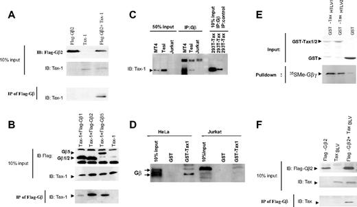
To gain insight into the molecular mechanisms that govern Tax functions, we performed yeast 2-hybrid screening and identified several Tax interactors.39 Two of these clones appeared to be related to the human Gβ2 coding sequence (accession no. XP005013). To confirm the binding specificity of Tax-1 to Gβ2, HEK293T cells were transfected with expression constructs for Tax-1 and Flag-tagged Gβ2. After cell lysis, Flag-Gβ2 was immunoprecipitated with an anti-Flag antibody. Immunoprecipitates then were resolved by SDS-PAGE and analyzed by Western blotting with an anti–Tax-1 antibody. As shown in Figure 1A, Tax-1 specifically copurified with Flag-Gβ2 when both proteins were expressed together. As control, no Tax-1 was detected in immunoprecipitates from cells expressing either Tax-1 or Flag-Gβ2 alone (Figure 1A, IP).
Tax oncoproteins interact with Gβ subunits. (A-B). HEK 293T cells were transfected with Flag-Gβ1, Flag-Gβ2, Flag-Gβ5, and HTLV-1 Tax expression constructs, as indicated. Forty-eight hours after transfection, cells were lysed and protein expression was verified by immunoblotting using anti–Tax-1 and anti-Flag–specific antibodies. Lysates were immunoprecipitated using the M2 Flag–specific antibody. Immunoprecipitated proteins were analyzed by SDS-PAGE and immunoblotting using an anti–HTLV-1 Tax antibody. (C) Lysates from MT4, Tesi, Jurkat, or Tax-1–transfected 293T cells were immunoprecipitated using an anti-Gβ or a control antibody. Immunoprecipitated proteins were analyzed by Western blot using an anti–HTLV-1 Tax antibody. (D) Lysates from HeLa or Jurkat cells were mixed with equal amounts of GST-TaxHTLV-1 or GST alone bound to glutathione-Sepharose beads. After incubation, the precipitated proteins were analyzed by Western blot using an anti-Gβ antibody. (E) The Gβ2γ4 complex was synthesized in vitro using rabbit reticulocyte lysates in the presence of 35S-labeled methionine and cysteine. Lysates were mixed with equal amounts of GST-TaxHTLV-1, GST-TaxHTLV-2 fusion proteins, or GST alone bound to glutathione-Sepharose beads. After incubation, the protein complexes were separated by SDS-PAGE, and 35S- Gβ2γ4 was visualized by autoradiography. (F) HEK 293T cells were transfected with Flag-Gβ2 and BLV Tax expression constructs, as indicated. Forty-eight hours after transfection, cells were lysed and protein expression was verified by immunoblotting using anti-Tax and anti-Flag–specific antibodies. Lysates were immunoprecipitated using the M2 Flag-specific antibody. Immunoprecipitated proteins were analyzed by SDS-PAGE and immunoblotting using an anti–BLV Tax antibody.
Tax oncoproteins interact with Gβ subunits. (A-B). HEK 293T cells were transfected with Flag-Gβ1, Flag-Gβ2, Flag-Gβ5, and HTLV-1 Tax expression constructs, as indicated. Forty-eight hours after transfection, cells were lysed and protein expression was verified by immunoblotting using anti–Tax-1 and anti-Flag–specific antibodies. Lysates were immunoprecipitated using the M2 Flag–specific antibody. Immunoprecipitated proteins were analyzed by SDS-PAGE and immunoblotting using an anti–HTLV-1 Tax antibody. (C) Lysates from MT4, Tesi, Jurkat, or Tax-1–transfected 293T cells were immunoprecipitated using an anti-Gβ or a control antibody. Immunoprecipitated proteins were analyzed by Western blot using an anti–HTLV-1 Tax antibody. (D) Lysates from HeLa or Jurkat cells were mixed with equal amounts of GST-TaxHTLV-1 or GST alone bound to glutathione-Sepharose beads. After incubation, the precipitated proteins were analyzed by Western blot using an anti-Gβ antibody. (E) The Gβ2γ4 complex was synthesized in vitro using rabbit reticulocyte lysates in the presence of 35S-labeled methionine and cysteine. Lysates were mixed with equal amounts of GST-TaxHTLV-1, GST-TaxHTLV-2 fusion proteins, or GST alone bound to glutathione-Sepharose beads. After incubation, the protein complexes were separated by SDS-PAGE, and 35S- Gβ2γ4 was visualized by autoradiography. (F) HEK 293T cells were transfected with Flag-Gβ2 and BLV Tax expression constructs, as indicated. Forty-eight hours after transfection, cells were lysed and protein expression was verified by immunoblotting using anti-Tax and anti-Flag–specific antibodies. Lysates were immunoprecipitated using the M2 Flag-specific antibody. Immunoprecipitated proteins were analyzed by SDS-PAGE and immunoblotting using an anti–BLV Tax antibody.
To generalize our findings, we then tested 2 other Gβ subunits, Gβ1 and Gβ5, for their ability to interact with Tax-1. As with Gβ2, coimmunoprecipitation experiments revealed a robust interaction of Tax-1 with Gβ5 and, to a lesser extent, with Gβ1 (Figure 1B).
We next analyzed the ability of endogenous Gβ proteins to interact with Tax in an HTLV-1–transformed cell line (MT4) and a Tax-transformed T-cell line (Tesi). To this end, MT4, Tesi, or Jurkat cells were lysed, and Gβ proteins were immunoprecipitated with an anti-Gβ antibody. Immunoprecipitates were then analyzed by Western blot with an anti–Tax-1 antibody. As shown on Figure 1C, Tax-1 coimmunoprecipitated with Gβ proteins in MT4 and Tesi cells. We also used GST–Tax-1 fusion proteins bound to Sepharose beads to precipitate endogenous Gβ subunits from HeLa and Jurkat cells. As shown on Figure 1D, Gβ proteins from HeLa or Jurkat cells coprecipitated with GST–Tax-1 but not with GST alone. These data demonstrated that physiological levels of Gβ subunits also interacted with HTLV-1 Tax.
G-protein complexes, made up of Gβγ heterotrimers, dissociate into Gα and Gβγ that separately activate or inhibit intracellular signaling molecules. To determine whether Tax-1 also could bind to the Gα subunit, we used G-protein subunits from rabbit reticulocyte lysate in vitro transcription/translation systems, which previously have been shown to be convenient for in vitro G-protein subunit assembly.43 Gβ2γ4 heterodimers and Gα15 proteins were incubated with GST–Tax-1 bound to Sepharose beads. After extensive washes, proteins were resolved by SDS-PAGE stained with Coomassie blue (Figure S1A, lower panel) and visualized by autoradiography. As shown on Figure S1A (middle panel), Gβ2γ4 heterodimer, but not Gα15, was pulled down by GST–Tax-1. Next, constant amounts of Gβ2γ4 heterodimer were incubated with increasing amounts of recombinant Gα15 and incubated together in a buffer favoring complex formation. In vitro reconstituted G-protein complexes then were pulled down by GST–Tax-1. As expected, Gβ2γ4 was pulled down specifically by GST–Tax-1. However, the association between Gβ2γ4 and Tax-1 was inhibited by increasing amounts of Gα (Figure S1B). Interestingly, Gα was not present in the GST–Tax-1 pull-down reaction (Figure S1B). These data suggested that Gα does not bind to Tax-1 but rather competes with the viral protein for association with Gβγ.
We next tested whether Gβ could associate with Tax-2B protein from HTLV-2. To this end, we used the GST pull-down assay and found that significant amounts of the in vitro–translated Gβ2 protein were pulled down by GST–TaxHTLV-1 and GST–TaxHTLV-2 fusion proteins compared with the GST control (Figure 1E). In addition, the Tax protein from bovine leukemia virus (BLV) also was associated with Flag-tagged Gβ2 in 293T cell lysates (Figure 1F). These data strongly suggested that binding to Gβ is a general property of deltaretrovirus-encoded Tax oncoproteins.
Tax-1 interacts with Gβ through its WD repeat domains
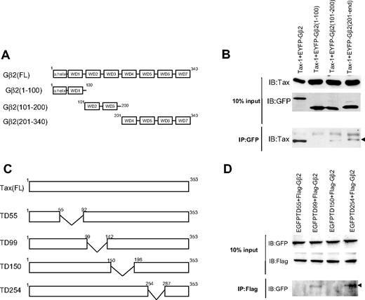
The crystal structure of Gβ revealed that the protein is made of 2 distinct regions: an amino-terminal α-helical segment and 7-repeat bladelike structures called WD repeats.44 To identify the region of Gβ required for Tax-1–binding, deletion mutants of Gβ2 corresponding to its amino-terminal region—including WD1 (aa 1-100) and the regions encompassing WD2 and WD3 (aa 101-200) and WD4 to WD7 (aa 201-340)—were tested in coimmunoprecipitation experiments (Figure 2A). Full-length Gβ2 and, to a lesser extent, polypeptides Gβ2 (101-200) and Gβ2 (201-340) interacted with Tax-1. However, the N-terminal α-helix region of Gβ did not interact with Tax (Figure 2B). These observations indicated that the Gβ WD repeat structures are essential for association with Tax-1.
Tax-1 and Gβ2 interaction domains. (A) Schematic representation of the Gβ2 subunit and deletion mutants. Gβ consists of an amino-terminal α-helical segment followed by 7-repeat bladelike structures called WD repeats. (B) HEK 293T cells were transfected with EYFP-Gβ2 (Fl), EYFP-Gβ2 (aa 1-100), EYFP-Gβ2 (aa 101-200), EYFP-Gβ2 (aa 201-340), and HTLV-1Tax expression constructs, as indicated. Forty-eight hours after transfection, cells were lysed and protein expression was verified by immunoblotting using anti-GFP and anti–Tax-1–specific antibodies. Lysates were immunoprecipitated with a GFP-specific antibody. Immunoprecipitated proteins were analyzed by SDS-PAGE and immunoblotting using an anti–Tax-1 antibody. (C) Schematic representation HTLV-1 Tax deletion mutants. Deleted regions are indicated by the wide V-like symbols. (D) HEK 293T cells were transfected with EGFP-TD55, EGFP-TD99, EGFP-TD150, EGFP-TD254, and Flag-Gβ2–expressing constructs, as indicated. Forty-eight hours after transfection, cells were lysed and protein expression was verified by immunoblotting using anti–Flag M2 and anti–GFP-specific antibodies. Lysates were immunoprecipitated with M2 Flag–specific antibody. Immunoprecipitated proteins were analyzed by SDS-PAGE and immunoblotting with anti–GFP antibody. Arrows indicate immunoprecipitated protein bands.
Tax-1 and Gβ2 interaction domains. (A) Schematic representation of the Gβ2 subunit and deletion mutants. Gβ consists of an amino-terminal α-helical segment followed by 7-repeat bladelike structures called WD repeats. (B) HEK 293T cells were transfected with EYFP-Gβ2 (Fl), EYFP-Gβ2 (aa 1-100), EYFP-Gβ2 (aa 101-200), EYFP-Gβ2 (aa 201-340), and HTLV-1Tax expression constructs, as indicated. Forty-eight hours after transfection, cells were lysed and protein expression was verified by immunoblotting using anti-GFP and anti–Tax-1–specific antibodies. Lysates were immunoprecipitated with a GFP-specific antibody. Immunoprecipitated proteins were analyzed by SDS-PAGE and immunoblotting using an anti–Tax-1 antibody. (C) Schematic representation HTLV-1 Tax deletion mutants. Deleted regions are indicated by the wide V-like symbols. (D) HEK 293T cells were transfected with EGFP-TD55, EGFP-TD99, EGFP-TD150, EGFP-TD254, and Flag-Gβ2–expressing constructs, as indicated. Forty-eight hours after transfection, cells were lysed and protein expression was verified by immunoblotting using anti–Flag M2 and anti–GFP-specific antibodies. Lysates were immunoprecipitated with M2 Flag–specific antibody. Immunoprecipitated proteins were analyzed by SDS-PAGE and immunoblotting with anti–GFP antibody. Arrows indicate immunoprecipitated protein bands.
Tax-1 has 2 sites of interaction with Gβ
To map the Tax-1 domains involved in Gβ-binding, HEK293T cells were cotransfected with Flag-Gβ2 and full-length GFP-tagged Tax-1 (GFP-Tax-1) or 4 different GFP-tagged Tax-1–deletion mutants (Figure 2C).45 Flag-Gβ2 was immunoprecipitated from transfected cell lysates, and coimmunoprecipitated GFP–Tax-1 proteins were revealed by immunoblotting. As shown in Figure 2D, the deletion mutants TD99 and TD254 associated with Gβ2, whereas the mutants TD55 and TD150 did not show any detectable binding. These results indicated that 2 distinct regions of Tax-1, amino acids 55-92 and a central domain (aa 150-198), are involved in Gβ-binding.
Gβ subunits colocalize with HTLV-1 Tax
The heterotrimeric G-protein complexes, through a prenylated cysteine residue on its Gγ subunit, are anchored at the inner side of the plasma membrane.25 Tax-1 has been shown to localize mainly in the nucleus because of the nuclear localization signal (NLS) present at its N-terminal region.46 Tax-1 also has a nuclear export signal (NES) and has been shown to accumulate in the cytoplasm, where it localizes to different organelles, including the endoplasmic reticulum and the Golgi complex.47 Phosphorylation, ubiquitination, and sumoylation also play roles in the subcellular localization of Tax.48-51
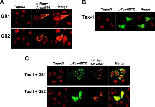
To examine the subcellular localization of Gβ and Tax-1 proteins, HeLa cells were transfected with constructs expressing Tax-1 or Flag-tagged versions of Gβ1 and Gβ2. The cells then were stained with an anti-Flag antibody followed by Alexa 546–conjugated secondary antibody (for Gβ subunits) or an anti–Tax-1 antibody followed by a fluorescein-conjugated secondary antibody. As expected, most Gβ subunit proteins were localized to the cytoplasm, with small fractions also detected in the nucleus (stained by TOPRO-3) and at the plasma membrane (Figure 3A). Tax predominantly localized to the nucleus, but substantial amounts of the protein also were found in the cytoplasm (Figure 3B). In addition, Tax-1 displayed a speckled pattern known as Tax-speckled structures (TSSs), which have been described as Tax-1 transcriptional regulation hot spots.52
Colocalization of Gβ subunits and Tax-1. (A-B) HeLa cells were transfected with Flag- Gβ1, Flag- Gβ2, or HTLV-1 Tax–expressing constructs. Twenty-four hours after transfection, cells were fixed, permeabilized, and stained with anti-Flag or anti–Tax-1 antibodies and Alexa 546 or fluorescein-conjugated secondary antibodies. Cells then were labeled with TOPRO-3 and analyzed under a confocal microscope. (C) HeLa cells were cotransfected with Flag-Gβ1+ HTLV-1 Tax– and Flag-Gβ2+ HTLV-1 Tax–expressing constructs. Twenty-four hours after transfection, cells were fixed, permeabilized, and stained with anti-Flag and anti–Tax-1 antibodies followed by Alexa 546– and fluorescein-conjugated secondary antibodies. Cells then were labeled with TOPRO-3 and analyzed under a confocal microscope.
Colocalization of Gβ subunits and Tax-1. (A-B) HeLa cells were transfected with Flag- Gβ1, Flag- Gβ2, or HTLV-1 Tax–expressing constructs. Twenty-four hours after transfection, cells were fixed, permeabilized, and stained with anti-Flag or anti–Tax-1 antibodies and Alexa 546 or fluorescein-conjugated secondary antibodies. Cells then were labeled with TOPRO-3 and analyzed under a confocal microscope. (C) HeLa cells were cotransfected with Flag-Gβ1+ HTLV-1 Tax– and Flag-Gβ2+ HTLV-1 Tax–expressing constructs. Twenty-four hours after transfection, cells were fixed, permeabilized, and stained with anti-Flag and anti–Tax-1 antibodies followed by Alexa 546– and fluorescein-conjugated secondary antibodies. Cells then were labeled with TOPRO-3 and analyzed under a confocal microscope.
We next assessed the localization of coexpressed Gβ and Tax-1 proteins by transfecting HeLa cells with equal amounts of Flag-Gβ and Tax-1 expression constructs (1 μg each vector) (Figures 3C, S2). In the presence of Gβ2, the subcellular localization of Tax-1 was identical to that seen with Tax-1 alone (compare αTax+FITC staining in Figure 3B and αTax+FITC staining in Figure 3C, Tax-1+Gβ2). Interestingly, in the presence of Gβ1, Tax-1 was localized exclusively to the cytoplasm (Figure 3C, αTax+FITC staining, Tax-1+Gβ1). Similar colocalization of Tax-1 was observed with Gβ5 (Figure S2; Tax-1+Gβ5). These experiments revealed that coexpression of Tax-1 dramatically altered the subcellular localization of Gβ proteins. In the presence of Tax-1, individual Gβ subunits were targeted to the TSS and completely lost their diffuse distribution in the cytoplasm and the plasma membrane (compare Figure 3C, αFlag+Alexa 546 staining with Figure 3A, αFlag+Alexa 546 staining). In addition, the Gβ subunits also colocalized with HTLV-2 and BLV Tax oncoproteins (Figure S3 and data not shown). We concluded that coexpression of Tax proteins and Gβ subunits changed their respective subcellular localization, suggesting that their association might have affected their individual functions.
Gβ regulates the Tax-1 transactivation of the HTLV-1 LTR promoter
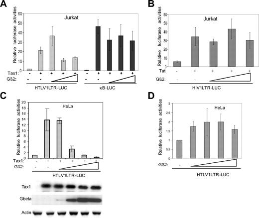
The transforming potential of HTLV-1 Tax has been attributed mainly to its ability to activate gene expression through the CREB/ATF and NF-κB pathways.5 To examine the functional consequences of association with Gβ proteins, we investigated Tax-1–induced transcriptional activation of the CREB/ATF and the NF-κB responsive elements in the presence of Gβ2. Jurkat cells were cotransfected with expression vectors for Tax-1 and Gβ2, together with the HTLV-1 LTR promoter (to measure the activation of the CREB/ATF pathway) or the NF-κB reporter constructs. As determined by luciferase reporter assays, low doses of Gβ2 significantly increased Tax-1–transactivation activity of the LTR promoter (Figure 4A). In contrast, overexpression of Gβ2 resulted in a 50% inhibition of Tax-1–dependent LTR promoter activation (Figure 4A, HTLV1LTR-LUC). Gβ2 did not significantly affect NF-κB activation by Tax-1 (Figure 4A, pκB-Luc) or HIV-1 LTR Tat–dependent activation (Figure 4B). As shown by Western blotting after similar cotransfection experiments in HeLa cells, the inhibition of transactivation was not caused by a decrease in the levels of Tax-1 protein (Figure 4C, lower panels). Finally, in the absence of Tax-1, Gβ2 did not significantly influence the LTR promoter (Figure 4D). Altogether, these data indicated that Gβ can interrupt Tax-1–dependent activation of the CREB/ATF pathway.
Gβ2 subunit inhibits Tax-1 transactivation activity. (A-B) Ten micrograms reporter plasmids pLTR1-Luc, pκB-Luc, or pHIV1-Luc and different amounts of effector vectors (1 μg pCMVTax-1 or pREP9Tat1 and 10, 100, or 1000 ng pcDNAFlag-Gβ2) were transfected into 107 Jurkat cells according to the DEAE dextran method. Forty-eight hours after transfection, cells were lysed and luciferase activities were determined. Luciferase data were normalized to protein content, and the data are reported as mean ± SD of 3 independent experiments in triplicate. (C-D) HeLa cells (3 × 10⁵) were cotransfected with 3 μg reporter construct pLTR1-Luc, increasing amounts of pcDNAFlag-Gβ2 (10, 100, 500, and 1000 ng) and with or without HTLV-1 Tax–expressing construct (300 ng). Twenty-four hours after transfection, cells were lysed and luciferase activities were determined. An aliquot from each lysate was separated by SDS-PAGE, and Western blot was performed using anti–Tax-1, anti-Flag, and antiactin antibodies.
Gβ2 subunit inhibits Tax-1 transactivation activity. (A-B) Ten micrograms reporter plasmids pLTR1-Luc, pκB-Luc, or pHIV1-Luc and different amounts of effector vectors (1 μg pCMVTax-1 or pREP9Tat1 and 10, 100, or 1000 ng pcDNAFlag-Gβ2) were transfected into 107 Jurkat cells according to the DEAE dextran method. Forty-eight hours after transfection, cells were lysed and luciferase activities were determined. Luciferase data were normalized to protein content, and the data are reported as mean ± SD of 3 independent experiments in triplicate. (C-D) HeLa cells (3 × 10⁵) were cotransfected with 3 μg reporter construct pLTR1-Luc, increasing amounts of pcDNAFlag-Gβ2 (10, 100, 500, and 1000 ng) and with or without HTLV-1 Tax–expressing construct (300 ng). Twenty-four hours after transfection, cells were lysed and luciferase activities were determined. An aliquot from each lysate was separated by SDS-PAGE, and Western blot was performed using anti–Tax-1, anti-Flag, and antiactin antibodies.
Tax-1 expression induces CXCR4 receptor activation
In vivo, heterotrimeric G proteins transduce an extracellular signal through GPCR. Activated GPCR facilitates the exchange of GDP for GTP in the Gα subunit and subsequent signal transmission to downstream effectors. Among GPCRs, chemokine receptors function in immune and inflammatory responses by regulating the activation and migration of blood cells.53,54 In the past 10 years, numerous studies in human retrovirology have focused on CXC chemokine receptor 4 (CXCR4) and CC chemokine receptor 5 (CCR5) because they are coreceptors for the entry of HIV into cells.55 Therefore, we used these 2 GPCR chemokine receptors as a means to demonstrate the functional relevance of the Tax-1–G-protein interaction.
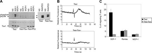
We used immortalized T-lymphocytes that conditionally expressed Tax-1 under the control of the tetracycline operon (Tesi cells).36,37 The Tesi-Tax control cells were generated by suppressing Tax-1 expression in Tesi cells with a 7-day period of propagation in the presence of tetracycline.36,37 Immunoblot analysis with an anti–Tax-1 monoclonal antibody demonstrated the suppression of Tax-1 protein expression in Tesi-Tax control cells (Figure 5A, bottom; compare Tesi and Tesi-Tax control lanes). Tesi cells then were treated with stromal cell-derived factor-1 (SDF-1; 30 ng/mL), which specifically binds to the CXCR4 receptor; monocyte chemoattractant protein-1 (MCP-1; 30 ng/mL), a ligand for CCR1 and CCR2 receptors; or regulated on activation normal T-cell expressed and secreted (RANTES; 50 ng/mL), which binds to CCR1, CCR3, and CCR5. Cells were evaluated for cellular activation. Given that these latter chemokines have been shown to activate the extracellular signal-regulated kinase (ERK) 1/2 mitogen-activated protein (MAP) kinase pathway, cell lysates were tested for the phosphorylation of ERK1/2 using a phosphospecific anti-p42/44 antibody. As shown in Figure 5A (top), stimulation of Tesi cells by SDF-1 resulted in the phosphorylation of ERK1/2 (Figure 5A; Tesi, SDF-1). The SDF-1 activation of ERK1/2 was both Tax-1 and Gi dependent because Tesi-Tax control cells, which do not express Tax-1, or Tesi-PTX cells, which had been treated with pertussis toxin for Gi inactivation, were not stimulated by SDF-1 (Figure 5A; SDF-1, Tesi-Tax control and Tesi-PTX). In contrast, MCP-1 did not activate chemokine receptors on Tesi cells (Figure 5A; MCP-1), whereas the phosphorylation of ERK1/2 after treatment of Tesi cells with RANTES was not Tax-1 dependent (Figure 5A, right; RANTES).
Activation of the SDF-1/CXCR4 pathway in Tesi cells. (A) Tesi, Tesi-Tax control (Tesi cells cultivated for 10 days in the presence of 1 μg tetracycline for Tax-1 suppression), and Tesi-PTX (Tesi cells cultivated for 24 hours in the presence of 100 ng/mL pertussis toxin) cells were stimulated for 5 minutes with 30 ng/mL SDF-1, 30 ng/mL MCP-1, or 50 ng/mL RANTES. Cells were lysed, and ERK1/2 activation was measured by Western blotting with an anti–phospho-p42/44 monoclonal antibody (top). The same blot was stripped, and immunoblotting was performed with an anti–Tax-1 antibody (bottom). Arrows indicate p42, p44, and protein bands. (B) Tesi and Tesi-Tax control cells were loaded with 5 μM Fura-2 for 30 minutes at 37°C in the dark. Loaded cells were washed twice, resuspended at 106 cells/mL, and kept for 30 minutes at 4°C in the dark. Ca2+ mobilization in response to 50 nM SDF-1 was measured using a luminescence spectrometer (LS50B; Perkin Elmer) by recording the ratio of fluorescence emitted at 510 nm after sequential excitation at 340 and 380 nm. Arrows indicate the injection of SDF-1. (C) Tesi and Tesi-Tax control migration assays were performed using Transwell culture chambers (5-μm pore size). Lower wells were filled with 500 μL medium (RPMI 1640, 1% FCS) containing 30 ng/mL SDF-1, 50 ng/mL RANTES, or 30 ng/mL MCP-1. Tesi or Tesi-Tax control cells (105 cells) suspended in 100 μL medium were loaded into the upper wells. After incubation for 2 hours at 37°C, the cells that had migrated to the lower wells were counted and are shown as percentages of the input cells. Data are reported as mean ± SD of 3 independent experiments in triplicate.
Activation of the SDF-1/CXCR4 pathway in Tesi cells. (A) Tesi, Tesi-Tax control (Tesi cells cultivated for 10 days in the presence of 1 μg tetracycline for Tax-1 suppression), and Tesi-PTX (Tesi cells cultivated for 24 hours in the presence of 100 ng/mL pertussis toxin) cells were stimulated for 5 minutes with 30 ng/mL SDF-1, 30 ng/mL MCP-1, or 50 ng/mL RANTES. Cells were lysed, and ERK1/2 activation was measured by Western blotting with an anti–phospho-p42/44 monoclonal antibody (top). The same blot was stripped, and immunoblotting was performed with an anti–Tax-1 antibody (bottom). Arrows indicate p42, p44, and protein bands. (B) Tesi and Tesi-Tax control cells were loaded with 5 μM Fura-2 for 30 minutes at 37°C in the dark. Loaded cells were washed twice, resuspended at 106 cells/mL, and kept for 30 minutes at 4°C in the dark. Ca2+ mobilization in response to 50 nM SDF-1 was measured using a luminescence spectrometer (LS50B; Perkin Elmer) by recording the ratio of fluorescence emitted at 510 nm after sequential excitation at 340 and 380 nm. Arrows indicate the injection of SDF-1. (C) Tesi and Tesi-Tax control migration assays were performed using Transwell culture chambers (5-μm pore size). Lower wells were filled with 500 μL medium (RPMI 1640, 1% FCS) containing 30 ng/mL SDF-1, 50 ng/mL RANTES, or 30 ng/mL MCP-1. Tesi or Tesi-Tax control cells (105 cells) suspended in 100 μL medium were loaded into the upper wells. After incubation for 2 hours at 37°C, the cells that had migrated to the lower wells were counted and are shown as percentages of the input cells. Data are reported as mean ± SD of 3 independent experiments in triplicate.
To confirm the role of Tax-1 in the activation of the SDF-1/CXCR4 axis, we analyzed calcium signaling in Tesi compared with Tesi-Tax control cells. To this end, cells were loaded with FURA-2, and calcium mobilization in response to SDF-1 was measured. As shown in Figure 5B, calcium signaling was activated by SDF-1 in Tesi but not in Tesi-Tax control cells, confirming the role of Tax-1 in the regulation of SDF-1/CXCR4 intracellular signaling. Chemotaxis of Tax-1–transformed T cells in response to SDF-1 also was examined using an in vitro migration assay. As shown in Figure 5C, migration of Tax-1–expressing Tesi cells toward SDF-1 was 12-fold more efficient than that of Tesi-Tax control cells. In addition, specific intracellular cascade activation and migration toward SDF-1 were confirmed with the MT4 HTLV-1–transformed cell line (Figures S4E-F).
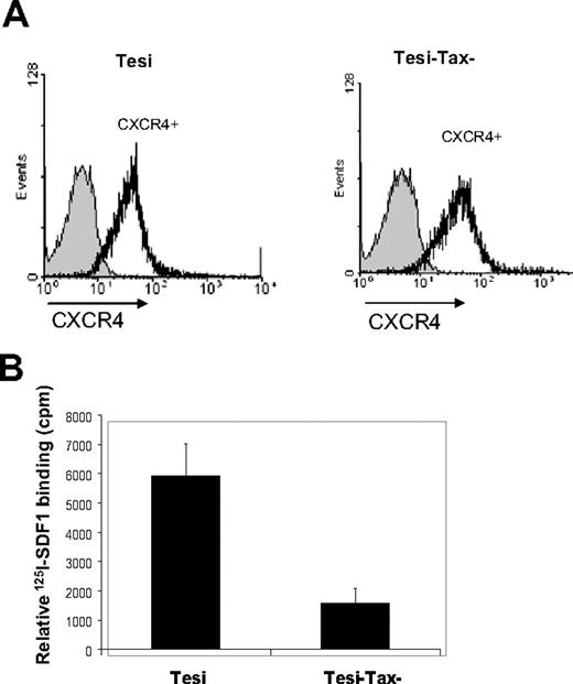
Tax-1 regulates the expression of several cellular genes involved in cellular activation, proliferation, and transformation. Furthermore, Tax-1 has been shown to be a relatively weak activator of the CXCR4 promoter.56 Therefore, we analyzed the surface expression of CXCR4 on Tesi and Tesi-Tax control lymphocytes by flow cytometry. Results presented in Figure 6A showed that surface CXCR4 receptor expression was similar between the Tesi and Tesi-Tax control cells. This demonstrated that the differences in intracellular cascade activation between Tesi and Tesi-Tax control cells were not caused by altered expression of the CXCR4 receptor.
Surface expression of CXCR4 on Tesi and Tesi-Tax control cells and binding properties of CXCR4 in the presence of Tax. (A) Tesi and Tesi-Tax control cells were incubated with a polyclonal rabbit anti-CXCR4 antibody followed by a PE-conjugated anti–rabbit secondary antibody. Flow cytometry analyses were performed with a FACScan. (B) Samples containing 5 μg membrane proteins from Tesi or Tesi-Tax control cells and 10 nM of 125I-SDF-1 in 100 μL final volume of assay buffer (50 mM HEPES, pH 7.4, 1 mM CaCl2, 5 mM MgCl2, and 0.5% BSA) were incubated for 90 minutes at 25°C. Bound SDF-1 was separated by filtration through GF/B filters presoaked in 0.5% polyethylenimine. Filters were counted in a Beckman β scintillation counter. Relative 125I-SDF-1 binding was calculated by subtracting nonspecific binding measured in the presence of a 100-fold excess of unlabeled SDF-1 from total 125I-SDF-1–binding data. Data are reported as the mean ± SD of 2 independent experiments in triplicate.
Surface expression of CXCR4 on Tesi and Tesi-Tax control cells and binding properties of CXCR4 in the presence of Tax. (A) Tesi and Tesi-Tax control cells were incubated with a polyclonal rabbit anti-CXCR4 antibody followed by a PE-conjugated anti–rabbit secondary antibody. Flow cytometry analyses were performed with a FACScan. (B) Samples containing 5 μg membrane proteins from Tesi or Tesi-Tax control cells and 10 nM of 125I-SDF-1 in 100 μL final volume of assay buffer (50 mM HEPES, pH 7.4, 1 mM CaCl2, 5 mM MgCl2, and 0.5% BSA) were incubated for 90 minutes at 25°C. Bound SDF-1 was separated by filtration through GF/B filters presoaked in 0.5% polyethylenimine. Filters were counted in a Beckman β scintillation counter. Relative 125I-SDF-1 binding was calculated by subtracting nonspecific binding measured in the presence of a 100-fold excess of unlabeled SDF-1 from total 125I-SDF-1–binding data. Data are reported as the mean ± SD of 2 independent experiments in triplicate.
Finally, we tested whether Tax-1 could affect the binding properties of the CXCR4 receptor. For this purpose, we compared the binding efficiency of 125I-SDF-1 to Tesi compared with Tesi-Tax control cell membrane proteins. Greater amounts of radiolabeled 125I-SDF-1 were bound specifically to Tesi membranes, suggesting that Tax-1 directly favors the SDF-1/CXCR4 interaction (Figure 6B). Together, these data indicate that Tax-1 expression in immortalized T-cell lines specifically regulates the activation of the SDF-1/CXCR4 ligand/receptor axis.
Discussion
In this study, we characterized a functional interaction between the HTLV-1 Tax oncoprotein and the G-protein signaling pathway.
The deltaretroviridae subgroup of retroviruses comprises bovine leukemia virus (BLV) and 3 types of primate T-cell leukemia viruses (PTLVs) namely, HTLV-1/STLV-1, HTLV-2/STLV-2, and HTLV-3/STLV-3.57,58 These viruses share similar genomic organization, and their tax genes seem to play a central role in the induced diseases. Indeed, studies using transgenic mice with Tax-1 expression restricted to developing thymocytes recently demonstrated that the TAX1 gene alone is able to induce leukemia and lymphoma identical to ATLL in humans.59 The transformation capacity of Tax-1 likely results from its interaction with numerous cellular proteins and the regulation of several cellular signaling and transcriptional control pathways.
We previously identified the Gβ2 subunit using a yeast 2-hybrid screen for Tax interactors.39 In this study, we used coimmunoprecipitation and GST pull-down assays to confirm the interactions and to extend our observations to HTLV-2 and BLV Tax oncoproteins (Figure 1). It appears that HTLV-1 and -2 and BLV Tax oncoproteins interact with the Gβ2 subunit. This observation suggests that a common mechanism is used by deltaretroviruses to subvert G-protein signaling. Furthermore, we showed that the Gβ subunit targets Tax-1 on the same domain (aa 55-92; Figure 2) required for p300/CBP-binding60 and suppresses the transcriptional activity of the Tax-1 protein on the HTLV-1 LTR promoter without affecting its NF-κB pathway regulation (Figure 4A). We speculate that the Gβ subunit interacts with Tax-1 and interferes with the recruitment of CREB and p300/CBP, resulting in the inhibition of viral mRNA synthesis.
Five known human Gβ subunit genes25,61 may form potential combinations with at least 12 Gγ subunits.62 The amino acid sequences of Gβ1, Gβ2, Gβ3, and Gβ4 are 80% to 90% identical, whereas Gβ5 has only 54% identity with other Gβ subunits.63,64 In addition, all Gβ subunits are composed of an α helix amino-terminal domain and the WD40 repeats. Thus, most Gβγ pairs exhibit little difference in receptor coupling and downstream effector activation. However, some examples support the in vivo specificity of Gβγ combinations in signaling pathway regulation.62,65,66 Our results indicated that Tax-1 interacts with Gβ1, Gβ2, and Gβ5 (Figure 1B) and that the strength of the Tax-1/Gβ interaction is related directly to the presence of WD repeat structures (Figure 2A-B). Together our observations suggest that Tax-1 may target all Gβγ pairs through the conserved WD40 repeats in the Gβ polypeptides.
Consistent with its transcriptional transactivation role, the Tax-1 protein localizes predominantly to the nucleus and contains a nuclear localization sequence (NLS) within its first 58-amino terminus.67 The cytoplasmic localization of Tax-1 is promoted by a nuclear export signal (NES) located between amino acids 188 and 200.68 With respect to its multiple nuclear and cytoplasmic functions, Tax-1 may shuttle constantly between the nucleus and the cytoplasm.69 The Gβγ complex is located at the interior surface of the plasma membrane, where it is anchored by the Gγ subunit.25 Our data showed that Tax-1 sequestrates Gβ subunits into transcriptional hot spot structures. Moreover, in the presence of Gβ subunits, Tax-1 is localized mostly to the cytoplasm. Given the overlap between 1 of the 2 regions of Tax-1 involved in Gβ-binding (aa 150-198) and the NES of Tax-1 (aa 188-200), we speculated about a potential role of Gβγ in cellular signaling mechanisms controlling the subcellular distribution of Tax-1. In accordance with our conclusion, several other WD40-containing proteins have been shown to control the dynamic distribution of their interactors.70-72 The molecular mechanisms that govern the nucleocytoplasmic shuttling of Tax-1 remain unclear. It will be important to examine more closely the role of WD40 proteins in this phenomenon.
Many viruses have developed strategies to hijack the cellular G-protein signaling transduction network. DNA viruses such as herpesviruses encode chemokine GPCRs critical for viral replication and viral-induced diseases in natural hosts.73 Among retroviruses, human immunodeficiency virus (HIV) uses 2 cellular host GPCRs, CC chemokine receptor 5 (CCR5) and CXC chemokine receptor 4 (CXCR4), as coreceptors for cell entry.74 Little is known about the role of G proteins and GPCRs in the HTLV-1 life cycle and pathogenesis. However, it has been shown that CCR4 and CCR7 frequently are expressed in ATL cells and may contribute to tissue infiltration by circulating leukemic cells and HTLV-1–infected T cells.75-77 In addition, chemokines such as MCP-1, RANTES, MIP-1–α, and SDF-1 also have been shown to modulate migration and tissue localization of HTLV-1–infected cells.78-80 Moreover, Swainson et al81 have shown that, in double-positive CD4 CD8 T cells, CXCR4 is coexpressed with the HTLV receptor glucose transporter-1, suggesting a possible role for CXCR4 in HTLV-1 infection and pathogenesis. It remains to be seen whether CXCR4 also may function as a coreceptor for HTLV-1.
The Tax1 protein is known as a potent transactivator that regulates the expression of several genes, and it was demonstrated previously that Tax-1 is able to activate the CXCR4 promoter after stimulation by PMA/ionomycin.56 Our study demonstrated that Tax-1 expression in primary T lymphocytes did not result in altered cell surface expression of CXCR4 (Figure 6A) but specifically increased the response to SDF-1 as measured by calcium mobilization, MAPK activation, or cell chemotaxis. In agreement with our functional data, we showed that the binding of radiolabeled SDF-1 was increased in membrane preparations from T cells expressing Tax-1 protein (Figure 6B). These results suggest that Tax-1 could modify the binding properties of the CXCR4 chemokine receptor.
Recently, it was reported that heterotrimeric G proteins are precoupled to GPCRs in the absence of agonist.82 It also is generally accepted that the active state of some GPCRs is stabilized by their association with a guanine nucleotide-free G-protein α subunit and that this activated/coupled form of the receptor displays the highest affinity for its agonists.83-85 We postulate that interaction with Tax-1 alters the conformation of the heterotrimeric G-protein and its association with specific receptors. Why such modification would affect CXCR4 and not other receptors, such as CCR2 and CCR5, is not understood.
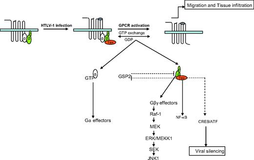
In conclusion, we provided molecular evidence showing a functional interaction between Tax oncoproteins and G-protein signaling. We propose a model in which, after T-cell infection by HTLV-1, Tax-1 would induce the expression of several proteins, including cytokines and chemokines. Tax-1 also would migrate to the cytoplasm and the plasma membrane, bind to the Gβγ heterodimer complex, and regulate specific ligand-receptor interactions. In addition, Tax-1 might affect intracellular Gβγ-effector activation by direct binding to the Gβ subunit (Figures 1, S1) or by inactivating the G-protein pathway suppressor 2.86 In a feedback loop, Gβγ would inhibit HTLV-1 expression and contribute to viral escape from immune surveillance (Figure 7). Our findings provide an additional clue to understanding the migration and infiltration of HTLV-1–infected cells into various tissues and suggest that the regulation of G proteins and GPCRs is a novel and important aspect of Tax function.
Model summarizing the Tax-1–G-protein interactions. After T-cell infection by HTLV-1, Tax-1 induced the expression of several genes, including chemokines, that bound to their specific GPCRs. Tax-1 also migrated to the cytoplasm and the plasma membrane, bound to the Gβγ heterodimer complex, and regulated specific ligand-receptor interaction, T-cell migration, and intracellular G-protein effector activation. Tax-1 also affected intracellular Gβγ-effector activation by direct binding to the Gβ subunit and by inactivating the G-protein pathway suppressor 2. As a feedback loop, Gβγ inhibits HTLV-1 expression and contributes to viral escape from immune surveillance.
Model summarizing the Tax-1–G-protein interactions. After T-cell infection by HTLV-1, Tax-1 induced the expression of several genes, including chemokines, that bound to their specific GPCRs. Tax-1 also migrated to the cytoplasm and the plasma membrane, bound to the Gβγ heterodimer complex, and regulated specific ligand-receptor interaction, T-cell migration, and intracellular G-protein effector activation. Tax-1 also affected intracellular Gβγ-effector activation by direct binding to the Gβ subunit and by inactivating the G-protein pathway suppressor 2. As a feedback loop, Gβγ inhibits HTLV-1 expression and contributes to viral escape from immune surveillance.
Authorship
Contribution: J.C.T. and J.-Y.S. designed and performed the research and analyzed the data. M.B., J.-F.D., J.D., and P.U. provided technical help on experiments presented in Figures 1 to 5. A.B. contributed critical reading of the manuscript. L.W. provided key reagents and contributed to research presented in Figure 3. F.D., D.P., C.V.L., P.L.G., R.M., and M.P. provided vital new reagents (antibodies, cell lines, and DNA constructions). J.-C.T., F.D., and R.K. wrote the paper.
Conflict-of-interest disclosure: The authors declare no competing financial interests.
Correspondence: Jean-Claude Twizere, Cellular and Molecular Biology, Faculty of Gembloux, 13 avenue Maréchal Juin, B-5030 Gembloux, Belgium; e-mail: twizere.jc@fsagx.ac.be.
The online version of this article contains a data supplement.
The publication costs of this article were defrayed in part by page charge payment. Therefore, and solely to indicate this fact, this article is hereby marked “advertisement” in accordance with 18 USC section 1734.
This work was supported by the Belgian program on Interuniversity Poles of Attraction, Prime Minister's Office, Science Policy Programming (R.K., M.P.), the Belgian Foundation against Cancer (R.K.), the Fonds National de la Recherche Scientifique (J.C.T., M.B., P.U., F.D., C.V.L., L.W., R.K.), the National Institutes of Health (grant CA100730) (P.L.G.), and the INSERM (R.M.).
We thank Dr S. Herlitze (Case Western Reserve University, Cleveland, OH), Dr B. Denker (Harvard Institute of Medicine, Boston, MA), Dr F. Bex (Free University of Brussels, Belgium), Dr H. Hamm (Department of Pharmacology, Vanderbilt University, Nashville, TN), Dr N. Gautam (Department of Anesthesiology, Washington University School of Medicine, St Louis, MO), Euroscreen S.A (Gosselies, Belgium), Dr R. Grassmann (Institute for Clinical and Molecular Virology, University of Erlangen-Nuremberg, Erlangen, Germany), Drs. K.T. Jeang and J-M. Peloponese (Molecular Virology Section, Laboratory of Molecular Microbiology, NIAID, National Institutes of Health, Bethesda, MD), the National Institutes of Health AIDS Research and Reference Reagent Program (Germantown, MD), and the University of Missouri cDNA Resource Center (University of Missouri, Rolla, MO) for providing reagents.

This feature is available to Subscribers Only
Sign In or Create an Account Close Modal