Abstract
Absence of shear stress due to disturbed blood flow at arterial bifurcations and curvatures leads to endothelial dysfunction and proinflammatory gene expression, ultimately resulting in atherogenesis. KLF2 has recently been implicated as a transcription factor involved in mediating the anti-inflammatory effects of flow. We investigated the effect of shear on basal and TNF-α–induced genomewide expression profiles of human umbilical vein endothelial cells (HUVECs). Cluster analysis confirmed that shear stress induces expression of protective genes including KLF2, eNOS, and thrombomodulin, whereas basal expression of TNF-α–responsive genes was moderately decreased. Promoter analysis of these genes showed enrichment of binding sites for ATF transcription factors, whereas TNF-α–induced gene expression was mostly NF-κB dependent. Furthermore, human endothelial cells overlying atherosclerotic plaques had increased amounts of phosphorylated nuclear ATF2 compared with endothelium at unaffected sites. In HUVECs, a dramatic reduction of nuclear binding activity of ATF2 was observed under shear and appeared to be KLF2 dependent. Reduction of ATF2 with siRNA potently suppressed basal proinflammatory gene expression under no-flow conditions. In conclusion, we demonstrate that shear stress and KLF2 inhibit nuclear activity of ATF2, providing a potential mechanism by which endothelial cells exposed to laminar flow are protected from basal proinflammatory, atherogenic gene expression.
Introduction
Atherosclerosis is a vascular disease with a clear focal nature, which has been shown to correlate with shear stress levels on the endothelium, resulting from specific blood flow patterns.1 Absence of shear stress due to oscillatory blood flow at arterial bifurcations and curvatures leads to endothelial dysfunction as characterized by a diminished barrier function and proinflammatory gene expression. These conditions facilitate the entry of lipids and inflammatory cells in the vascular wall, ultimately leading to the formation of an atherosclerotic plaque. On the other hand, endothelial cells exposed to high levels of shear stress maintain an atheroprotective gene expression profile and have a differentiated, quiescent phenotype.2 Transcription factors, being the integrators of various mechanical and biologic stimuli, play a pivotal role in the regulation of gene expression and determine the resulting biologic effect. A possible candidate that could be critical in the protection from atherogenesis is the shear-inducible transcription factor Krüppel-like factor 2 (KLF2). It has become clear in recent publications that KLF2 plays a significant role in maintaining an atheroprotective, quiescent endothelial phenotype.3–5 Protective genes like endothelial nitric oxide synthase (NOS3) and thrombomodulin (TM) are induced by KLF2, whereas expression of the proatherogenic monocyte chemoattractant protein 1 (CCL2) and endothelin (EDN1) is reduced
The inflammatory component in atherosclerotic pathology suggests that inflammatory gene expression is activated in the atherosclerotic vascular wall, which is mediated by transcription factors associated with inflammation. Inflammatory gene expression has been detected at vascular sites prone to atherosclerotic plaque formation.6 Furthermore, the transcription factor nuclear factor–κB (NF-κB) and its inhibitor IκB were shown to be present at elevated levels in the cytoplasm of endothelial cells at sites exposed to disturbed blood flow.7 These cells, however, do not show increased nuclear levels of NF-κB, which only becomes transcriptionally active when translocated to the nucleus after liberation from its inhibitor IκB. This translocation was only observed after inflammatory activation by a secondary stimulus like LPS or atherogenic diet, which then indeed occurred much more prominently at the low-flow regions.7 Thus, endothelial cells at sites with disturbed blood flow should not exhibit inflammatory gene expression to the same magnitude in the absence of induction by cytokines. Indeed, a moderate induction of proinflammatory gene expression was observed at the disturbed flow regions of the porcine carotid artery bifurcation.8 A potent induction of inflammatory gene expression by inflammatory cytokines depends on the actively promoted formation of a transcriptional complex, usually composed of NF-κB, activator protein-1 (AP-1), and coactivators like CREB-binding protein CBP/p300.9 The transcriptional effects of TNF-α on human umbilical vein endothelial cells (HUVECs) are considered a physiological representative of atherogenesis10 and are indeed mediated by both NF-κB and the AP-1–activating p38 mitogen-activated protein kinase (MAPK).11 Pharmacologic interventions and the protective effects of short-term flow preconditioning in vitro suggested a dominant role of the former transcription factor.11,12 The anti-inflammatory action of shear-induced KLF2, however, was shown to be mainly dependent on cofactor modulation rather than on directly affecting NF-κB activation and nuclear translocation,13 consistent with the documented effects of flow in vivo.
Given the absence of translocated, nuclear NF-κB at noninflamed atheroprone sites in vivo and the complex effects of long- versus short-term shear and KLF2 on NF-κB activity, we decided to reevaluate prolonged shear and KLF2 modulation of proinflammatory gene expression. In the present study, we demonstrate that shear stress and KLF2 can modulate transcription factor activity and basal inflammatory gene expression of HUVECs. We show that shear stress inhibits, in a KLF2-dependent manner, the nuclear activation of activating transcription factor 2 (ATF2), one of the heterodimeric components of AP-1. Moreover, elevated levels of phosphorylated ATF2 protein are shown in endothelial cells overlying early atherosclerotic plaques compared with healthy endothelium. This provides a novel mechanism by which shear stress might protect endothelial cells from a proatherogenic phenotype.
Materials and methods
Cell culture and shear stress experiments
HUVECs were isolated and cultured in medium-199 (M199; Invitrogen, Carlsbad, CA) supplemented with 20% (vol/vol) fetal bovine serum (FBS), 50 μg/mL heparin (Sigma, St Louis, MO), 6 to 25 μg/mL endothelial-cell growth supplement (ECGS; Sigma), and 100 U/mL penicillin/streptomycin (Invitrogen) as described.10 The 24-hour shear stress experiments were performed in a parallel platetype flow chamber with pulsatile flow (12 ± 7 dynes/cm2) as described,4,14 using a CellMax Quad positive-displacement pump (Cellco, Germantown, MD). Long-term shear stress exposure (6 days) was as described.4,14 In brief, HUVECs were seeded in fibronectin-coated artificial capillary cartridges (Polypropylene 70, Cellco; DIV-BBB cartridge, Flocel, Cleveland, OH) in medium containing 10 μg/mL ECGS. Cells were allowed to adhere and reach confluency overnight, with medium flowing through the extracapillary space using the CellMax Quad pump system to provide oxygen and nutrients. Next, flow was guided through the capillaries and gradually increased to correspond to a pulsatile shear stress of 19 ± 12 dynes/cm2, which was maintained over the next 6 days with intermediary medium changes. For static controls, HUVECs from the same isolate were seeded in fibronectin-coated cell-culture flasks and grown to confluency. After indicated treatments, either total RNA was obtained using Trizol reagent (Invitrogen) or nuclear extracts were made.
Inflammatory cytokine stimulation during shear stress experiments
TNF-α (R&D Systems, Abingdon, United Kingdom) was reconstituted in PBS supplemented with 1% (wt/vol) BSA and used at a final concentration of 25 ng/mL in the culture medium during the final 6 hours of shear stress exposure or static culture.
Real-time RT-PCR
cDNA from 0.5 to 1 μg total RNA was synthesized according to the manufacturer's protocol (Invitrogen) and diluted 10× for gene-specific analysis with real-time reverse transcriptase–polymerase chain reaction (RT-PCR). All RT-PCR reactions were performed in a 15 μL reaction on an iCycler thermal cycler system (Bio-Rad Laboratories, Veenendaal, Netherlands). Measured mRNA level were expressed as normalized ratios compared with ribosomal phosphoprotein P0 expression levels. Gene-specific primers were designed using Beacon Designer 3 software (Premier Biosoft International, Palo Alto, CA), and optimal melting temperature was obtained using a temperature gradient reaction.
Microarray probe synthesis and hybridization
A human oligonucleotide library containing 18.659 gene-specific 65-mer sequences was purchased from Sigma/Compugen (Sigma, Saint Louis, MO; Compugen, San Jose, CA) and spotted on glass slides by the Microarray Department of the University of Amsterdam. Microarrays and coverslips were pretreated for 1 hour at 40°C in a buffer containing 25% formamide (vol/vol), 5× SSC, 0.2% (wt/vol) SDS, and 0.1% (wt/vol) BSA. All microarray experiments were performed using a common reference RNA composed of a pool of RNA from HUVECs, the monocytic cell line THP-1, and wholemount human carotid and aortic arteries. Up to 1 μg total RNA from samples or common reference was amplified in a single round using the T7-based Ambion MessageAmp kit (Ambion, Huntingdon, United Kingdom), with 50% of rUTP ribonucleotides replaced by aminoallyl-rUTP (Sigma). Aminoallyl-modified amplified RNA (aRNA) was labeled with either Cy3 (common reference) or Cy5 (samples) monoreactive dyes (GE Healthcare, Uppsala, Sweden). Next, labeled probes were fragmented followed by purification using the RNeasy mini kit (Qiagen, Hilden, Germany). RNA concentration as well as dye incorporation was measured using the Nanodrop Spectrophotometer (Nanodrop Technologies, Wilmington, DE). Equivalent amounts of labeled aRNA were applied to pretreated oligonucleotide microarrays in duplicate and hybridized for 16 hours at 40°C. After hybridization, slides were washed and subsequently scanned using an Agilent-II Scanner (Agilent Technologies, Palo Alto, CA). Feature extraction was done using Arrayvision 8.0 software (GE Healthcare Europe, Diegem, Belgium), and background subtracted intensities were subsequently normalized using locally weighted linear regression smoothing (LOESS) in the R software environment (LIMMA package; Bioconductor software15 ). Normalized data were imported into Rosetta Resolver (Rosetta Biosoftware, Seattle, WA).
Microarray data analysis
Re-ratio–based experiment definitions were constructed in Rosetta Resolver, followed by initial analysis consisting of marker gene verification and hierarchical clustering. For promoter analysis, data were exported from Rosetta Resolver and analyzed using whole genome rVista software.16 These calculations used a database of all transcription factor binding sites (TFBSs) conserved in the human to mouse whole genome alignment of May 2004. Locus link identifications of the genes from each group (Table S1, available on the Blood website; see the Supplemental Materials link at the top of the online article) were used as input. Calculated were the TFBSs overrepresented in 500 base pair upstream regions of these genes using all upstream regions of human genes in release 3 of the Reference Sequence (RefSeq) database (http://www.ncbi.nlm.nih.gov/RefSeq/) as outgroup (Table S2).
Immunohistochemistry
Human vascular tissue specimens were collected from organ donors after obtaining informed consent with approval of the Academic Medical Center (AMC) Medical Ethical Committee, and procedures conformed to the Declaration of Helsinki. Paraffin sections were deparaffinized and dried, followed by antigen retrieval by boiling the slides for 10 minutes in a 10-mM citrate buffer at pH 6.0. Primary antibody incubation with Thr71-phosphorylated ATF2 antibody was performed overnight at 4°C. Next, biotinylated secondary antibodies were used for 1 hour at room temperature, followed by incubation with streptavidin-biotin complexes conjugated to horseradish peroxidase (Dako, Glostrup, Denmark). Peroxidase substrate coloring with the VECTOR NovaRED substrate kit (Vector Laboratories, Burlingame, CA) was allowed to proceed for 10 minutes. Sections were examined using a Zeiss Axiophot microscope equipped with 2.5×/0.075 numeric aperture (NA), 5×/0.15 NA, 10×/0.3 NA, 20×/0.5 NA, 40×/0.65 NA, and 63×/0.8 NA Plan Neofluar objective lenses (Zeiss, Oberkochen, Germany) and photographed using a Sony DXC-950P digital camera (Sony, Tokyo, Japan) operated with Leica QWin software (Leica Imaging Systems, Cambridge, United Kingdom). Images were processed using Adobe Photoshop CS2 9.0 software (Adobe Systems, San Jose, CA). Overview images of entire vessels were obtained by scanning the slides on an Epson EU-35 flatbed scanner (Seiko Epson, Nagano, Japan) with a resolution of 6400 dpi and importing the images into Adobe Photoshop CS2 9.0 (Adobe Systems, San Jose, CA) using Epson TWAIN Pro software (Seiko Epson).
Lentiviral KLF2 overexpression and knockdown
Nuclear extract preparation and transcription factor enzyme-linked immunosorbent assay (ELISA)
Nuclear proteins were prepared with the nuclear extract kit in accordance with the manufacturer's protocol (Active Motif Europe, Rixensart, Belgium). Transcription factor activity was determined with TransAM MAPK family kit (Active Motif Europe). In brief, 2 to 20 μg nuclear extract was added to each microtiter plate well into which an oligonucleotide with an ATF2 or NF-κB consensus binding site had been immobilized. Transcription factors bound to their cognate DNA binding site were detected using specific horseradish peroxidase–conjugated antibodies for p65 or Thr71-phosphorylated ATF2 supplied in the kit. After substrate coloring, absorption at 450 nm was measured on an EL808 microplate reader (BioTek, Winooski, VT).
Immunofluorescence
For immunofluorescence, mock- and KLF2-transduced HUVECs were grown on gelatin-coated glass coverslips and fixed with 4% (vol/vol) formaldehyde. Primary antibody incubation using Thr71-phosphorylated ATF2 antibody (Cell Signaling Technology, Danvers, MA) was performed overnight at 4°C. Alexa 488–labeled secondary antibodies were used for 1 hour at room temperature, followed by Hoechst nuclear staining, mounting, and fluorescence microscopy analysis. Photomicrographs were acquired using a Zeiss Axioplan 2 microscope equipped with 10×/0.3 NA, 20×/0.5 NA, 63×/1.4 NA, and 100×/1.5 NA Plan Neofluar objective lenses (Zeiss) and a Coolsnap HQ digital camera (Roper Scientific, Ottobrunn, Germany). Images were processed with Adobe Photoshop C52 9.0 software.
RNA interference with duplex siRNA and Western blotting
HUVECs from 3 different isolates were grown to 80% confluency according to cell-culture methods described in “Cell culture and shear stress experiments.” Cells were then changed to 1 mL per well of Optimem reduced serum medium (Invitrogen) and transfected with 325 pmol nonspecific (5′-CAGUCGCGUUUGCGACUGG-3′ synthesized small interfering RNA [siRNA]; Ambion) or ATF2 siRNA (Silencer predesigned siRNA no. 16704; Ambion) using the Oligofectamine reagent (Invitrogen) according to manufacturer's protocol. After 4 hours, 2 mL M199 supplemented with 20% (vol/vol) FBS, 50 μg/mL heparin, 12.5 μg/mL ECGS, and 100 U/mL penicillin/streptomycin were added to the Optimem medium in each well. After 24 hours, medium was changed to 2 mL of full M199 containing 12.5 μg/mL ECGS, and RNA or protein was harvested after 48 hours. Where applicable, cells were stimulated with 10 ng/mL of TNF-α or vehicle (PBS + 1% BSA) during the final 6 hours of the experiment. Western blotting was performed as described.17 Total ATF2 protein levels were detected using a monoclonal ATF2 antibody (Cell Signaling Technology), and an α-tubulin staining was performed as a control for equal loading. Densitometric quantification of the Western blot was performed using cyQuant software version 2003.03 (Amersham, Piscataway, NJ).
Statistical analysis
Experimental data are shown as mean of normalized ratios ± standard error of the mean (SEM) for the indicated number of experiments. The paired or unpaired Student t test was used to calculate statistical significance of expression ratios or optical densities versus controls. P values of less than .05 were considered statistically significant.
Results
Shear stress inhibits basal but not TNF-α–induced expression of inflammatory genes
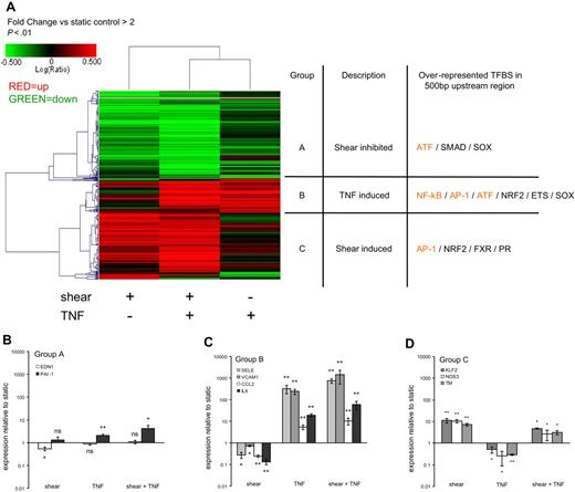
The artificial capillary system14 was used to obtain gene expression profiles of endothelial cells stimulated by inflammatory cytokines, comparing their inflammatory response under prolonged, unidirectional pulsatile laminar flow to static conditions. We studied the effects of these conditions using genomewide expression profiling. Figure 1A shows a hierarchical clustering of a selection of genes modulated more than 2-fold by shear, TNF-α, or shear and TNF-α combined, with all 3 treatments relative to static control conditions. Three main cluster groups can be discriminated: Group A contains genes whose expression is down-regulated by shear but that are unaffected by TNF-α (Table S1A); group B contains genes whose expression is up-regulated by TNF-α under both static and shear conditions (Table S1B); and group C contains genes that are up-regulated by shear stress and are unaffected or down-regulated by TNF-α (Table S1C). Validation of microarray expression data by real-time PCR was performed for a selection of genes from each group and showed that the expression of 8 of 9 genes agreed with the microarray data (Figure 1B-D; Table S1). In endothelial cells under prolonged pulsatile flow compared with static conditions, EDN1 and plasminogen activator inhibitor 1 (PAI-1) from group A were down-regulated or unchanged, respectively, whereas PAI-1 was only slightly induced after 6 hours of TNF-α, which did not match the microarray expression data (Figure 1B; Table S1A). In contrast, several adhesion molecules and chemokines were potently induced by TNF-α and moderately suppressed by shear stress (Figure 1C). In line with our previous reports, mRNA levels of KLF2, NOS3, and TM were found to be highly up-regulated by prolonged shear stress and to be inhibited by TNF-α (Figure 1D).
Analysis of the effect of shear stress on basal and TNF-α–induced gene expression in endothelial cells. (A) Hierarchical clustering of a selection of genes modulated more than 2-fold by shear, TNF-α, or shear and TNF-α combined; all 3 treatments relative to static control conditions. Three main cluster groups can be discriminated, which were analyzed for the presence of overrepresented TFBSs in the promoters of the genes in each group compared with the whole genome in conserved 500 base pair upstream regions in the human-mouse alignment of May 2004. A selection of transcription factors with overrepresented binding sites is shown for each cluster group, and highlighted in orange are transcription factors that are well described to be involved in inflammatory signaling. (B-D) Real-time PCR validation of gene expression as obtained by microarray. PCR analysis was done for a selection of genes from each of the 3 cluster groups as identified by microarray data analysis. Real-time PCR was performed in duplicate on HUVEC cDNA from 3 independent isolates, comparing static conditions with either 7 days of pulsatile shear stress (19 ± 12 dynes/cm2), a 6-hour treatment of TNF-α (25 ng/mL), or both. Expression levels relative to P0 housekeeping gene were obtained for a selection of genes from each cluster group and represented relative to static controls. Error bars indicate SEM. Significant difference versus static control conditions is indicated (*P < .05; **P < .01); ns indicates not significant.
Analysis of the effect of shear stress on basal and TNF-α–induced gene expression in endothelial cells. (A) Hierarchical clustering of a selection of genes modulated more than 2-fold by shear, TNF-α, or shear and TNF-α combined; all 3 treatments relative to static control conditions. Three main cluster groups can be discriminated, which were analyzed for the presence of overrepresented TFBSs in the promoters of the genes in each group compared with the whole genome in conserved 500 base pair upstream regions in the human-mouse alignment of May 2004. A selection of transcription factors with overrepresented binding sites is shown for each cluster group, and highlighted in orange are transcription factors that are well described to be involved in inflammatory signaling. (B-D) Real-time PCR validation of gene expression as obtained by microarray. PCR analysis was done for a selection of genes from each of the 3 cluster groups as identified by microarray data analysis. Real-time PCR was performed in duplicate on HUVEC cDNA from 3 independent isolates, comparing static conditions with either 7 days of pulsatile shear stress (19 ± 12 dynes/cm2), a 6-hour treatment of TNF-α (25 ng/mL), or both. Expression levels relative to P0 housekeeping gene were obtained for a selection of genes from each cluster group and represented relative to static controls. Error bars indicate SEM. Significant difference versus static control conditions is indicated (*P < .05; **P < .01); ns indicates not significant.
To gain insight into the coordinate regulation of genes within a specific group, a search for transcription factor binding elements in upstream regions of constituent genes was performed using whole genome rVISTA. This software package evaluates which TFBSs, conserved between pairs of species, are statistically significantly overrepresented in upstream regions in a group of genes.18 Using the human-mouse alignment of May 2004 and setting a 500 bp upstream search region, we found overrepresentation of distinct TFBSs in each of the 3 main groups (Table S2). Importantly, the TNF-α–responsive group B is enriched in binding sites for NF-κB and the AP-1 family of transcription factors (indicated in Table S2 as AP1FJ). Group B also shows enrichment of an ATF4 binding site. Furthermore, ATF3 and ATF4 binding sites are overrepresented in group A, which is composed of genes down-regulated by shear stress but not modulated by TNF-α. The latter is also evident from a lack of enrichment of NF-κB binding sites in group A. Thus, it seems that group A and B share ATF binding site enrichment as well as down-regulation of unstimulated basal expression by shear stress. In group C, composed predominantly of shear induced genes, enrichment for AP-1 and NRF2 binding sites is detected.
Evidently, there seems to be a clear distinction in identity of different members of the AP-1/ATF families in the different clusters. Detailed inspection showed this to be based on subtle sequence differences, because homodimers or heterodimers of the ATF family bind the cAMP response element (CRE) 5′-TGAC′GTCA-3′, which differs by 1 nucleotide from the consensus AP-1 binding site 5′-TGAC′GTCA-3′.19,20 One of the most studied members of the ATF transcription factor family, ATF2, is crucial for cytokine-induced expression of E selectin in endothelial cells.21,22 Furthermore, the ATF2 transcription factor has been described to be constitutively expressed,19 including in HUVECs,23 which makes it a prime target through which down-regulation of unstimulated basal expression of group A and B genes by shear stress could be mediated. In contrast to this, key members of the AP-1 family, c-Jun and c-Fos, are known to be inducible transcription factors at the expression level in response to growth factors, cytokines, and stress.24 ATF3 and ATF4 also are inducible transcription factors, acting mostly through increased expression in response to endoplasmic reticulum (ER) stress,25 with ATF3 having almost no detectable levels in unstimulated endothelial cells.23 We validated this in our microarray expression profiles from unstimulated endothelial cells. Signal intensity levels for ATF3 and ATF4 are around or below reliable detection levels, whereas the ATF2 signal is well above this threshold. The constitutive expression of ATF2 in unstimulated endothelial cells and its key role in proinflammatory signaling prompted us to further investigate its role in the atheroprotective effect of shear stress.
Human lesional endothelial cells are positive for phosphorylated ATF2
To our knowledge, ATF2 has not been described in the context of shear stress and/or atherosclerosis; therefore, its potential physiological relevance was first assessed. The presence of active, phosphorylated ATF2 in endothelial cells from healthy or early atherosclerotic lesions was probed by immunohistochemistry (Figure 2). After determining the presence of a continuous layer of endothelial cells by CD31 staining in human iliac and carotid arteries, a clear and consistent positive signal for phosphorylated ATF2 could be seen in endothelial cells overlying early atherosclerotic lesions. In contrast, endothelium overlying morphologically healthy vessel wall is completely devoid of phosphorylated ATF2, although a strong positive signal for phosphorylated ATF2 was present in the media, presumably in smooth muscle cells.
Phosphorylated ATF2 is expressed specifically in lesional endothelium. (A) Human donor tissue (iliac artery, male, age 43, died from subdural hematoma after fall) was probed for the presence of a continuous layer of endothelial cells by immunohistochemical CD31 staining. After confirmation of CD31 positivity, adjacent sections were stained for Thr71-phosphorylated ATF2 (pATF2). The left part of the panel shows an overview of the entire vessel, indicating the lesional and lesionfree areas that are enlarged in the composite images on the right, which show pATF2 or CD31 staining as red coloration. (B-E) Enlarged parts of lesional and lesionfree areas stained for pATF2 or CD31, taken from panel A as indicated by the red-lined boxes. Similar enlargements are shown for 2 other vascular specimens, obtained from human donor ([F-I] iliac artery, female, age 35, died in falling accident) or human obduction tissue ([J-M] internal carotid artery, male, age 85, died from liver failure). All sections were counterstained for nuclei with hematoxylin. The enlargements in panels A and B were obtained using a magnification of × 63.
Phosphorylated ATF2 is expressed specifically in lesional endothelium. (A) Human donor tissue (iliac artery, male, age 43, died from subdural hematoma after fall) was probed for the presence of a continuous layer of endothelial cells by immunohistochemical CD31 staining. After confirmation of CD31 positivity, adjacent sections were stained for Thr71-phosphorylated ATF2 (pATF2). The left part of the panel shows an overview of the entire vessel, indicating the lesional and lesionfree areas that are enlarged in the composite images on the right, which show pATF2 or CD31 staining as red coloration. (B-E) Enlarged parts of lesional and lesionfree areas stained for pATF2 or CD31, taken from panel A as indicated by the red-lined boxes. Similar enlargements are shown for 2 other vascular specimens, obtained from human donor ([F-I] iliac artery, female, age 35, died in falling accident) or human obduction tissue ([J-M] internal carotid artery, male, age 85, died from liver failure). All sections were counterstained for nuclei with hematoxylin. The enlargements in panels A and B were obtained using a magnification of × 63.
Shear stress suppresses nuclear levels of activated ATF2 via KLF2
Based on the promoter analysis and immunohistochemical data described in the 2 previous headings of “Results,” we investigated the role of ATF2 in the modulation of gene expression by shear stress in more detail. For this purpose, we used an enzyme-linked immunosorbent assay (ELISA)–based assay that measures nuclear levels of activated transcription factors that are able to bind to oligonucleotides containing their cognate DNA binding sites. Nuclear extracts from HUVECs exposed to pulsatile flow show a clear reduction in levels of phosphorylated ATF2, most prominently after 5 days of shear (Figure 3A). Stable lentiviral overexpression of KLF2 for 7 days resulted in a suppression of nuclear activated ATF2 to a level similar to that reached by prolonged shear (Figure 3B). The reduced nuclear levels of phosphorylated ATF2 were not due to a change in total ATF2 levels, because neither shear stress nor KLF2 overexpression altered ATF2 mRNA expression compared with static or mock controls, as measured by RT-PCR (Figure S1). Knockdown of KLF2 using lentivirally delivered siRNA abrogates the shear stress–mediated inhibition of ATF2, implicating a direct dependence on KLF2 in this observation (Figure 3C). Even under static conditions, KLF2 siRNA increases ATF2 levels significantly compared with a control siRNA against FLUC. The latter observation correlates with an increase in expression levels of several proinflammatory genes in static cells transduced with KLF2 siRNA compared with mock siRNA (Figure S2).
Functional analysis of nuclear ATF2 activity in endothelial cells exposed to prolonged shear and its dependence on KLF2. Nuclear extracts from HUVECs exposed to 5 days of pulsatile flow (A) or HUVECs overexpressing KLF2 (B) were assayed for the presence of functional Thr71-phosphorylated ATF2 protein. Data from 3 independent isolates are expressed relative to static or mock controls. (C) HUVECs containing lentiviral-delivered double-stranded siRNA directed against KLF2 or a control siRNA against FLUC were exposed for 5 days to shear stress or to static conditions and assayed for nuclear activated ATF2. The means of 3 different isolates are expressed relative to static FLUC. Error bars indicate SEM. Significant difference versus static control conditions is indicated (*P < .05; **P < .01).
Functional analysis of nuclear ATF2 activity in endothelial cells exposed to prolonged shear and its dependence on KLF2. Nuclear extracts from HUVECs exposed to 5 days of pulsatile flow (A) or HUVECs overexpressing KLF2 (B) were assayed for the presence of functional Thr71-phosphorylated ATF2 protein. Data from 3 independent isolates are expressed relative to static or mock controls. (C) HUVECs containing lentiviral-delivered double-stranded siRNA directed against KLF2 or a control siRNA against FLUC were exposed for 5 days to shear stress or to static conditions and assayed for nuclear activated ATF2. The means of 3 different isolates are expressed relative to static FLUC. Error bars indicate SEM. Significant difference versus static control conditions is indicated (*P < .05; **P < .01).
KLF2 overexpression inhibits TNF-α–induced nuclear activation of ATF2
To further investigate the mechanism by which shear stress mediates its effects on ATF2, we measured nuclear activation of ATF2 in cells overexpressing lentiviral KLF2. KLF2 has been shown to be the causal factor in shear-mediated inhibition of several genes involved in inflammation and vascular tone.3–5,13 Figure 4A shows that nuclear ATF2 was potently suppressed by KLF2 compared with mock-transduced cells in the unstimulated control. Furthermore, TNF-α–induced activation of nuclear ATF2, as seen in mock-transduced cells, was completely abolished by KLF2. Conversely, it appeared that TNF-α–induced activation of the NF-κB component p65 was only partly inhibited by KLF2, whereas basal levels showed no difference between mock- and KLF2-transduced cells (Figure 4B).
Effect of TNF-α on nuclear transcription factor activity in mock- and KLF2-tranduced HUVECs. Nuclear activation of ATF2 (A) or p65 (B) was determined in mock- and KLF2-overexpressing HUVECs activated for 0 to 3 hours with TNF-α (10 ng/mL). Basal and TNF-α–induced ATF2 levels were strongly suppressed by KLF2, whereas basal p65 levels were not changed and TNF-α–induced p65 levels were only partially inhibited. Data are represented as mean ratios of 2 to 3 different isolates relative to the unstimulated mock condition. Error bars indicate SEM. Significant difference between mock and KLF2 values is indicated (*P < .05; **P < .01); ‡, significant difference (P < .01) between TNF-α–stimulated and unstimulated control values.
Effect of TNF-α on nuclear transcription factor activity in mock- and KLF2-tranduced HUVECs. Nuclear activation of ATF2 (A) or p65 (B) was determined in mock- and KLF2-overexpressing HUVECs activated for 0 to 3 hours with TNF-α (10 ng/mL). Basal and TNF-α–induced ATF2 levels were strongly suppressed by KLF2, whereas basal p65 levels were not changed and TNF-α–induced p65 levels were only partially inhibited. Data are represented as mean ratios of 2 to 3 different isolates relative to the unstimulated mock condition. Error bars indicate SEM. Significant difference between mock and KLF2 values is indicated (*P < .05; **P < .01); ‡, significant difference (P < .01) between TNF-α–stimulated and unstimulated control values.
Phosphorylated ATF2 is excluded from the nucleus by KLF2
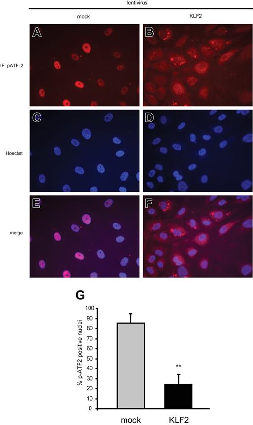
Because the activated transcription factor assay cannot discern between the degree of nuclear localization and the degree of phosphorylation, immunofluorescence was used to clarify this issue. Fluorescent staining of phosphorylated ATF2 in HUVECs showed a clear and predominant cytoplasmic localization of phosphorylated ATF2 in KLF2-overexpressing cells (Figure 5B), whereas mock transduction resulted in exclusive nuclear staining of phosphorylated ATF2 (Figure 5A). These observations were confirmed by an additional nuclear staining with Hoechst (Figure 5C-D), which led to merged pictures (Figure 5E-F) clearly identifying the major effect of KLF2 on the localization of phosphorylated ATF2. Quantification of the immunofluorescence data from 2 independent experiments show a clear reduction in pATF2-positive nuclei from 85% in mock cells to 25% in KLF2 transduced cells (Figure 5G).
KLF2 suppresses nuclear localization of phosphorylated ATF2. HUVECs transduced with mock (A,C,E) or KLF2 (B,D,F) lentivirus were fixed with paraformaldehyde and stained for Thr71-phosphorylated ATF2 protein by immunofluorescence (A-B). Nuclei were made visible with a Hoechst nuclear staining (C-D), and the pictures were merged (E-F). Photographs representative for 2 independent experiments were obtained by fluorescence microscopy using a magnification of × 63. (G) Quantification was performed by counting nuclei with strong nuclear pATF2 positivity in 3 representative microscopic fields from mock- and KLF2-transduced cells taken from the 2 independent experiments, with each field containing an average of about 350 cells. **Significant difference in pATF2-positive nuclei between mock and KLF2 values (P < .01).
KLF2 suppresses nuclear localization of phosphorylated ATF2. HUVECs transduced with mock (A,C,E) or KLF2 (B,D,F) lentivirus were fixed with paraformaldehyde and stained for Thr71-phosphorylated ATF2 protein by immunofluorescence (A-B). Nuclei were made visible with a Hoechst nuclear staining (C-D), and the pictures were merged (E-F). Photographs representative for 2 independent experiments were obtained by fluorescence microscopy using a magnification of × 63. (G) Quantification was performed by counting nuclei with strong nuclear pATF2 positivity in 3 representative microscopic fields from mock- and KLF2-transduced cells taken from the 2 independent experiments, with each field containing an average of about 350 cells. **Significant difference in pATF2-positive nuclei between mock and KLF2 values (P < .01).
ATF2 knockdown leads to reduction of basal and TNF-induced proinflammatory gene expression levels
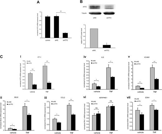
Having shown that ATF2 is inhibited by shear stress via KLF2, we next investigated the effect of direct inhibition of ATF2 on proinflammatory gene expression employing duplex siRNA against ATF2. A solid knockdown of ATF2 mRNA and protein levels by about 80% was achieved in HUVECs 48 hours after addition of ATF2 siRNA (Figure 6A-C). The siRNA-mediated inhibition of ATF2 was equally potent in vehicle- and TNF-α–treated cells, even though TNF-α caused a minor 1.5-fold increase in ATF2 expression in the nonspecific siRNA control (Figure 6Di). Knockdown of ATF2 caused a reduction in both basal and TNF-α–induced expression levels of proinflammatory genes from cluster groups A (EDN1) and B (SELE, CCL2, VCAM1, IL-8) could be confirmed by real-time PCR (Figure 6Dii-vii). The expression of PAI-1 was not significantly reduced by siATF2 in either vehicle or TNF-α–treated cells. Interestingly, the inducibility of proinflammatory genes by TNF-α as seen in cells treated with the nonspecific control was preserved when ATF2 was knocked down.
ATF2 knockdown reduces basal and TNF-α–induced expression of proatherogenic genes. Total cell mRNA and protein were harvested from HUVECs that were untransfected (control) or transfected with nonspecific (siNS) or ATF2 siRNA (siATF2). (A) ATF2 mRNA expression levels represented relative to ribosomal protein P0, as measured by real-time PCR. (B) Total ATF2 protein levels were measured using Western blot, and equal loading was verified with α-tubulin. (C) These protein levels were quantified as ratios versus siNS, corrected for α-tubulin. (D) Inhibition of basal and TNF-α–induced expression of genes from cluster groups A and B by knockdown of ATF2 compared with a nonspecific control. ATF2 expression was inhibited by siATF2 during both vehicle and TNF-α treatment (i), resulting in reduction of basal and TNF-α–induced expression of SELE (ii), CCL2 (iii), IL-8 (iv), VCAM1 (v), and EDN1 (vii) but not PAI-1 (vi). Represented are the mean P0-corrected relative mRNA expression levels from 3 different HUVEC isolates measured in duplicate by RT-PCR. Error bars indicate SEM. Significant difference versus nonspecific siRNA is indicated (*P < .05; **P < .01).
ATF2 knockdown reduces basal and TNF-α–induced expression of proatherogenic genes. Total cell mRNA and protein were harvested from HUVECs that were untransfected (control) or transfected with nonspecific (siNS) or ATF2 siRNA (siATF2). (A) ATF2 mRNA expression levels represented relative to ribosomal protein P0, as measured by real-time PCR. (B) Total ATF2 protein levels were measured using Western blot, and equal loading was verified with α-tubulin. (C) These protein levels were quantified as ratios versus siNS, corrected for α-tubulin. (D) Inhibition of basal and TNF-α–induced expression of genes from cluster groups A and B by knockdown of ATF2 compared with a nonspecific control. ATF2 expression was inhibited by siATF2 during both vehicle and TNF-α treatment (i), resulting in reduction of basal and TNF-α–induced expression of SELE (ii), CCL2 (iii), IL-8 (iv), VCAM1 (v), and EDN1 (vii) but not PAI-1 (vi). Represented are the mean P0-corrected relative mRNA expression levels from 3 different HUVEC isolates measured in duplicate by RT-PCR. Error bars indicate SEM. Significant difference versus nonspecific siRNA is indicated (*P < .05; **P < .01).
Discussion
There is still no definitive explanation for the cause of the moderate proinflammatory status of endothelium in the absence of flow in vitro or at disturbed flow regions of the vasculature in vivo. In this study we focused on noncytokine-induced gene expression as modulated by prolonged shear stress. The promoters of genes that are down-regulated by prolonged exposure to shear stress show a clear enrichment for ATF binding sites (Figure 1A-D). Our present results show that both shear stress and the shear-induced transcription factor KLF2 inhibit the activity of the constitutively expressed proinflammatory transcription factor ATF2 by inhibiting its nuclear translocation in HUVECs (Figures 3 and 5). Furthermore, constitutive and cytokine-induced expression of the panel of inflammatory genes, several adhesion molecules, and chemokines5 is indeed sensitive to ATF2 activity as shown by siRNA-mediated knockdown (Figure 6C), and their expression is suppressed by both shear stress and KLF2 (Figures 1 and 3). Interestingly, knockdown of residual KLF2 results in a further increase of constitutive expression of these genes under static conditions (Figure S2). Together, these data strongly suggest that blunting of ATF2 activity is one prominent mechanism by which shear stress and KLF2 inhibit the constitutive proinflammatory gene expression observed in the absence of biomechanical stimulation of endothelium. The physiological relevance of these in vitro data is supported by our novel observation that endothelial cells overlying human early atherosclerotic plaques have vastly increased levels of phosphorylated ATF2 compared with healthy endothelium (Figure 2). Interestingly, we consistently observed a much stronger staining for phosphorylated ATF2 in medial smooth muscle cells, located directly underneath the endothelial layer in plaquefree areas, compared with neointimal smooth muscle cells. This finding demonstrates for the first time a differential expression of activated, phosphorylated transcription factor ATF2 between medial and intimal smooth muscle cells, suggesting that it would be one of the molecular mediators of the phenotypic change undergone by smooth muscle cells during plaque formation. Together, these findings warrant further detailed investigation into the role of ATF2 during in vivo atherosclerosis.
The effect of shear stress on basal and TNF-α–induced gene expression was investigated by genomewide expression profiling and cluster-based promoter analysis showing 3 general cluster groups. Promoters of genes in cluster group B, composed of TNF-α–induced genes, showed enrichment in TFBSs for NF-κB and AP-1 (Figure 1A), confirming that both transcription factors are crucial for a potent transcriptional response to cytokine stimulation.11 It is well documented that potent induction of inflammatory gene expression depends on formation of a transcriptional complex, the enhanceosome, composed of NF-κB, AP-1, and cofactor CBP/p300.9 ATF2, together with Jun and Fos subfamilies, collectively constitute the family of AP-1 transcription factors, which are homodimers and heterodimers composed of basic-region leucine zipper (bZIP) proteins.19 Jun proteins form stable homodimers or heterodimers with Fos that bind the AP-1 DNA recognition element 5′-TGAG′CTCA-3′. However, ATF2 also forms homodimers or heterodimers with Jun that bind preferentially to the slightly different sequence of the CRE, 5′-TGACGTCA-3′. The latter response element is also the preferential binding site for other members of the CRE-binding family (CREB), including ATF3 and ATF4. However, involvement of ATF3 and ATF4 in shear stress–mediated inhibition of basal proinflammatory transcription is less likely because, like c-Jun and c-Fos, they are normally expressed at low levels in resting cells and are usually induced at the expression level.9,22,26 Still, a role for other ATF members in prolonged inflammatory signaling is evident, because Gargalovic and coworkers recently showed that both basal and ox-PAPC–induced expression of CCL2, IL-6, and IL-8 was partly dependent on ATF4.27,28 Furthermore, TNF-α apparently overrides the suppressive effects on ATF2 by highly inducing NF-κB, as shown by induction of genes containing NF-κB binding sites under stimulated conditions in both static and sheared endothelial cells (Figure 1) and by increased nuclear NF-κB protein even during overexpression of KLF2 (Figure 4). Indeed, several reports have directly shown that NF-κB prevails over p38/AP-1–driven expression after TNF-α activation.11,12 In line with this, the actual induction of proinflammatory genes by TNF-α was preserved in cells in which specifically ATF2 had been knocked down by siRNA, even though ATF2 knockdown did attenuate both basal and TNF-α–stimulated proinflammatory transcription levels (Figure 6). This finding seemingly contradicts previous reports that show a decrease in cytokine-induced inflammatory gene expression in endothelial cells after up to 24 hours of preexposure to laminar flow.29,30 It has become increasingly evident, however, that these time points may still be regarded as preconditioning and are not sufficient for termination of transient shear effects or for establishing the full KLF2 effect.5,14 We now indeed show that a full suppression of activated ATF2 in nuclear extracts from HUVECs requires more than 24 hours of arterial-level shear exposure to reach the same suppression level as caused by overexpressing KLF2 (Figure 3).
KLF2 has been shown to control multiple processes that maintain a healthy, functional endothelium and confer protection from initiation of atherosclerosis.3,5,14 For cluster group C, real-time PCR validated that shear stress increased KLF2, NOS3, and TM mRNA expression compared with static conditions and that these genes were inhibited by TNF-α (Figure 1D). Both observations are in accordance with previous results.3,14 KLF2 was also shown to dampen IL-1β–induced proinflammatory gene expression.3,13 Paradoxically, we and others report that TNF-α and IL-1 inhibit KLF2 expression14,31 and KLF2 moderately inhibits NF-κB activation13 (Figure 4B). It is likely that prolonged cytokine stimulation through NF-κB and AP-1 will therefore lift KLF2-mediated inhibition of proinflammatory genes, allowing a transient but potent inflammatory response. Along these lines, we argued that the protective effects of shear stress and KLF2 are more likely due to suppression of basal inflammatory gene expression depending on ATF2. In support of this view, the absence of shear stress indeed elevates basal expression levels of adhesion molecules and chemokines, an effect that is KLF2 dependent 3,4 (Figures 1 and 3). Additionally, knockdown of KLF2 in static cells elevated basal transcription of these proinflammatory genes (Figure S2).
Recruitment of CBP/p300 has recently been implicated in the KLF2-mediated inhibition of cytokine-induced gene expression via NF-κB.13,31 Another recent study in monocytes has shown that KLF2 can also recruit the PCAF cofactor away from the NF-κB complex.32 Interestingly, ATF2 has intrinsic histone acetyltransferase (HAT) activity and might recruit other HATs, like cofactors CBP/p300 and PCAF.33 Suppression of phosphorylated ATF2 levels in the nucleus by shear stress would result in decreased HAT activity, reduced cofactor recruitment and, ultimately, decreased constitutive transcription of genes (partially) dependent on ATF2. Thus, a straightforward explanation is supplied for the observed decreased CBP/p300 and PCAF activities and the effects on NF-κB–dependent gene expression reported previously.13,31 The next challenge is to explain the mechanism by which shear stress and KLF2 modulate nuclear ATF2 levels, especially since overall ATF2 phosphorylation levels do not seem to be affected in HUVECs overexpressing KLF2 (Figure 5). It could be that the nuclear import and export machinery is directly involved, possibly through c-Jun as a prerequisite nuclear anchor for ATF2,34 or that modulation of upstream MAP kinase pathways is affected.29 Further investigation is currently underway to find the detailed mechanism underlying the crucial observation of suppression of ATF2 nuclear localization by KLF2.
In conclusion, our in vitro results show a prominent role for the activated transcription factor ATF2 in basal but also inducible inflammatory transcription in endothelial cells, with endothelial activated ATF2 being present in vivo only at lesional areas of the human vasculature. This proinflammatory status is repressed by both shear stress and KLF2, which are also able to inhibit the nuclear activity of the transcription factor ATF2 in vitro. This strongly suggests that shear stress through KLF2 inhibits proinflammatory gene expression in regions of the vasculature that appear to be protected against the focal initiation of atherosclerosis.
Authorship
Contribution: J.O.F. designed and performed research, analyzed data, and wrote the manuscript; J.V.v.T. and R.J.D. performed research and contributed vital experimental tools; R.A.B. and O.L.V. performed research; J.R. contributed vital experimental tools; A.-P.J.J.B., M.J.A.P.D., J.K., T.J.C.v.B., and H.P. designed research; and A.J.G.H. designed research, analyzed data, and edited manuscript.
Conflict-of-interest disclosure: The authors declare no competing financial interests.
Correspondence: A. J. G. Horrevoets, Rm K1-114, Department of Biochemistry, Academic Medical Center, Meibergdreef 15, 1105 AZ, Amsterdam, The Netherlands; e-mail: a.j.horrevoets@amc.uva.nl.
The publication costs of this article were defrayed in part by page charge payment. Therefore, and solely to indicate this fact, this article is hereby marked “advertisement” in accordance with 18 USC section 1734.
An Inside Blood analysis of this article appears at the front of this issue.
The online version of this article contains a data supplement.
This work was supported by the Netherlands Organisation for Scientific Research (NWO), The Hague (grant 050-10-014); The Netherlands Heart Foundation, The Hague (grant M93.007); and the European Union (European Vascular Genomics Network grant LSHM-CT-2003-503254).


![Figure 2. Phosphorylated ATF2 is expressed specifically in lesional endothelium. (A) Human donor tissue (iliac artery, male, age 43, died from subdural hematoma after fall) was probed for the presence of a continuous layer of endothelial cells by immunohistochemical CD31 staining. After confirmation of CD31 positivity, adjacent sections were stained for Thr71-phosphorylated ATF2 (pATF2). The left part of the panel shows an overview of the entire vessel, indicating the lesional and lesionfree areas that are enlarged in the composite images on the right, which show pATF2 or CD31 staining as red coloration. (B-E) Enlarged parts of lesional and lesionfree areas stained for pATF2 or CD31, taken from panel A as indicated by the red-lined boxes. Similar enlargements are shown for 2 other vascular specimens, obtained from human donor ([F-I] iliac artery, female, age 35, died in falling accident) or human obduction tissue ([J-M] internal carotid artery, male, age 85, died from liver failure). All sections were counterstained for nuclei with hematoxylin. The enlargements in panels A and B were obtained using a magnification of × 63.](https://ash.silverchair-cdn.com/ash/content_public/journal/blood/109/10/10.1182_blood-2006-07-036020/4/m_zh80100701110002.jpeg?Expires=1769086233&Signature=UybR5Oraj08foWDlr7P4sjyXDX~Y-k-~ymGI2G72eSkIw155Vn4xP1gBCPMniCvHQIMATDoMQQnPJ7TjeshT9YcKWSPHNRdA5goMjI1oumUWxPhF2-2RpVerJuo4sSLVqPYt~k-XF~wrIBrjfCpTc7lyvXplXCukZ0eCh7TFiOVAF3aJ-5soYhlMlje2pDZeX--eEKRwBNodVPIe4Fw~SmcDmwGNtGm1IzJb-4Txu45B3mkAi1F2fuj6Mf2B-MkZLih58bIqy5CiEm479Zis9hir4~InT9LeYRyGtmVZjn4oOZ7ZqyfP55eSGIS~BoaYULGbZDiO8kfE4oQAbZJ8Hg__&Key-Pair-Id=APKAIE5G5CRDK6RD3PGA)


This feature is available to Subscribers Only
Sign In or Create an Account Close Modal