Abstract
Pbx1, a homeodomain transcription factor that was originally identified as the product of a proto-oncogene in acute pre-B–cell leukemia, is a global regulator of embryonic development. However, embryonic lethality in its absence has prevented an assessment of its role in B-cell development. Here, using Rag1-deficient blastocyst complementation assays, we demonstrate that Pbx1 null embryonic stem (ES) cells fail to generate common lymphoid progenitors (CLPs) resulting in a complete lack of B and NK cells, and a partial impairment of T-cell development in chimeric mice. A critical role for Pbx1 was confirmed by rescue of B-cell development from CLPs following restoration of its expression in Pbx1-deficient ES cells. In adoptive transfer experiments, B-cell development from Pbx1-deficient fetal liver cells was also severely compromised, but not erased, since transient B lymphopoiesis was detected in Rag-deficient recipients. Conditional inactivation of Pbx1 in pro-B (CD19+) cells and thereafter revealed that Pbx1 is not necessary for B-cell development to proceed from the pro-B–cell stage. Thus, Pbx1 critically functions at a stage between hematopoietic stem cell development and B-cell commitment and, therefore, is one of the earliest-acting transcription factors that regulate de novo B-lineage lymphopoiesis.
Introduction
Hematopoiesis is a dynamic life-long process that is initiated and maintained by hematopoietic stem cells (HSCs), which differentiate through a series of committed progenitors to generate mature blood elements of diverse hematopoietic lineages. The molecular programs that are required for generation of specific progenitors and that orchestrate their critical transitions to more developmentally restricted progeny remain poorly defined. Some of the candidate factors that serve these master regulatory roles are transcription factors that are activated by chromosomal translocations in acute leukemia, which result in dramatic perturbations of normal hematopoiesis. In support of this, the translocated genes AML1 (Runx1/CBF), LMO2 (Rbtn2, Ttg2), SCL/TAL1, TAN1/Notch1, and MLL1 have all been shown to be critical for embryonic hematopoiesis,1,2 their loss blocking or significantly perturbing hematopoiesis through a variety of pathways
Pbx1 is also a proto-oncogene with a role in hematopoiesis. It was originally discovered at the site of chromosomal translocations in pre-B–cell acute leukemia3,4 and codes for a TALE (3-amino acid loop extension) class homeodomain transcription factor, which is a component of hetero-oligomeric protein complexes that regulate developmental gene expression. Lack of Pbx1 results in embryonic lethality at E15 and is associated with multiple patterning malformations, including homeotic transformations, and hypoplasia or aplasia of most internal organs.5–8 In addition, reduced numbers and impaired functions of committed hematopoietic progenitors in the fetal liver result in inadequate maintenance of definitive hematopoiesis and severe anemia.9 Finally, several of the embryonic defects partially phenocopy those associated with loss of various Hox and orphan homeodomain proteins, indicating an in vivo role for Pbx1 in multiple transcriptional programs as a DNA-binding cofactor for a large subset of homeodomain transcription factors, some of which are implicated in the regulation of hematopoietic progenitor expansion.
In leukemia, PBX1 mutations are restricted to a subset with pre-B–cell features and result in in-frame fusions with the E2A gene,3,4 which codes for critical regulators of B-lineage development.10,11 These features suggest that Pbx1 may be required for normal B-lineage lymphopoiesis. Because Pbx1 null embryos die at E15, we have used Rag1-deficient chimera analyses and cell transfer studies to evaluate this possibility. We demonstrate here that Pbx1 null embryonic stem (ES) cells fail to generate common lymphoid progenitors (CLPs), resulting in the complete lack of B and natural killer (NK) cells in the chimeric mice and a partial impairment of T-cell development. B-cell development from null fetal liver cells in adoptive Rag-deficient recipients is similarly compromised, although B and T cells are partially reconstituted in a subset of mice. Because conditional inactivation of Pbx1 at the pro-B–cell stage and thereafter does not prevent B-cell development, our studies indicate that Pbx1 is required very early in B-cell commitment and acts at a stage between the development of HSCs and the origin of CLPs.
Materials and methods
Animals
Four- to 8-week-old C57BL/6 Rag1−/− mice (Jackson Laboratories, Bar Harbor, ME), C57BL/6 Rag2−/− mice (Taconic, Hudson, NY), and C57BL/6 (Stanford Animal Facility) were used for experiments. All animals were maintained according to Public Health Service policy for Humane Care and Use of Laboratory Animals.
Antibodies
The following fluorochrome-conjugated antibodies were produced in our laboratories: M1/70 (anti–Mac-1/CD11b), 8C5 (anti–Gr-1), 6B2 (anti-B220), 145-2C11 (anti-CD3), GK1.5 (anti-CD4), 53-6.7 (anti-CD8), A7R34 (anti–IL-7R/CD127), 2B8 (anti–c-Kit/CD117), 3C11 (anti–c-Kit/CD117), E13-161-7 (anti–Sca-1), 331 (anti-IgM), 11-26 (anti-IgD), 6C31BP1 (anti–BP-1), and B3-B4 (CD23) (Table S1, available on the Blood website; see the Supplemental Materials link at the top of the online article). Monoclonal antibodies against Ly9.1 (CD229), B220, CD19, CD43, CD24, CD5, CD21, IgM (Igh-6a), IgM (Igh-6b), TCRβ, CD4, CD8, NK1.1, CD11b, CD11c, and Gr-1 and isotype controls were purchased from BD PharMingen (San Diego, CA). For visualization of biotinylated antibodies, streptavidin-conjugated FITC, PE, Cy5.5-PE, and Alexa 594 were used (BD PharMingen). The lineage cocktail included unconjugated rat antibodies specific for CD3 (KT31.1), CD4 (GK1.5), CD8 (53-6.7) B220 (6B2), Mac-1 (M1/70), Gr-1 (bC5), and TER119, which were visualized with Cy5-PE–conjugated goat anti–rabbit IgG polyclonal antibodies (Caltag, Burlingame, CA).
Targeting vector construction and generation of Pbx1−/− ES cells
The establishment of Pbx1+/− ES cell lines and mice is described elsewhere.8 For the generation of Pbx1−/− ES cell lines, the remaining wild-type allele was floxed by transfection of Pbx1+/−ES cells with the targeting construct illustrated in Figure S1. Correct gene targeting in 6 of 400 clones was confirmed by Southern blot analysis (3′ external probe on Ssp1-digested DNA). The floxed exon 3 was removed by Cre recombination. Floxed (6567 bp) and Cre-deleted (3268 bp) Pbx1 alleles were distinguished by long-distance polymerase chain reaction (PCR) analysis (primers: 5′-TGCTACTTCCATTTGTCACGTCCTGCACGA-3′ and 5′CCATCAGAAGCGGTCGACTAGAGCTTGCGG-3′). Homozygous mutation of Pbx1 was confirmed by Southern blot analysis. Two homozygous Pbx1 mutant ES cell lines (A11 and D2) were used for Rag1-deficient blastocyst complementation assays and yielded similar results.
Restoration of Pbx1 expression in Pbx1−/− ES cells
Pbx1b cDNA was introduced into Pbx1−/− ES cells (clones A11 and D2) using a lentiviral vector to restore Pbx1 expression (Figure S2). Lentivirus-expressing Pbx1b under control of the PGK promoter was produced using a conditional packaging system12 and 293FT cells (Invitrogen, Carlsbad, CA) as previously described13 and concentrated 100-fold. Single-cell suspensions of 1 × 10⁵ ES cells were infected overnight with the lentivirus in a volume of 500 μL.14
Rag1-deficient blastocyst complementation assay
Rag1−/− blastocysts (E3.5) were microinjected with either of 2 Pbx1−/− ES cell clones, the parental Pbx1+/− ES cells, or the original Pbx1+/+ ES cells using standard procedures, and then transferred into pseudopregnant recipients. Peripheral blood from the chimeric offspring was analyzed for reconstitution of mature lymphocytes by flow cytometry. Fluorescence-activated cell sorting (FACS)–purified Ly9.1+ cells were analyzed for the presence of wild-type and mutant Pbx1 alleles by PCR.
Fetal liver reconstitution of lymphoid system in Rag-deficient mice
Single-cell suspensions of 1 × 10⁶ whole fetal liver mononuclear cells harvested from E14.5 Pbx1−/− donor embryos8 were injected intravenously into the lethally irradiated (850 cGy, using a 200 kV x-ray source) 8-week-old male Rag2−/− recipient mice. Peripheral blood from the chimeric mice was analyzed for B and T cells, 6, 12, and 18 weeks after transplantation.
Generation of Pbx1f/−CD19Cre mice
Pbx1+/− mice were bred with CD19Cre knock-in mice expressing Cre recombinase under control of the endogenous CD19 promoter15 or with mice containing a floxed Pbx1 gene to obtain Pbx1+/−CD19Cre and Pbx1f/−offspring, respectively, which were then interbred to obtain Pbx1f/−CD19Cre mice and control littermates. Pbx1 and Cre genotypes were determined by PCR.
Southern blot analysis
Genotype analysis was performed on DNA extracted from targeted ES cells or FACS-sorted B (CD19+) cells. Following digestion with SspI, DNAs were subjected to Southern blot analysis using the Pbx1 3′ external probe.8
Immunocytochemistry
Cytospin preparations of 1 × 10⁴ flow-sorted CLPs were fixed in cold acetone, blocked, and permeabilized (10% normal goat serum, 0.5% Triton-X 100) at room temperature, and incubated overnight with mouse anti-Pbx1b antibody at 4°C. For visualization, Texas red–conjugated anti–mouse IgG (Jackson ImmunoResearch, West Grove, PA) was used. The cells were mounted in a medium containing DAPI (Vector Laboratories, Burlingame, CA). Spleens were processed for cryosections and stained with Texas red–conjugated anti-B220 antibody. Microscopic images were obtained using an Eclipse E800M microscope (Nikon, Melville, NY) with a SPOT RT digital camera and acquisition software (Diagnostic Instruments, Sterling Heights, MI) with a final magnification of 600 (objective, 20×/0.45 numerical aperture). Images were processed using Photoshop 7 (Adobe Systems, San Jose, CA).
Western blot analysis
Double FACS-sorted pro-B (B220+CD43+), pre-B (B220+CD43−), and mature B cells (B220+IgM+) were lysed, electrophoresed, and subjected to immunoblotting using monoclonal antibodies specific for Pbx1a, Pbx1b, or β-actin as described previously.16 The presence of Pbx1 isoforms was also analyzed in Pbx1+/+, Pbx1+/−, Pbx1−/−, and Pbx1−/− (rescued) ES cells and Pbx1+/+ and Pbx1−/− mouse embryonic fibroblasts (MEFs).
High-dimensional 11-color FACS analysis
Peripheral blood (7-10 drops) was collected in 500 μL 10 mM EDTA in phosphate-buffered saline (PBS) and incubated with 1 mL 2% dextran in PBS, for 2 hours at 37°C. The supernatant was then transferred to a fresh tube for red cell lysis. Bone marrow cells were harvested by flushing femurs and tibias with PBS. Spleen, thymus, and lymph nodes were harvested in PBS and pressed through nylon mesh. To obtain peritoneal cells, PBS was injected into the peritoneal cavity of a recently killed mouse, and the fluid was withdrawn. The single-cell preparations were incubated with 150 mM ammonium chloride for 5 minutes for red cell lysis, washed, and resuspended in PBS containing 2% fetal calf serum. Cells were stained with cocktails of fluorochrome-conjugated antibodies.17 Propidium iodide was added to all samples to exclude dead cells. High-dimensional FACS data were collected on a modified triple-laser FACS instrument. FlowJo (Tree Star, San Carlos, CA) software was used for subsequent analysis.
Real-time PCR analysis
RNA was prepared from double FACS-sorted cell populations using TRIzol reagent (Invitrogen,), treated with DNase, and converted to single-strand cDNA using the SuperScript II kit (Invitrogen). cDNA equivalent to 200 cells/reaction were mixed with FastStart Master SYBR Green polymerase mix (Roche, Indianapolis, IN) and primers (Pbx1a: ACAATAAAGCAGGCCCAAGTT and ATACATGACTTGCCCAACAGC, Pbx1b: GAGGTTGGCAGGATGCTACTAC and ACTGCCAGGGCCTTCTGTA, Pbx1: AGGACATCGGGGACATTTTAC and CATTAAACAAGGCAGGCTTCA), and real-time PCR was performed using an ABI 7000 Sequence Detection System (Applied Biosystems, Foster City, CA) according to the manufacturer's instructions. Results of triplicate analyses were normalized based on expression of 18S RNA.
Assessment of immune response
Fetal liver chimeras were immunized by intraperitoneal injection of 100 μg DNP-conjugated keyhole limpet hemocyanin (DNP-KLH) with complete Freund adjuvant. Preimmune and postimmune (14 days) sera were collected, and the titers of DNP-specific IgM and IgG measured by enzyme-linked immunosorbent assay (ELISA) using DNP-BSA as capture antigen.
Results
Pbx1 is expressed at the earliest stages of B-cell development
Two alternative isoforms of Pbx1, designated Pbx1a (47 kDa) and Pbx1b (38 kDa), arise from differential splicing of the Pbx1 transcript. Pbx1b, but not Pbx1a, is present in the mesenchyme throughout the mid-gestation embryo18 and at E11.5 in the aorta-gonad-mesonephros (AGM), the primary site for intraembryonic hematopoiesis. Notably, Pbx1b is present in committed myeloid progenitors and HSCs of the fetal liver at E14.5 as well as postnatally in the adult bone marrow.9
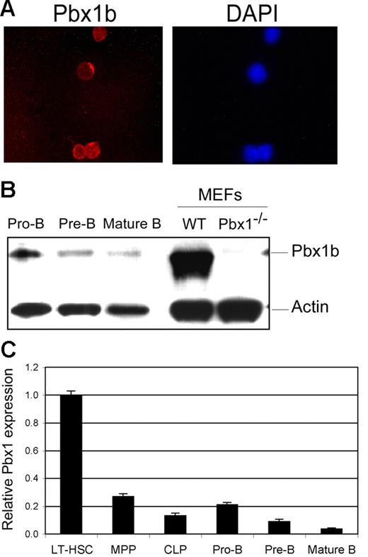
We investigated Pbx1 expression during B-cell development starting with CLPs, isolated by flow cytometry from adult bone marrow as Lin−CD127+c-KitintSca-1int cells.19 Immunocytochemical analysis showed that Pbx1b was present in CLPs (Figure 1A). Western blot analysis of FACS-purified B-cell progenitors from the adult bone marrow revealed Pbx1b in pro-B (B220+CD43+), pre-B (B220+CD43−), and mature B (B220+IgM+) cells (Figure 1B). Quantitative reverse transcription PCR (RT-PCR) analysis of purified progenitors and B-cell precursors showed that Pbx1 expression levels generally decreased as cells progressed toward maturity (Figure 1C). Pbx1a, in contrast, was not detected in any of these populations. Thus, all cells with B-lineage differentiation potential (HSCs, CLPs, and pro-B, pre-B, and mature B cells) selectively express Pbx1b, which is significantly down-regulated with maturation.
Pbx1 is present throughout B-cell development. (A) FACS-purified CLPs (Lin−CD127intc-kitintSca-1+) were analyzed by immunofluorescence using a Pbx1b-specific monoclonal antibody (left panel). Red staining (Texas red) that was confined to the nucleus (blue staining right panel, DAPI) indicates the presence of Pbx1b. (B) Western blot analysis of double-sorted B-cell populations shows presence of Pbx1b protein in pro-B (B220+CD43+), pre-B (B220+CD43−), and mature B (B220+IgM+) cells that progressively decreases with increasing maturation. Pbx1+/+ and Pbx1−/− MEFs were used as positive and negative controls, respectively. Faint cross-reactive band in last lane is not representative of residual Pbx1b protein. (C) Pbx1 transcripts were assessed in purified progenitors and B-cell populations by quantitative RT-PCR analysis. Relative levels of expression are compared to long-term HSCs. Error bars represent SEMs for triplicate analyses.
Pbx1 is present throughout B-cell development. (A) FACS-purified CLPs (Lin−CD127intc-kitintSca-1+) were analyzed by immunofluorescence using a Pbx1b-specific monoclonal antibody (left panel). Red staining (Texas red) that was confined to the nucleus (blue staining right panel, DAPI) indicates the presence of Pbx1b. (B) Western blot analysis of double-sorted B-cell populations shows presence of Pbx1b protein in pro-B (B220+CD43+), pre-B (B220+CD43−), and mature B (B220+IgM+) cells that progressively decreases with increasing maturation. Pbx1+/+ and Pbx1−/− MEFs were used as positive and negative controls, respectively. Faint cross-reactive band in last lane is not representative of residual Pbx1b protein. (C) Pbx1 transcripts were assessed in purified progenitors and B-cell populations by quantitative RT-PCR analysis. Relative levels of expression are compared to long-term HSCs. Error bars represent SEMs for triplicate analyses.
Failure of in situ B-cell development in Pbx1−/−Rag1−/− blastocyst-complemented chimeric mice
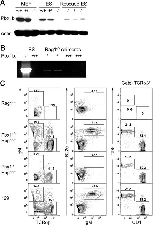
The embryonic lethality of Pbx1−/− embryos at E15 prior to establishment of a lymphoid system prevented an analysis of their in situ lymphoid development. Therefore, the role of Pbx1 in successive stages of the B-cell developmental pathway was examined in chimeric mice using the Rag1-deficient blastocyst complementation assay.20,21 Initially, Pbx1−/− ES cell lines were generated by gene targeting (Figure S1 presents targeting strategy), and the lack of Pbx1 protein in appropriately targeted ES clones was confirmed by Western blot analysis (Figure 2A). Somatic chimeras were then generated by injecting Pbx1−/− (or control) ES cells into Rag1-deficient (Rag1−/−) blastocysts. Analysis of the offspring mice revealed that Pbx1+/+ and Pbx1+/− ES cells contributed to high levels (85%-95%) of coat chimerism in most of the progeny, whereas only low levels of chimerism (10%-15%) were observed in a minority of mice resulting from Pbx1−/− ES cell blastocyst injections, likely reflecting the essential role of Pbx1 in embryogenesis. Nevertheless, a subset of Pbx1−/−Rag1−/− coat color chimeras (20%) contained Pbx1 null cells in the peripheral blood as detected by PCR analysis (Figure 2B).
Absence of B lymphocytes in the peripheral blood of Pbx1−/−Rag1−/− blastocyst-complemented mice. (A) Western blot analysis of ES cells and MEFs (genotypes indicated at the top of the panel) demonstrates the absence of Pbx1b protein in homozygous null cells and the presence of exogenous Pbx1b in Pbx1−/− ES cells that were transfected with a lentiviral vector expressing Pbx1b cDNA under the control of the PGK promoter. Actin was used as control for protein loading. (B) PCR analysis confirmed the presence of Pbx1−/− ES cell-derived cells in the blood of Pbx1−/−Rag1−/− chimeric mice. (C) FACS analysis using B and T cell-specific fluorochrome-conjugated antibodies was performed on cells from the peripheral blood mononuclear cells (PBMCs) of chimeric and control mice. No IgM+ or IgM+B220+ B cells were detected in the Pbx1−/−Rag1−/− adult chimeric mice (third row panels). Conversely, TCRαβ+ CD4 and CD8 single-positive cells were detected in the blood of Pbx1−/−Rag1−/− mice. The ratio of CD4+CD8− to CD4−CD8+ cells was not significantly altered. Pbx1+/+Rag1−/− chimeric control mice had normal numbers of B and T cells in their PBMCs (second row panels). Analyses from age-matched Rag1−/− and 129 mice are also shown.
Absence of B lymphocytes in the peripheral blood of Pbx1−/−Rag1−/− blastocyst-complemented mice. (A) Western blot analysis of ES cells and MEFs (genotypes indicated at the top of the panel) demonstrates the absence of Pbx1b protein in homozygous null cells and the presence of exogenous Pbx1b in Pbx1−/− ES cells that were transfected with a lentiviral vector expressing Pbx1b cDNA under the control of the PGK promoter. Actin was used as control for protein loading. (B) PCR analysis confirmed the presence of Pbx1−/− ES cell-derived cells in the blood of Pbx1−/−Rag1−/− chimeric mice. (C) FACS analysis using B and T cell-specific fluorochrome-conjugated antibodies was performed on cells from the peripheral blood mononuclear cells (PBMCs) of chimeric and control mice. No IgM+ or IgM+B220+ B cells were detected in the Pbx1−/−Rag1−/− adult chimeric mice (third row panels). Conversely, TCRαβ+ CD4 and CD8 single-positive cells were detected in the blood of Pbx1−/−Rag1−/− mice. The ratio of CD4+CD8− to CD4−CD8+ cells was not significantly altered. Pbx1+/+Rag1−/− chimeric control mice had normal numbers of B and T cells in their PBMCs (second row panels). Analyses from age-matched Rag1−/− and 129 mice are also shown.
Flow cytometric analysis showed that chimeric mice generated from blastocysts injected with Pbx1+/+ or Pbx1+/− ES cells contained mature B (IgM+B220+) and T (TCRαβ+) lymphocytes in the peripheral blood. Since Rag1−/− mice do not produce mature lymphocytes (Figure 2C) due to a deficiency in V(D)J recombination,22 circulating B and T cells in these somatic chimeras must have exclusively derived from the microinjected ES cells indicating that they complemented the Rag1 deficiency in lymphopoiesis. The chimeras generated from Pbx1−/− ES cells displayed a complete absence of B cells (IgM+ or IgM+B220+) in the peripheral blood (Figure 2C) demonstrating that Pbx1 was required for B-lineage lymphopoiesis. In contrast to the lack of circulating B cells, TCRαβ+ cells comprised of mature CD4 and CD8 single-positive T cells (Figure 2C) were present in the blood of Pbx1−/−Rag1−/− chimeric mice suggesting a differential requirement of Pbx1 for B- versus T-lineage development.
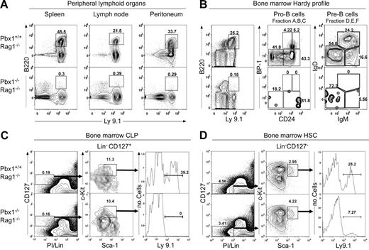
The absence of B cells in the peripheral blood prompted an analysis for abnormalities in the B-cell compartments of secondary lymphoid organs in Pbx1−/−Rag1−/− chimeric mice. Expression of the Ly9.1 alloantigen was used to distinguish cells derived from recipient blastocyst (C57BL/6 background) versus microinjected ES cells (129 background). First described as an alloantigenic marker of lymphocyte differentiation,23,24 Ly9 is a surface glycoprotein (Lgp-100, CD229) that is expressed on most thymocytes, peripheral T and B cells, bone marrow lymphoid cells, and hematopoietic progenitors. In chimeras generated from Pbx1−/−ES cells, no B220+ cells were detected in the Ly9.1+ fraction of the spleen and lymph node (Figure 3A), demonstrating the absence of Pbx1−/− conventional (B2) B lymphocytes in these secondary lymphoid locations. Analysis of the peritoneal cells revealed an absence of detectable B2 as well as B1 cells (Figure 3A). Thus, the peripheral lymphoid tissues were completely devoid of mature B220+ B cells representative of either the B1 or B2 lineages.
Absence of B cells in the secondary lymphoid tissues and impaired B-cell development in the bone marrow of Pbx1−/−Rag1−/− chimeric mice. (A) FACS analysis was performed on cells harvested from the spleen, lymph node, and peritoneal cavity of chimeric mice. Staining with anti-B220 (B cells) and anti-Ly9.1 (donor) antibodies showed that all sites in Pbx1−/−Rag1−/− mice were devoid of donor-derived B cells (Ly9.1+B220+). (B) Hardy profile analysis25 was performed on bone marrow cells of chimeric mice. Donor-derived (Ly9.1+) cells from fractions A through C (B220+CD43+) and D through F (B220+CD43−) were completely absent in the bone marrow of the Pbx1−/−Rag1−/− mice. (C) FACS analysis of bone marrow cells for CLPs19 revealed the absence of Ly9.1+ CLPs in Pbx1−/−Rag1−/− chimeras. (D) FACS analysis of bone marrow cells for HSCd revealed the presence of Ly9.1+ HSCs in Pbx1−/−Rag1−/− chimeras.
Absence of B cells in the secondary lymphoid tissues and impaired B-cell development in the bone marrow of Pbx1−/−Rag1−/− chimeric mice. (A) FACS analysis was performed on cells harvested from the spleen, lymph node, and peritoneal cavity of chimeric mice. Staining with anti-B220 (B cells) and anti-Ly9.1 (donor) antibodies showed that all sites in Pbx1−/−Rag1−/− mice were devoid of donor-derived B cells (Ly9.1+B220+). (B) Hardy profile analysis25 was performed on bone marrow cells of chimeric mice. Donor-derived (Ly9.1+) cells from fractions A through C (B220+CD43+) and D through F (B220+CD43−) were completely absent in the bone marrow of the Pbx1−/−Rag1−/− mice. (C) FACS analysis of bone marrow cells for CLPs19 revealed the absence of Ly9.1+ CLPs in Pbx1−/−Rag1−/− chimeras. (D) FACS analysis of bone marrow cells for HSCd revealed the presence of Ly9.1+ HSCs in Pbx1−/−Rag1−/− chimeras.
The bone marrow was investigated to localize the stage where B-cell development was blocked by Pbx1 deficiency. B-cell progenitor analysis (Hardy profile)25 revealed that Hardy fractions A through F representing all stages of B-cell development from pro-B through mature B cells were completely missing in the Ly9.1+ bone marrow fraction of Pbx1−/−Rag1−/− chimeric mice (Figure 3B). This contrasted with the normal course of B-lymphocyte development that was present in the bone marrows of chimeric mice generated from Pbx1+/+ ES cells. These observations indicate that in situ B-lineage lymphopoiesis fails in Rag1−/− chimeras in the absence of Pbx1 and is blocked at an early stage of development prior to Hardy fraction A.
Development of CLPs is dependent on Pbx1
B cells differentiate from CLPs.19 Because Pbx1−/− ES cells did not contribute to the earliest stages of B-cell development in the bone marrow, the chimeric mice were analyzed by FACS for potential alterations of CLPs. Despite significant chimerism in secondary lymphoid tissues and the bone marrow of Pbx1−/−Rag1−/− chimeric mice, no Ly9.1+ CLPs were detected in their bone marrows (Figure 3C). By comparison, a substantial proportion (up to 39% of total) of CLPs in the bone marrows of Pbx1+/+Rag1−/− mice was ES cell-derived (Ly9.1+). We further analyzed upstream of CLPs for the presence of HSCs. Pbx1−/−Rag1−/− chimeras produced donor-derived (Ly9.1+) HSCs; however, the percentage was lower compared to the control chimeras (Figure 3D). Therefore, development of CLPs in Rag1−/− chimeras requires Pbx1, and the failure of B-lineage lymphopoiesis in the absence of Pbx1 is likely due to a lack of upstream CLP.
T-lineage developmental potential is partially preserved in the absence of Pbx1
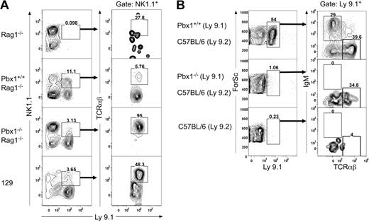
CLPs have been reported to differentiate into lymphocytes of the T-, B-, and NK-cell lineages.19 Consistent with the absence of CLPs and B lymphocytes, very few Pbx1−/−ES cell-derived NK1.1+ cells were detected in the Pbx1−/−Rag1−/− chimeric mice (Figure 4A). Nonetheless, despite the absence of detectable CLPs in Pbx1−/−Rag1−/− chimeric mice, substantial numbers of TCRαβ+ T cells were present in the peripheral blood (Figure 2C) and secondary lymphoid tissues (data not shown) at 5 to 20 weeks of age. The few NK1.1+ cells detected in the Ly9.1+ fraction of these mice were exclusively NKT cells (NK1.1+TCRαβ+; Figure 4A). Furthermore, the ratios of CD4+ to CD8+ cells appeared to be normal in Pbx1−/−Rag1−/− chimeric mice, similar to those in the control Pbx1+/+ ES-complemented mice.
Poor lymphoid reconstitution in transplant recipients of Pbx1−/−Rag1−/− bone marrow cells. (A) FACS analysis using NK and T cell-specific fluorochrome-conjugated antibodies was performed on cells from the peripheral blood of chimeric and control mice.26 Very few Ly9.1+NK1.1+ cells were detected in the Pbx1−/−Rag1−/− adult chimeric mice. All NK1.1+ cells in Pbx1−/−Rag1−/− chimeric mice were TCRαβ+ (NKT cells) in contrast to Pbx1+/+Rag1−/− mice. Analyses of Rag1−/− and 129 mice are shown for comparison. (B)Equal numbers of donor-derived (Ly9.1+) Pbx1−/− or Pbx1+/+ cells were FACS purified from Rag1−/− blastocyst-complemented mice and transplanted into lethally irradiated C57BL/6 recipients along with 2 × 10⁶Rag1−/− bone marrow cells. No donor-derived B cells were detected in the peripheral blood of recipients receiving Pbx1−/−Rag1−/− bone marrow in contrast to recipients of control Pbx1+/+Rag1−/− bone marrow. Reconstitution of TCRαβ cells was present, but poor, in recipients receiving Pbx1−/−Rag1−/− bone marrow.
Poor lymphoid reconstitution in transplant recipients of Pbx1−/−Rag1−/− bone marrow cells. (A) FACS analysis using NK and T cell-specific fluorochrome-conjugated antibodies was performed on cells from the peripheral blood of chimeric and control mice.26 Very few Ly9.1+NK1.1+ cells were detected in the Pbx1−/−Rag1−/− adult chimeric mice. All NK1.1+ cells in Pbx1−/−Rag1−/− chimeric mice were TCRαβ+ (NKT cells) in contrast to Pbx1+/+Rag1−/− mice. Analyses of Rag1−/− and 129 mice are shown for comparison. (B)Equal numbers of donor-derived (Ly9.1+) Pbx1−/− or Pbx1+/+ cells were FACS purified from Rag1−/− blastocyst-complemented mice and transplanted into lethally irradiated C57BL/6 recipients along with 2 × 10⁶Rag1−/− bone marrow cells. No donor-derived B cells were detected in the peripheral blood of recipients receiving Pbx1−/−Rag1−/− bone marrow in contrast to recipients of control Pbx1+/+Rag1−/− bone marrow. Reconstitution of TCRαβ cells was present, but poor, in recipients receiving Pbx1−/−Rag1−/− bone marrow.
Transplantation experiments were performed to further interrogate the lymphoid differentiation potential of Pbx1-deficient bone marrow progenitors. Lin− cells (Ly9.1+) were purified by flow cytometry from chimeric mice and transplanted into lethally irradiated C57BL/6 (Ly9.2) recipient mice along with a rescue dose of Rag1-deficient bone marrow cells. Pbx1−/− cells engrafted very poorly as evidenced by the low percentage of Ly9.1+ cells in the recipient bone marrow when compared to the high levels of Ly9.1+ cells present in the control mice given transplants with equal numbers of Pbx1+/+ cells (Figure 4B). Pbx1−/− cells did not contribute to development of B cells; however, a low level of TCRαβ+ T cells was detected in the peripheral blood of the transplant recipients (Figure 4B). Similar reduced levels of chimerism and lack of B cells or their early progenitors were observed in all lymphoid organs and the bone marrow of the recipients of transplants with Pbx1−/− cells (data not shown). Therefore, Pbx1 deficiency severely compromises hematopoietic differentiation potential along the B- and NK-cell lineages initiating from CLPs, but permits at least partial long-term T-lineage lymphopoiesis.
B-cell development is restored by constitutive expression of Pbx1b
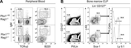
Rescue of the Pbx1 lymphoid deficiency in chimeric mice was attempted to confirm the specific role of Pbx1b in CLPs and B-lineage development. Pbx1−/− ES cells were transduced with a lentiviral vector constitutively expressing Pbx1b cDNA under control of the PGK promoter (Figure S2). Clones (Pbx1−/−R) with the highest level of re-expressed Pbx1b protein by Western blot analysis (Figure 2A) were selected for injection into Rag1−/− blastocysts. FACS analysis of the resulting chimeric mice showed the presence of IgM+ and B220+ cells in the peripheral blood at levels comparable to the control Pbx1+/+Rag1−/− chimeric mice (Figure 5A), indicating successful rescue of the B-cell deficiency observed in Pbx1−/−Rag1−/− chimeras. Donor ES cell-derived B220+ cells were also present in the spleen, lymph nodes, and peritoneal cavity, and complete restoration of the B-cell differentiation pathway from Hardy fractions A through F was observed in the bone marrow (not shown). Similarly, reintroduction of Pbx1b restored the loss of CLPs in the bone marrow (Figure 5B). These data demonstrate phenotypic rescue of adult B lymphopoiesis and further confirm a critical role for Pbx1b in specification of the B-lymphoid lineage.
Reintroduction of Pbx1b in Pbx1−/− ES cells restores B-cell and CLP development in Rag1-deficient blastocyst-complemented mice. (A) FACS analysis shows the presence of IgM+B220+ B cells and normal numbers of TCRαβ+ cells in the peripheral blood of mice chimeric for Pbx1b-rescued ES cells at levels comparable with Pbx1+/+Rag1−/− control mice. (B) FACS analysis shows that donor-derived (Ly9.1+) CLPs are present in Pbx1−/−RRag1−/− chimeras.
Reintroduction of Pbx1b in Pbx1−/− ES cells restores B-cell and CLP development in Rag1-deficient blastocyst-complemented mice. (A) FACS analysis shows the presence of IgM+B220+ B cells and normal numbers of TCRαβ+ cells in the peripheral blood of mice chimeric for Pbx1b-rescued ES cells at levels comparable with Pbx1+/+Rag1−/− control mice. (B) FACS analysis shows that donor-derived (Ly9.1+) CLPs are present in Pbx1−/−RRag1−/− chimeras.
Transient B-cell development occurs when Pbx1-deficient fetal liver cells are transferred to Rag-deficient mice
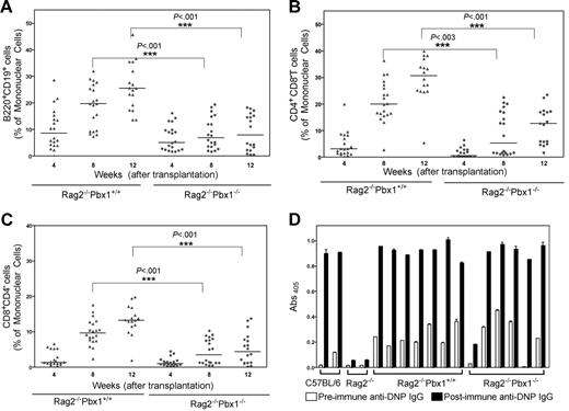
Pbx1−/− embryos die by E15 with a significant defect in myeloid/erythroid development.9 The fetal liver is small (about 20% of normal size) but yields cells that support long-term (> 9 months) reconstitution and persistent erythroid development. To assess their B-cell developmental potential, fetal liver mononuclear cells from E14.5 Pbx1−/− and control (Pbx1+/+) embryos were transplanted into lethally irradiated Rag-deficient adult mice and monitored for reconstitution of lymphocytes by analyzing peripheral blood at 4, 8, and 12 weeks after transfer. B cells (B220+CD19+) in control recipients increased steadily with time, whereas B cells in Pbx1−/− fetal liver recipients, although initially detectable, did not increase, resulting in a significant difference between B-cell numbers in the peripheral blood in control and Pbx1−/− recipients at 8 and 12 weeks after transfer (P < .001 and P < .001, respectively; Figure 6A). Examination of the T-cell compartment in recipients of fetal liver from Pbx1−/− embryos indicated that T-cell development was significantly decreased but that mature CD4+ and CD8+ T cells were detectable in these mice (Figure 6B-C) and followed similar kinetics of reconstitution as B cells. Pbx1−/− fetal liver recipients displayed disorganized lymphoid follicles (Figure S3) and very few immature B cells (B220+IgMhighIgDlow) in the spleen, but nevertheless mounted comparable antigen-specific IgM (not shown) and IgG (Figure 6D) antibody responses against a T-dependent antigen (KLH-DNP). Thus, the fetal liver transfer experiments suggest that there was a transient wave of lymphocyte development in the Pbx1−/− fetal liver chimeras in contrast to the continued lymphocyte development that occurred in wild-type chimeras. However, B cells in the absence of Pbx1 mount comparable immune responses.
A transient and quantitatively reduced lymphoid development occurs in Pbx−/− fetal liver transplant recipients. (A) Wild-type or Pbx1−/− fetal liver cells (1 × 10⁶) were transferred to lethally irradiated Rag2−/− recipients. At the indicated time points, FACS analysis was performed to assess B-cell reconstitution in peripheral blood from chimeric mice using fluorochrome-conjugated B cell-specific (B220 and CD19) antibodies. Very few B220+CD19+ cells were observed in Pbx1−/− as well as Pbx1+/+ fetal liver recipients at 4 weeks after transplantation. The numbers of B cells steadily increased at later time points in Pbx1+/+Rag2−/− fetal liver chimeras (▴), but not in the Pbx1−/− chimeras (•). Median values (of 20 recipients) are represented by a solid horizontal line at each time point. (B) FACS analysis was performed to monitor T- cell reconstitution in the peripheral blood of chimeric mice using fluorochrome-conjugated anti-CD4 and CD8 antibodies. Reconstitution of CD4+CD8− T cells in the Pbx1++ and Pbx1−/− fetal liver chimeras followed similar patterns as B cells. (C) The kinetics of reconstitution of CD8+CD4− was also similar to B cells indicating significantly reduced lymphopoiesis in the Pbx1−/−Rag2−/− fetal liver chimeras. (D) Chimeric mice were immunized with KLH-DNP (100 μg, intraperitoneally) and serum anti-DNP IgG antibodies were measured by ELISA. Pbx1−/− chimeras mounted comparable DNP-specific IgG response with wild-type chimeras. Error bars represent SEMs for triplicate analyses.
A transient and quantitatively reduced lymphoid development occurs in Pbx−/− fetal liver transplant recipients. (A) Wild-type or Pbx1−/− fetal liver cells (1 × 10⁶) were transferred to lethally irradiated Rag2−/− recipients. At the indicated time points, FACS analysis was performed to assess B-cell reconstitution in peripheral blood from chimeric mice using fluorochrome-conjugated B cell-specific (B220 and CD19) antibodies. Very few B220+CD19+ cells were observed in Pbx1−/− as well as Pbx1+/+ fetal liver recipients at 4 weeks after transplantation. The numbers of B cells steadily increased at later time points in Pbx1+/+Rag2−/− fetal liver chimeras (▴), but not in the Pbx1−/− chimeras (•). Median values (of 20 recipients) are represented by a solid horizontal line at each time point. (B) FACS analysis was performed to monitor T- cell reconstitution in the peripheral blood of chimeric mice using fluorochrome-conjugated anti-CD4 and CD8 antibodies. Reconstitution of CD4+CD8− T cells in the Pbx1++ and Pbx1−/− fetal liver chimeras followed similar patterns as B cells. (C) The kinetics of reconstitution of CD8+CD4− was also similar to B cells indicating significantly reduced lymphopoiesis in the Pbx1−/−Rag2−/− fetal liver chimeras. (D) Chimeric mice were immunized with KLH-DNP (100 μg, intraperitoneally) and serum anti-DNP IgG antibodies were measured by ELISA. Pbx1−/− chimeras mounted comparable DNP-specific IgG response with wild-type chimeras. Error bars represent SEMs for triplicate analyses.
B-cell development occurs in the absence of Pbx1 from the pro-B cell (CD19+) stage onward
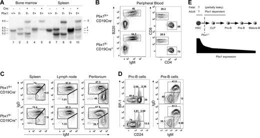
The foregoing results obtained from blastocyst complementation assays and fetal liver transplantation experiments demonstrated that Pbx1b is required at the earliest stages of B-cell development. To assess its potential continued requirement at later stages, a conditional knockout approach was used to specifically inactivate the Pbx1 gene at the pro-B–cell stage of differentiation. Mice containing a floxed Pbx1 gene were bred with Pbx1+/− mice and then with mice that express the Cre recombinase under control of the CD19 locus15 to obtain Pbx1f/−CD19Cre mice. Southern blot analysis confirmed specific and complete deletion of Pbx1 in CD19+ B-cell populations in the spleen and bone marrow of Pbx1f/−CD19Cre mice (Figure 7A). FACS analysis demonstrated that mice lacking Pbx1 at the pro-B stage (CD19+, Hardy fraction B) had normal percentages of B220+IgM+ cells in the peripheral blood (Figure 7B) and secondary lymphoid organs (Figure 7C). In the bone marrow, normal numbers and ratios of cells in Hardy fractions A through F indicated that B-cell differentiation proceeded in mice lacking Pbx1 from CD19+ pro-B cells onward (Figure 7D). Therefore, the requirement for Pbx1 in specification of the B-cell lineage occurs prior to the pro-B–cell stage of differentiation consistent with the lack of CLPs in Pbx1-deficient chimeras.
Conditional inactivation of Pbx1 in pro-B cells using CD19Cre does not prevent B-cell development. (A) Southern blot analysis of Pbx1 gene configurations in FACS-purified B cells (CD19+) in the bone marrow (left) and spleen (right) was performed on mice with the genotypes indicated above the gel lanes. The respective migrations of Pbx1 DNA fragments corresponding to the deleted (Δ), floxed (f), wild-type (+), and null (−) alleles are indicated to the right of the panel. The absence of floxed and wild-type alleles in CD19+ cells of Pbx1f-−CD19Cre mice indicates complete Pbx1 deletion in B cells (lanes 3 and 8). (B) FACS analysis shows normal numbers of B220+IgM+ B cells and CD4+ and CD8+ single-positive T cells in the peripheral blood of Pbx1f/−CD19Cre transgenic mice. (C) FACS analysis demonstrates that IgMhiIgDlo and IgMloIgDhi B-cell subsets are present at normal levels in the secondary lymphoid organs of Pbx1f/−CD19Cre mice. (D) B-cell developmental subsets (Hardy fractions A-F) are normally represented in the bone marrow of Pbx1f/−CD19Cre mice. (E) Role of Pbx1 during B-cell development. The B-cell developmental pathway originating from upstream HSCs and CLPs is shown schematically. Relative Pbx1 expression levels are illustrated below and depict the marked down-regulation of Pbx1 with progressive differentiation along the B-cell lineage. Genetic analyses in this report define a Pbx1-dependent stage of B-cell development, with a critical requirement for Pbx1 function at a point between HSCs and B-cell commitment.
Conditional inactivation of Pbx1 in pro-B cells using CD19Cre does not prevent B-cell development. (A) Southern blot analysis of Pbx1 gene configurations in FACS-purified B cells (CD19+) in the bone marrow (left) and spleen (right) was performed on mice with the genotypes indicated above the gel lanes. The respective migrations of Pbx1 DNA fragments corresponding to the deleted (Δ), floxed (f), wild-type (+), and null (−) alleles are indicated to the right of the panel. The absence of floxed and wild-type alleles in CD19+ cells of Pbx1f-−CD19Cre mice indicates complete Pbx1 deletion in B cells (lanes 3 and 8). (B) FACS analysis shows normal numbers of B220+IgM+ B cells and CD4+ and CD8+ single-positive T cells in the peripheral blood of Pbx1f/−CD19Cre transgenic mice. (C) FACS analysis demonstrates that IgMhiIgDlo and IgMloIgDhi B-cell subsets are present at normal levels in the secondary lymphoid organs of Pbx1f/−CD19Cre mice. (D) B-cell developmental subsets (Hardy fractions A-F) are normally represented in the bone marrow of Pbx1f/−CD19Cre mice. (E) Role of Pbx1 during B-cell development. The B-cell developmental pathway originating from upstream HSCs and CLPs is shown schematically. Relative Pbx1 expression levels are illustrated below and depict the marked down-regulation of Pbx1 with progressive differentiation along the B-cell lineage. Genetic analyses in this report define a Pbx1-dependent stage of B-cell development, with a critical requirement for Pbx1 function at a point between HSCs and B-cell commitment.
Discussion
Studies presented here demonstrate that Pbx1, a homeodomain transcription factor and pre-B–cell oncoprotein, is required in chimeric mice for the development of B cells initiating at CLPs. The lack of Pbx1 completely depletes CLPs and results in the absence of detectable numbers of B and NK cells. T-cell numbers are also decreased, but residual T cells are clearly detectable in the absence of Pbx1.
These conclusions are based on the inability of Pbx1-deficient ES cells to generate CLPs and their progeny lymphocytes in chimeric Rag1-deficient mice. The experimental approach using chimeric mice was necessitated by the embryonic lethality of Pbx1 null embryos prior to formation of a lymphoid compartment. A similar deficit of B-cell development was observed in Pbx1−/− fetal liver recipients where transient lymphocyte development was observed and also suggesting that the requirement is not absolute. Thus, Pbx1 joins a small cohort of transcriptional regulators that are cell intrinsic factors specifically required for development of the B-lymphoid lineage. However, our results distinguish Pbx1 from these critical regulators as a more upstream B-lineage requirement and one of the first factors known to contribute to development of CLPs.
Specification of the B-cell lineage and differentiation of committed pro-B cells depend critically on the coordinated and sequential actions of transcription factors E2A, EBF, and Pax5.27 The E2a protein isoforms E12 and E4728 appear to be the most upstream acting of these B-lineage regulators and together with HEB and E2-2 constitute a family of related basic helix-loop-helix (bHLH) proteins (known as E proteins). B lymphopoiesis is dependent on the combined dosage of E proteins, because pro-B–cell development at or prior to the Hardy fraction A stage is severely affected in the absence of either E12 or E47,10,11 whereas it is reduced 2-fold in mice lacking HEB or E2-2.29 E proteins regulate expression of several B cell-specific genes including EBF, which together with E2a proteins, is required for transition of pro-B cells to the Hardy fraction B stage. EBF null mice accumulate B lineage-committed progenitors that express the IL-7 receptor and germline μ transcripts.30 Pax5 appears to regulate a slightly later stage transition as evidenced by maturation arrest of progenitors (CD19−λ5+VpreB+) just prior to the pre-B–cell stage in Pax5 null mice.31
Pbx1, in contrast, is required upstream of all 3 of the described transcription factors during B lymphopoiesis since Pbx1-deficient chimeric mice lack all (B220+) B-lineage intermediates as well as CLPs, the upstream progenitors of the earliest B-cell progenitors. Our demonstration in this study that Pbx1 is required for CLP development introduces this transcription factor as a key regulator in B-cell development. Recently, the zinc-finger transcription factor Bcl11a (Evi9) was shown to serve an essential role in early B lymphopoiesis upstream of EBF and Pax5.32 Bcl11a mutant embryos lack B cells and have alterations in several types of T cells. Effects on CLPs remain to be determined. Ikaros-deficient mice,33 which also show severe defects in B-cell development, may also lack CLPs.34 However, this study used AA4.1 to phenotypically identify CLPs, a marker that has been reported to be expressed mainly on B cell-restricted lymphoid cells. Thus, Pbx1 is one of the first transcriptional regulators whose function is clearly required for CLP development.
CLPs were originally identified and defined as precursors of both B- and T-lineage lymphocytes as well as NK and dendritic cells.19 Consistent with lack of CLPs we also show the chimeras that are generated from Pbx1 null ES cells lack NK cells. NK cells are bone marrow-derived lymphocytes distinct from B and T cells and can be distinguished from other lymphocytes by the absence of B- and T-cell antigen receptors, and their development does not require events that are necessary for antigen receptor gene rearrangement.35 Indeed, they are present in scid and Rag-1– or Rag-2–deficient mice (Figure 4A).22,36,37 Thus, lack of NK cells in Pbx1-deficient chimeras cannot be attributed to a defective bone marrow microenvironment, and the chimeras generated from control wild-type ES cells produced significant percentages of NK cells (Figure 4A). The lack of NK cells in Pbx1−/−Rag1−/− chimeras could be due to a competitive disadvantage of Pbx1−/− NK cells compared to Rag1−/− NK cells. However, the complete absence of CLPs in Pbx1−/−Rag1−/− chimeras suggests that NK cells may not have developed at all as is the case for B cells and conventional T cells. In mouse adult bone marrow, NK cells develop from CLPs through an intermediate step identified as NK precursor (NKP).26 A subset of transcription factors, which includes Ikaros, ETS family members, Id2, and Id3 have been shown to be involved with development of NK cells.38–41 In our study, we propose that the absence of NK cells in Pbx1−/− chimeras is a consequence of lack of CLP development.
Dendritic cells can be derived from both lymphoid as well as myeloid precursors.42,43 Analysis of dendritic cell lineage in our chimeras was limited by the fact that the Ly9.1 marker is not very well expressed on these cells. Nevertheless, we have shown that despite the absence of detectable CLP, B, and NK cells, HSCs along with T- and NKT-lineage cells developed from Pbx1−/− ES cells in Rag1-deficient mice. This supports the existence of alternative pathways for T-cell generation, including extrathymic pathways,44,45 or pathways independent of CLPs.34 We cannot rule out the possibility that T cells arose during fetal development and persisted postnatally; however, the presence of T cells in mice receiving transplants of Pbx1−/− bone marrow cells is most consistent with de novo T-cell production independent of CLPs. Thus, although CLPs are capable of generating B-, T-, and NK-lineage cells, they are likely not the exclusive progenitor for T cells, which may also arise from HSCs or other upstream multipotent progenitors. The substantial reduction of T-cell numbers in the absence of Pbx1 supports the hypothesis that CLPs represent a major pathway and a crucial intermediate in the development of both B and T cells. The development of T cells in conjunction with the presence of a significant percent of donor-derived (Ly9.1+) cells in all lymphoid locations in Pbx1−/−Rag1−/− chimeric mice strongly suggests that HSCs generated in the absence of Pbx1 are functional. Thus, the failure of B-lineage lymphopoiesis is unlikely to simply reflect a general HSC failure, although our studies cannot exclude HSC defects as a possible contributing factor to the failure of B-cell development. Conversely, tissue-specific inactivation of Pbx1 at the pro-B–cell stage demonstrated that this transcription factor is not required at later stages (beyond Hardy fraction B) of B-cell development. Taken together, the data are most consistent with a Pbx1 requirement between hematopoietic stem cells and commitment to B-cell development at or upstream of CLPs (Figure 7E). However, this requirement is partially leaky in fetal lymphopoiesis.
Our studies provide a developmental context for considering the oncogenic contributions of PBX1, a proto-oncogene that was originally discovered by virtue of its location at the site of chromosomal translocations in acute pre-B–cell leukemia. Interestingly, chromosomal translocations result in the fusion of PBX1 with the E2A gene, which is also required for B-cell development, and resultant production of E2a-Pbx1 chimeric proteins that retain the DNA-binding homeodomain of Pbx1.3,4 The oncogenic fusion of 2 transcriptional regulators that are required for early-stage B-lineage development raises the intriguing possibility that they may normally function collaboratively in B-lineage commitment.
There is precedent for physical and functional interactions of E proteins and TALE homeodomain proteins. Pbx1 binds DNA cooperatively in vitro with the myogenic hHLH proteins MyoD, myf5, and others.46 During muscle differentiation, MyoD regulates expression of myogenin through a divergent E box element in its promoter as a complex with Pbx and Meis TALE proteins.47 Because Pbx-Meis heterodimers occupy this element in undifferentiated cells in the absence of MyoD, it has been proposed that they act as a DNA-bound marker for subsequent recruitment of MyoD.
The generality of the model described is not clear. However, we propose that a similar functional relationship may serve a critical role in establishment of the B lineage by facilitating the recruitment and targeting of E proteins to initiate B-cell commitment. Forced expression of Pbx1 as a fusion protein under control of the E2A promoter may perturb their normal regulated interactive roles and prevent down-regulation of Pbx1 expression that normally accompanies progressive differentiation of B-cell progenitors. The characterization of B cell-specific enhancers occupied by Pbx1 or E proteins in CLPs may support this hypothetical model and provide further insights into the transcriptional regulatory networks that underlie B-cell commitment and are corrupted in a subset of B-cell precursor leukemia that harbor Pbx1 and E2A mutations.
Authorship
Contribution: M.S. designed and performed experiments, analyzed data, and wrote the manuscript; J.W.T. performed experiments and analyzed data; H.K. designed and performed experiments, analyzed data, and edited the manuscript; H.Z. performed experiments; L.S. designed and performed experiments; I.L.W. designed and directed experiments; L.A.H. designed and directed experiments and edited the manuscript; M.C. designed and directed experiments and edited the manuscript.
Conflict-of-interest disclosure: The authors declare no competing financial interests.
Correspondence: Michael L. Cleary, Department of Pathology, Stanford University of School of Medicine, 300 Pasteur Dr, Stanford, CA 94305; e-mail: mcleary@stanford.edu.
The online version of the article contains a data supplement.
The publication costs of this article were defrayed in part by page charge payment. Therefore, and solely to indicate this fact, this article is hereby marked “advertisement” in accordance with 18 USC section 1734.
We thank Richard Hardy for critical reading of the manuscript; Francesca Ficara for helpful discussions; Michael White for assistance in establishing Rag-deficient blastocyst complementation techniques; Patricia Labosky for providing the TL1 cell line; Laurie Ailles for providing lentiviral vectors; Yanru Chen-Tsai, Jennifer Lin, and Denise Leong for blastocyst injections; Cita Nicolas and Maria Ambrus for expert technical assistance; and Omita Herman and Sue Sheppard for tissue processing for flow cytometric analysis.
This work was supported by National Institutes of Health grants CA42971 and CA90735 (M.L.C.), HD43997 (L.S.), and AI047458 (I.L.W.), as well as grant 6-FY03-071 from the March of Dimes and Birth Defects Foundation (L.S.). L.S. is an Irma T. Hirschl Scholar.

This feature is available to Subscribers Only
Sign In or Create an Account Close Modal