Abstract
Human CD32B (FcγRIIB), the low-affinity inhibitory receptor for IgG, is the predominant Fc receptor (FcR) present on B cells. Immunohistochemical and expression studies have identified CD32B expression in a variety of B-cell malignancies, suggesting that CD32B is a potential immunotherapeutic target for B-cell malignancies. A high-affinity monoclonal antibody (mAb 2B6), from a novel panel of anti–human CD32B–specific mAbs, was chimerized (ch2B6) and humanized (hu2B6-3.5). Both ch2B6 and hu2B6-3.5 were capable of directing cytotoxicity by peripheral blood mononuclear cells and monocyte-derived macrophages against B-lymphoma lines in vitro. In a human B-cell lymphoma mouse xenograft model, administration of ch2B6 or hu2B6-3.5 reduced tumor growth rate and improved tumor-free survival. Both the in vitro and in vivo activities of 2B6 required an intact Fc, suggesting an FcR-mediated mechanism of action. These data support the hypothesis that CD32B is a viable target for mAb treatment of B-cell lymphoproliferative disorders.
Introduction
Non-Hodgkin lymphoma (NHL) is the fifth most common form of cancer in the United States, with approximately 55 000 new cases diagnosed annually.1 B-cell malignancies comprise more than 85% of diagnosed lymphomas, with diffuse large B-cell lymphoma (DLBCL) and follicular lymphoma (FL) being the most common (30% and 22%, respectively) of all diagnosed NHLs.2 The anti-CD20 chimeric antibody, rituximab (Rituxan) has been used successfully to improve the outcome of certain groups of patients with NHL.3-5 The improvements in treatment gained with rituximab have been remarkable; however, the limitations of this therapy in certain B-cell malignancies, including those that show limited or no expression of CD20, make a strong case for identifying additional monoclonal antibody (mAb) therapeutics.
The inhibitory Fc-γ receptor IIB (FcγRIIB, CD32B), a type I transmembrane receptor with low affinity for monomeric IgG (Kd ∼106 M–1), is a critical regulatory element in B-cell homeostasis. CD32B controls the threshold and the extent of cell activation by counterbalancing the stimulatory activity of a variety of receptors, including the B-cell antigen receptor.6,7 Consistent with its regulatory function, CD32B is found on several hematopoietic cell types, which also express activating FcRs, and represents the predominant FcR expressed by the B-cell lineage.6,8
The generation of mAbs specific for the extracellular region of human CD32B has been hindered by its homology to CD32A (FcγRIIA), with 96% identity to CD32B within the extracellular region.9 CD32A is highly expressed by myeloid cells and is absent in B cells.8,10 Currently available anti–human CD32 mAbs show variable degrees of cross-reactivity with both receptors or react selectively with CD32A.11 A therapeutic mAb that engages only CD32B, and spares CD32A-expressing cells, may have utility in the treatment of B-cell malignancies. By immunizing CD32A transgenic mice, we have succeeded in generating a set of high-affinity mAbs against human CD32B that do not cross-react with CD32A (M.-C.V., S.G., H.L., S.B., L.H., S.J., J.S., Jeffrey V. Ravetch, Kathryn E. Stein, E.B., and S.K., manuscript in preparation). One mAb, 2B6, demonstrated high affinity and selectivity for CD32B in vitro and was chosen for further study. Here we report that chimeric and fully humanized versions of this mAb can mediate in vitro antibody-dependent cellular cytotoxicity (ADCC) against B-cell lymphoma lines and display potent antitumor activity in vivo in a xenograft mouse model of B-cell lymphoma. These results suggest that targeting CD32B may represent a novel approach to the immunotherapy of B-cell malignancies.
Materials and methods
Chimerization and humanization of the anti-CD32B murine mAb, 2B6
A panel of antibodies specific for human CD32B was generated by immunization of transgenic mice expressing human CD32A with recombinant soluble human CD32B protein (M.-C.V., S.G., H.L., S.B., L.H., S.J., J.S., Jeffrey V. Ravetch, Kathryn E. Stein, E.B., and S.K., manuscript in preparation). Based on affinity and specificity measurements, one antibody, 2B6, was chosen for further study. cDNAs corresponding to the 2B6 variable heavy (VH) and light (VL) chains were amplified by polymerase chain reaction (PCR) using the RLM-RACE kit (Ambion, Austin, TX) with total RNA from the 2B6 hybridoma as template. Gene-specific primers for the VH were SJ15R (5′-GGT CAC TGT CAC TGG CTC AGG G-3′) and SJ16R (5′-agg cgG ATC CAG GGG CCA GTG GAT AGA C-3′). Gene-specific primers for the VL were SJ17R (5′-GCA CAC GAC TGA GGC ACC TCC AGA TG-3′) and SJ18R (5′-cgg cgg atc cGA TGG ATA CAG TTG GTG CAG CAT C-3′). The RACE products were inserted into the plasmid pCR2.1-TOPO using a TOPO TA cloning kit (Invitrogen, Carlsbad, CA). The resulting plasmids were sequenced to determine the VH and VL sequences for 2B6. From these sequences, the framework (FR) and complementarily determining regions (CDR) were identified as defined by Kabat et al.12 For chimerization,13 the mouse VH and VL were then joined to a human C-γ1 constant region, or a human Cκ segment, respectively, and an immunoglobulin leader sequence and inserted into pCI-neo (Promega, Madison, WI) for mammalian expression. Humanization of the 2B6 antibody was carried out as previously described.14 Briefly, murine 2B6 VH and VL FR sequences were replaced by human germline FR sequences, combined with a leader sequence and C-γ1orC-κ constant region segment, respectively, and cloned into the pCI-neo mammalian expression vector. The hu2B6-3.5 VH consists of the FR segments from the human germline VH segment VH1-18 and JH6, and the CDR regions of the 2B6 VH. The h2B6-3.5 VL consists of the FR segments of the human germline VL segment VK-A26 and JK4, and the CDR regions of 2B6 VL. In addition, the following changes were introduced into the hu2B6-3.5 VH and VL by site-directed mutagenesis using the QuickChange kit (Stratagene, La Jolla, CA): VH-M48I and T71V; VL-I21F and N50E.
Reagents and cell lines
Cell lines. Daudi, Raji, and BL41 cell lines were purchased from the American Type Culture Collection ([ATCC], Manassas, VA). Cell lines were grown at 37°C, 5% CO2, in RPMI 1640 media supplemented with 10% fetal bovine serum, 4 mM l-glutamine, 10 mM HEPES (N-2-hydroxyethylpiperazine-N′-2-ethanesulfonic acid), 1 mM sodium pyruvate, 0.1 mM nonessential amino acids, 50 μg/mL penicillin, and 100 μg/mL streptomycin. The human embryonic kidney cell line, 293-H (Invitrogen), and the Chinese hamster ovary cell line, CHO-K1 (ATCC), were grown at 37°C, 5% CO2 in Dulbecco modified Eagle media (high glucose) supplemented with 10% fetal bovine serum, 2 mM l-glutamine, 1 mM sodium pyruvate, 50 μg/mL penicillin, and 100 μg/mL streptomycin.All media, serum, and supplements were purchased from Invitrogen.
Antibodies. Hybridomas expressing anti-CD64 (clone 32.2) and anti-CD32A (IV.3) antibodies were purchased from ATCC. All fluorochrome-labeled isotype control antibodies were purchased from BD Phar Mingen (San Diego, CA). Chimeric and humanized versions of 2B6 were expressed transiently in 293-H cells using Lipofectamine 2000 (Invitrogen). Antibodies were purified by protein Aaffinity and size exclusion chromatography. For flow cytometry, murine 2B6, ch2B6-N297Q, or hu2B6-3.5-N297Q was labeled with fluorescein-isothiocyanate (FITC) or Alexa Fluor-488 (Invitrogen-Molecular Probes, Carlsbad, CA), and purified by size exclusion chromatography
Soluble CD32A(131R) and CD32B proteins. Full-length CD32A(131R) and CD32B1 cDNAs were kind gifts from Dr Jeffrey V. Ravetch (Rockefeller University, New York, NY). Soluble, monomeric CD32B was generated by insertion of the CD32B extracellular domain9 into pcDNA3.1 (Invitrogen; and M.-C.V., S.G., H.L., S.B., L.H., S.J., J.S., Jeffrey V. Ravetch, Kathryn E. Stein, E.B., and S.K., manuscript in preparation). Expression plasmids containing soluble CD32A(131R) or CD32B Fc fusion proteins were generated by combining the extracellular portion of CD32A or CD32B9 with a human IgG2 Fc region mutated at position 297 (N297Q) to eliminate residual FcR binding (M.-C.V., S.G., H.L., S.B., L.H., S.J., J.S., Jeffrey V. Ravetch, Kathryn E. Stein, E.B., and S.K., manuscript in preparation). For expression, soluble receptor constructs were transfected into 293H cells using Lipofectamine 2000 (Invitrogen).
ELISA
For direct-binding enzyme-linked immunosorbent assay (ELISA), plates were coated with 100 ng/well soluble CD32A(131R) or CD32B Fc fusion proteins in phosphate-buffered saline (PBS) overnight at 4°C. After 3 washes with PBS containing 0.1% Tween-20 (PBS-T), plates were incubated with PBS containing 1% bovine serum albumin (BSA), fraction V (Sigma-Aldrich, St Louis, MO) for 1 hour at room temperature. Ten-fold dilutions of the purified antibodies were incubated on the plates coated with the receptors for 1 hour at room temperature. Detection of the bound antibodies was performed with a F(ab′)2 fragment of goat anti–mouse IgG horseradish peroxidase-conjugated antibody (dilution 1:10 000) or F(ab′)2 fragment of goat anti–human IgG F(ab′)2 fragment-specific horseradish peroxidase-conjugated antibody (dilution 1:10 000; Jackson ImmunoResearch Labs, West Grove, PA). After incubation at room temperature for 30 minutes, and 3 washes with PBS-T, the colorimetric reaction was developed with TMB peroxidase substrate and the absorbance was measured at 450 nm using Versamax microplate reader (Molecular Devices, Sunnyvale, CA).
Surface plasmon resonance analysis
Monovalent antibody binding to CD32B was analyzed by surface plasmon resonance using a Biacore 3000 biosensor (Biacore, Uppsala, Sweden). The capturing antibody, a goat F(ab′)2 fragment specific for the Fc fragment of mouse or human IgG1 (Jackson Immuno Research Labs), was immobilized on the CM-5 sensor chip using the amino-coupling kit (Biacore) following the procedure provided by the manufacturer. Binding experiments were performed in HBS-P buffer containing 10 mM HEPES, pH 7.4, 150 mM NaCl, and 0.005% P20 surfactant. Murine, chimeric, or humanized 2B6 was captured by injecting an antibody solution at a flow rate of 5 μL/min, followed by an injection of the monomeric soluble CD32B receptor in duplicates at concentrations of 0 (blank control), 3.13, 6.25, 12.5, 25, 50, and 100 nM, at a flow rate of 50 μL/min for 90 seconds, and a dissociation time of 120 seconds.
Reference curves were obtained by injection of each soluble receptor over the surface with no captured antibody. Experimental data were corrected for the reference and blank control, normalized to the same level of captured antibody, and analyzed using BIAevaluation 3.0 software (Biacore, Uppsala, Sweden). The kinetic association, Ka, and dissociation, Kd, constants were estimated by global fitting analysis of the binding curves to the 1:1 Langmurian interaction model. The equilibrium dissociation constant (KD) was calculated as KD = Kd/Ka.
Cytotoxicity assays
Peripheral-blood mononuclear cell–mediated ADCC assay. Cytotoxicity was measured using an indium 111 (111In) release assay.15 Peripheral-blood mononuclear cells (PBMCs) were purified from whole human blood (BRT Laboratories, Baltimore, MD) by Ficoll-Hypaque (Amersham Biosciences, Piscataway, NJ) density gradient centrifugation following the manufacturer's instructions. Burkitt lymphoma target cells, Daudi, Raji or BL41, (5 × 10⁶ in 0.5 mL) were labeled with 100 μCi (3.7 MBq) 111In-oxine (GE Healthcare, Silver Spring, MD) at room temperature for 30 minutes. Unincorporated 111In was removed through 4 sequential washes with culture media. For effector-target (E/T) ratio titrations (100:1-0.78:1) labeled target cells (5000/well) were coated with antibody at 2 μg/mL for 30 minutes at 37°C, and then combined with an equal volume of PBMCs. For antibody titrations, target cells were opsonized with antibody at various concentrations (30 000-0.01 ng/mL) for 30 minutes at 37°C, 5% CO2. Labeled target cells (5000/well), opsonized with anti-CD32B antibody, were combined with PBMCs at an E/T ratio of 75:1, in round-bottom 96-well plates. Following an 18-hour incubation at 37°C, 5% CO2, cell supernatants were harvested using the Skatron supernatant collection system (Molecular Devices), and released radioactivity was quantified using a Wallac 1470 γ counter (Perkin Elmer, Shelton, CT). Maximal release and spontaneous release were determined by incubation of target cells with 2% Triton X-100 and media alone, respectively. Antibody-independent cellular cytotoxicity was measured by incubation of target and effector cells in the absence of antibody. Each assay was performed in triplicate. The mean percentage specific lysis was calculated from the formula: % specific lysis = (experimental cpm – antibody-independent cpm)/(maximal release cpm – spontaneous release cpm) × 100.
MDM cytotoxicity assay. Elutriated monocytes (Advanced Biotechnologies, Columbia, MD) were plated in 96-well microtiter plates at 100 000 cells/well in macrophage serum-free medium (Invitrogen) in the presence of the following cytokines: human granulocyte-macrophage colony-stimulating factor (GM-CSF, 10 ng/mL; Biosource International, Camarillo, CA), human macrophage colony-stimulating factor (M-CSF, 25 ng/mL; R&D Systems, Minneapolis, MN), human interleukin 10 (IL-10, 25 ng/mL; R&D Systems), human interleukin-1β (IL-1β, 25 ng/mL; R&D Systems), and human IL-6 (25 ng/mL, R&D Systems); cells were grown for 6 days at 37°C, 7% CO2. Media and cytokines (M-CSF, IL-10, IL-1β, and IL-6) were replaced, along with the addition of human interferon γ (IFN-γ, 25 ng/mL; Biosource International) on day 3 and day 5 of culture. Daudi target cells were labeled with 111In according to the protocol used with the PBMC-mediated cytotoxicity assay and then combined with antibodies at various concentrations (10 000-0.1 ng/mL) and monocyte-derived macrophages (MDMs; E/T ratio = 20:1). Following an 18-hour incubation at 37°C, 7% CO2, supernatants were harvested and analyzed as indicated for the PBMC-mediated cytotoxicity assay.
Flow cytometry analysis
Flow cytometry experiments were performed using a FACSCalibur (BD Biosciences-Immunocytometry Systems, San Jose, CA) and analyzed using Cell Quest software (BD Biosciences-Immunocytometry Systems).
CHO cell-binding assays. CHO cell lines expressing CD32A(131R) and CD32B were generated by transfection of CD32A(131R) and CD32B cDNAs and selection with G418 (800 μg/mL; M.C.-V., S.G., H.L., S.B., L.H., S.J., J.S., Jeffrey V. Ravetch, Kathryn E. Stein, E.B., and S.K., manuscript in preparation). For flow cytometry analysis, cells were removed using trypsin-EDTA (Invitrogen) and resuspended in PBS containing 1% BSA, 0.1% sodium azide, (PBS-1% BSA-SA) and 10% AB human serum (NABI Biopharmaceuticals, Rockville, MD). Samples were incubated with ch2B6, hu2B6-3.5, FLI8.26, or IV.3 antibodies (5 μg/mL) for 30 minutes on ice, and washed with PBS-1% BSA-SA. Bound antibodies were detected by incubation for 30 minutes on ice with Cy5-labeled goat F(ab′)2 fragment specific for the Fcγ fragment of either mouse or human IgG (Jackson Immuno Research Laboratories), followed by washing twice with PBS-1% BSA-SA prior to analysis.
CD32B expression in PBLs. Peripheral blood leukocytes (PBLs) were isolated from whole human blood (BRT Laboratories) by lysis of red blood cells in ACK buffer (155 mM ammonium chloride,10 mM potassium bicarbonate, 0.1 mM EDTA, pH 7.4), washed twice in PBS, and resuspended in PBS-1% BSA-SA with 10% AB human serum. PBLs were incubated with Alexa Fluor-488–labeled anti-CD32B antibodies (ch2B6-N297Q, 1 μg/mL or hu2B6-3.5-N297Q, 1 μg/mL), or anti–CD32-FITC (clone FLI8.26, 1 μg/mL, Research Diagnostics, Flanders, NJ), and a lineage-specific marker, anti–CD20-PE (clone L27, BD Bioscience-PharMingen, San Diego, CA), anti–CD3-PE (clone UCHT1, BD Bioscience-PharMingen), anti–CD56-PE (clone MY31, BD-Bioscience-PharMingen), anti–CD14-PE (clone M5E2, BD Bioscience-PharMingen), or anti–CD16-FITC (clone Dj130c, Dako Cytomation, Carpinteria, CA). Samples were incubated on ice for 30 minutes and washed in PBS-1%BSA-SA.
Fc receptor expression in MDMs
MDMs were plated in 6-well dishes (2 × 10⁶/well) and cultured with cytokines as indicated (see “MDM cytotoxicity assay”). MDMs were collected by incubation of cells for 20 to 30 minutes at 37°C, 5% CO2 in PBS/0.53 mM EDTA/glucose (1 g/L). MDMs were resuspended in PBS-1%BSA-SA containing 10% human serum and incubated with the following FITC-labeled antibodies (1-2 μg/mL): anti-CD64 (clone 32.2), anti-CD32A (clone IV.3), anti-CD32B (clone 2B6), or PE-labeled anti-CD16 (clone 3G8, BD Bioscience-PharMingen), for 30 to 60 minutes on ice. Samples were washed twice in PBS-1%BSA-SA prior to analysis.
Mouse xenograft models
Female athymic BALB/c nude (Foxn1nu/nu) mice, 7 to 11 weeks old, were purchased from Taconic (Germantown, NY) and maintained under pathogen-free conditions. Daudi cells (5 × 10⁶/mouse) were resuspended in PBS (5 × 10⁷cells/mL, 100 μL/mouse), combined with Matrigel (100 μL/mouse, BD Biosciences, Discovery Labware, Bedford, MA), and injected subcutaneously (200 μL total volume) into the right flank of BALB/c nude mice. Tumor development was monitored twice per week, using calipers, and tumor weight was estimated by the following formula: tumor weight = (length × width2)/2. For tumor-prevention studies, intraperitoneal injections of antibodies at various concentrations (0.25-250 μg/mouse) were performed weekly for 8 weeks, starting at day 0. In established tumor experiments, implanted Daudi cells were allowed to grow for 7 days prior to intraperitoneal injection of 4 daily consecutive doses of antibody (4 μg/g, 100 μg/mouse). Animal studies were conducted in accordance with the Public Health Service Policy on the Humane Care and Use of Laboratory Animals.
CD32B antigen expression in the presence of anti-CD32B antibodies
In vitro. Daudi cells were cultured in the presence of ch2B6 (1 μg/mL) or hu2B6-3.5 (1 μg/mL) for 24 hours at 37°C, 5% CO2, and analyzed for expression of CD32B or the presence of anti-CD32B antibodies by flow cytometry. Samples were resuspended in PBS-1%BSA-SA, incubated with an FITC-labeled anti-CD32 antibody, FLI8.26 (2 μg/mL), or PE-conjugated, F(ab′)2 goat anti–human Fc–specific secondary antibody (1:500 dilution). Samples were washed twice in PBS-1%BSA-SA and analyzed by flow cytometry using a FACSCalibur.
In vivo. Daudi tumors were generated in vivo as described for the tumor prevention xenograft model. Mice received 8 weekly doses of hu2B6-3.5 (25 μg/mouse), rituximab (25 μg/mouse), or PBS and tumors present following treatment were allowed to grow in vivo for an additional week. Residual tumors were then excised and dissociated into single-cell suspensions using cell growth media supplemented with collagenase IV (3 mg/mL) and DNase (200 μg/mL). Dissociated tumor cells were washed twice in Daudi cell growth media and resuspended in PBS-1%BSA-SA. For flow cytometry analysis, samples were incubated for 30 to 60 minutes at room temperature with FITC-labeled anti-CD32B (murine 2B6, 1 μg/mL) or FITC-labeled anti-CD20 (clone L27, BD Bioscience-PharMingen), washed in PBS-1%BSA-SA, and analyzed using a FACSCalibur.
Statistical methods
Results
Affinity and specificity of chimeric and humanized forms of the 2B6 antibody
mAb 2B6 is a murine IgG1 directed against human CD32B (M.-C.V., S.G., H.L., S.B., L.H., S.J., J.S., Jeffrey V. Ravetch, Kathryn E. Stein, E.B., and S.K., manuscript in preparation).18,19 To investigate the activity of 2B6 mediated by human effector cells, mouse-human chimeric (ch2B6) and fully humanized (hu2B6-3.5) versions of the mAb were generated. Comparison of the relative affinities of murine 2B6, ch2B6, and hu2B6-3.5 for the extracellular domain of CD32B was performed by surface plasmonresonance analysis using a BIAcore 3000. The 1:1 Langmurian interaction data-fitting model was used to predict dissociation constants (KD) as shown in Table 1. The original subnanomolar affinity of murine 2B6 (0.30 ± 0.09 nM) was well preserved in the ch2B6 (0.38 ± 0.14 nM) and hu2B6-3.5 (0.23 ± 0.03 nM) antibodies. Aglycosylated versions of ch2B6 and hu2B6-3.5 were constructed by mutation of an asparagine at position 297 in the Fc region to glutamine (N297Q) to explore the FcR-dependence of the activities triggered by these antibodies. This mutation removes the Fc glycosylation site and eliminates FcR binding.20 The affinity constants of the aglycosylated chimeric and humanized 2B6 antibodies for CD32B were identical to that of the corresponding 2B6 versions containing wild-type Fc regions (data not shown).
Surface plasmon resonance analysis of anti-CD32B antibodies
. | CD32B monovalent binding constants . | . | . | ||
|---|---|---|---|---|---|
| Antibody . | Ka, M-1s-1 . | Kd, s-1 . | KD, nM . | ||
| 2B6 | 26.1 × 10⁵ ± 12.4 × 10⁵ | 6.9 × 10⁻⁴ ± 0.9 × 10⁻⁴ | 0.30 ± 0.09 | ||
| ch2B6 | 31.0 × 10⁵ ± 10.3 × 10⁵ | 10.9 × 10⁻⁴ ± 1.7 × 10⁻⁴ | 0.38 ± 0.14 | ||
| hu2B6-3.5 | 51.2 × 10⁵ ± 9.6 × 10⁵ | 11.4 × 10⁻⁴ ± 1.5 × 10⁻⁴ | 0.23 ± 0.03 | ||
Values are the mean ± SD of at least 3 independent experiments.
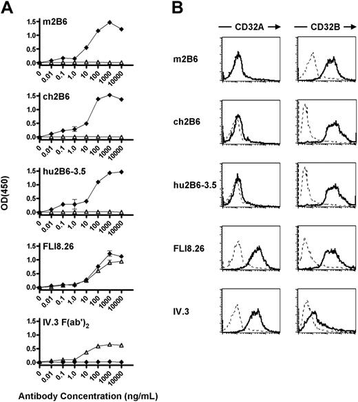
Specificity of ch2B6 and hu2B6 antibody binding to recombinant CD32B. (A) Microtiter plates were coated with soluble CD32A (▵) or CD32B (♦) Fc fusion proteins. Purified 2B6 antibodies or CD32A reactive antibodies (FLI8.26, IV.3 F(ab′)2) were added at the indicated concentrations (10 000-0.01 ng/mL). Measurements were performed in duplicate and results were normalized to a control mouse or human antibody. Error bars represent SD. (B) Stably transfected CHO cells expressing CD32A-R131 or CD32B were incubated with 5 μg/mL 2B6, ch2B6, hu2B6-3.5, FLI8.26, or IV.3 antibodies (bold line), or the appropriate isotype control antibody (dashed line) in PBS-1% BSA-SA with 10% human AB serum, followed by staining with Cy5-labeled goat F(ab′)2 fragment specific for the Fc portion of either mouse or human IgG.
Specificity of ch2B6 and hu2B6 antibody binding to recombinant CD32B. (A) Microtiter plates were coated with soluble CD32A (▵) or CD32B (♦) Fc fusion proteins. Purified 2B6 antibodies or CD32A reactive antibodies (FLI8.26, IV.3 F(ab′)2) were added at the indicated concentrations (10 000-0.01 ng/mL). Measurements were performed in duplicate and results were normalized to a control mouse or human antibody. Error bars represent SD. (B) Stably transfected CHO cells expressing CD32A-R131 or CD32B were incubated with 5 μg/mL 2B6, ch2B6, hu2B6-3.5, FLI8.26, or IV.3 antibodies (bold line), or the appropriate isotype control antibody (dashed line) in PBS-1% BSA-SA with 10% human AB serum, followed by staining with Cy5-labeled goat F(ab′)2 fragment specific for the Fc portion of either mouse or human IgG.
Specificity of ch2B6 and hu2B6-3.5 for CD32B was confirmed by ELISA and flow cytometry using the murine 2B6 as a reference. Binding of ch2B6 or hu2B6-3.5 to CD32B in ELISA was maximal at approximately 100 ng/mL, with negligible binding to CD32A (Figure 1A). In contrast, a pan-CD32 mAb, FLI8.26,21 recognized either form of the receptor, whereas a CD32A-specific antibody, IV.3,22 reacted exclusively with CD32A, confirming the specificity of the ELISA. To further test the specificity of ch2B6 and hu2B6-3.5 against the cell surface-expressed antigen, CHO cell lines that stably express full-length human CD32A or CD32B were generated and used in flow cytometry analysis. All 2B6 versions reacted with human CD32B-expressing CHO cells, but did not bind CD32A-expressing CHO cells, confirming that ch2B6 and hu2B6-3.5 retain the specificity for human CD32B observed with the original murine mAb (Figure 1B). In addition, aglycosylated versions of chimeric and humanized 2B6 bound CD32B-expressing but not CD32A-expressing CHO cells, confirming that the Fc region is not involved in 2B6 binding or its specificity (Figure S1, available on the Blood website; see the Supplemental Figure link at the top of the online article).
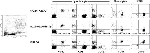
Reactivity of ch2B6 and hu2B6 with human PBLs. Flow cytometry analysis of human PBLs using Alexa Fluor-488–labeled, anti-CD32B antibodies: ch2B6-N297Q (1 μg/mL), hu2B6-3.5-N297Q (1 μg/mL), or FLI8.26-FITC (1 μg/mL), which reacts with both CD32A and CD32B, and PE-labeled antibodies against lineage-specific markers: CD19 (B cells), CD3 (T cells), CD56 (NK cells), CD14 (monocytes), and CD16 (PMNs).
Reactivity of ch2B6 and hu2B6 with human PBLs. Flow cytometry analysis of human PBLs using Alexa Fluor-488–labeled, anti-CD32B antibodies: ch2B6-N297Q (1 μg/mL), hu2B6-3.5-N297Q (1 μg/mL), or FLI8.26-FITC (1 μg/mL), which reacts with both CD32A and CD32B, and PE-labeled antibodies against lineage-specific markers: CD19 (B cells), CD3 (T cells), CD56 (NK cells), CD14 (monocytes), and CD16 (PMNs).
Flow cytometry analysis of peripheral human blood using Alexa Fluor-488–labeled, aglycosylated, chimeric or humanized 2B6 demonstrated robust reactivity with B cells, minimal staining of polymorphonuclear neutrophils (PMNs), and no staining of T cells, natural killer (NK) cells (Figure 2), and platelets (data not shown). Staining of monocytes was donor dependent, with a majority of donors having low levels of staining (Figure 2) with the remainder demonstrating moderate levels of CD32B expression (M.C.-V., S.G., H.L., S.B., L.H., S.J., J.S., Jeffrey V. Ravetch, Kathryn E. Stein, E.B., and S.K., manuscript in preparation). In contrast, staining of human peripheral blood cells with the pan-CD32 reactive antibody, FLI8.26, recognized CD32B-expressing B cells as well as neutrophils and monocytes, which express CD32A (Figure 2). These results further confirm that ch2B6 and hu2B6-3.5 recognize CD32B expressed on normal human PBLs without cross-reactivity with CD32A.
In vitro cytotoxicity of 2B6 against human B-lymphoma cell lines
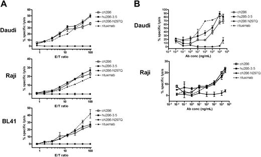
The ability of chimeric and humanized versions of 2B6 to direct ADCC against CD32B-expressing target cells was tested using purified human PBMCs as effector cells. Chimeric and humanized 2B6 antibodies mediated ADCC against the Burkitt lymphoma cell lines, Daudi, Raji, and BL41 (Figure 3A). The ADCC activity of ch2B6 and hu2B6-3.5 was observed at multiple E/T ratios and was dose dependent, with activity observed at concentrations just exceeding 1 ng/mL (Figure 3B). The cell-mediated cytotoxic activity of the anti-CD32B antibodies was dependent on Fc-FcR interactions, as shown by the observation that the aglycosylated form of ch2B6 was devoid of ADCC activity at all concentrations tested (Figure 3A-B).
PBMC-mediated cytotoxicity of ch2B6 and hu2B6-3.5 antibodies against human B-cell lines in vitro. (A) Burkitt lymphoma cell lines, Daudi (top), Raji (middle), and BL41 (bottom) were opsonized with the antibodies at 2 μg/mL: ch2B6 (□), hu2B6-3.5 (▪), ch2B6-N297Q (•), or rituximab (♦), and used as targets for lysis by PBMCs in an 18-hour 111In-release assay. (B) Daudi (top) and Raji (bottom) target cells were used as targets for PBMC-mediated lysis in an 18-hour 111In-release assay with a fixed E/T ratio of 75:1, and a titration of antibodies: ch2B6 (□), hu2B6-3.5 (▪), ch2B6-N297Q (•), or rituximab (♦), at the designated concentrations. All measurements were performed in triplicate. Error bars represent SD.
PBMC-mediated cytotoxicity of ch2B6 and hu2B6-3.5 antibodies against human B-cell lines in vitro. (A) Burkitt lymphoma cell lines, Daudi (top), Raji (middle), and BL41 (bottom) were opsonized with the antibodies at 2 μg/mL: ch2B6 (□), hu2B6-3.5 (▪), ch2B6-N297Q (•), or rituximab (♦), and used as targets for lysis by PBMCs in an 18-hour 111In-release assay. (B) Daudi (top) and Raji (bottom) target cells were used as targets for PBMC-mediated lysis in an 18-hour 111In-release assay with a fixed E/T ratio of 75:1, and a titration of antibodies: ch2B6 (□), hu2B6-3.5 (▪), ch2B6-N297Q (•), or rituximab (♦), at the designated concentrations. All measurements were performed in triplicate. Error bars represent SD.
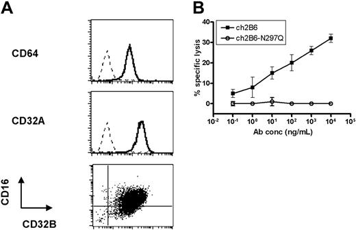
At least 2 different types of cells capable of exerting cytotoxic activity, NK cells and monocytes, are present in unfractionated PBMCs. The contribution of monocytes, which typically require activation to exert cytotoxic activity,23 is likely to be negligible under the conditions of our assay, leaving NK cells as the major cytotoxic players in unfractionated PBMCs. Activated monocytes/macrophages, however, can be efficient cytotoxic cells.23 Because macrophages can express the inhibitory receptor8 and themselves be targeted by an anti-CD32B mAb, it was of interest to ascertain their effective tumoricidal capacity in anti-CD32B–mediated ADCC. MDMs were generated by culturing elutriated monocytes for 6 days in the presence of cytokines (GM-CSF, M-CSF, IL-6, IL-10, IL-1β, and IFN-γ). Flow cytometry analysis of cultured MDMs detected the expression of activating (CD64, CD32A, and CD16) and inhibitory (CD32B) FcRs (Figure 4A). In addition, concurrent expression of CD16 and CD32B on a majority of cultured MDMs was observed (Figure 4A). MDMs were capable of mediating dose-dependent cytolytic activity against Daudi cells in the presence of ch2B6 (Figure 4B). As with PBMC-mediated ADCC, ch2B6-N297Q was devoid of ADCC activity, implying an FcR-dependent mechanism for MDM-mediated killing as well (Figure 4B). These data suggest that CD32B-directed antibodies can recruit different effector cell populations and mediate cytotoxicity against lymphoma cell lines.
In vivo antitumor activity of 2B6 in B-lymphoma xenografts
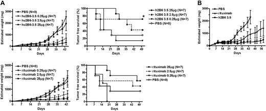
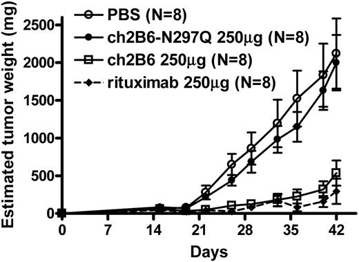
To test the ability of anti-CD32B mAbs to mediate antitumor activity in vivo, xenograft studies were performed using Burkitt lymphoma Daudi cells implanted into immunocompromised mice as a tumor model. Athymic BALB/c nude (Foxn1nu/nu) mice were injected subcutaneously with 5 × 10⁶ Daudi cells per mouse. Weekly administration of hu2B6-3.5 reduced the rate of tumor growth when compared to vehicle controls (Figure 5A). The reduction in tumor growth and improvement in tumor-free survival observed with hu2B6-3.5 were both dose dependent. The activity of hu2B6-3.5 in this xenograft model was comparable to that observed with the anti-CD20 antibody, rituximab (Figure 5A). In addition, antitumor activity was observed when therapy was delayed for 1 week to allow tumor establishment prior to intraperitoneal antibody injection (Figure 5B). Compared to ch2B6, the aglycosylated, ch2B6-N297Q version of the antibody had no protective effect in vivo, with tumor growth rates in this animal group comparable to those of control animals even at the high dose (Figure 6). Therefore, Fc-dependent interactions are necessary to prevent tumor growth in this model, consistent with the Fc requirement for 2B6-mediated cytotoxicity in vitro.
Cytotoxicity mediated by CD32B+ MDMs against the Burkitt lymphoma cell line, Daudi, in vitro. (A) Elutriated monocytes were cultured and collected as described in “Materials and methods.” MDMs were incubated with FcR-specific antibodies (1-2 μg/mL): anti–CD64-FITC (clone 32.2, top panel), anti–CD32A-FITC (IV.3, middle panel), anti–CD32B-FITC (2B6), and anti–CD16-PE (3G8; bottom panel), washed, and analyzed by flow cytometry. The isotype control is represented by the dashed line and the specific antibody is represented by the bold line. (B) Cytotoxicity of CD32B-expressing MDMs against Daudi target cells (E/T ratio = 20:1) using ch2B6 (▪) or ch2B6-N297Q (○), at the indicated antibody concentrations in an 18-hour 111In-release assay. Measurements were performed in triplicate and error bars represent SD.
Cytotoxicity mediated by CD32B+ MDMs against the Burkitt lymphoma cell line, Daudi, in vitro. (A) Elutriated monocytes were cultured and collected as described in “Materials and methods.” MDMs were incubated with FcR-specific antibodies (1-2 μg/mL): anti–CD64-FITC (clone 32.2, top panel), anti–CD32A-FITC (IV.3, middle panel), anti–CD32B-FITC (2B6), and anti–CD16-PE (3G8; bottom panel), washed, and analyzed by flow cytometry. The isotype control is represented by the dashed line and the specific antibody is represented by the bold line. (B) Cytotoxicity of CD32B-expressing MDMs against Daudi target cells (E/T ratio = 20:1) using ch2B6 (▪) or ch2B6-N297Q (○), at the indicated antibody concentrations in an 18-hour 111In-release assay. Measurements were performed in triplicate and error bars represent SD.
hu2B6-3.5 exert in vivo antitumor activity against B-lymphoma xenografts. (A) Daudi cells (5 × 10⁶/mouse) in Matrigel were injected subcutaneously into the right flank of 8- to 11-week-old BALB/c nude female mice (N = number of mice per group). Intraperitoneal injections of antibodies hu2B6-3.5 (top panels) or rituximab (bottom panels) at the indicated total mouse doses were performed weekly for 8 weeks, starting at day 0. Mean tumor volumes are plotted and error bars represent the SE of measurement (left panels). Tumor-free survival was plotted using the Kaplan-Meier method (right panels) and analyzed for significance (*P < .001) using the log-rank test. (B) The experiment was conducted as in panel A except that Daudi cells (5 × 10⁶/mouse) were allowed to establish for 7 days prior to treatment with 4 consecutive daily intraperitoneal injections of PBS or the indicated antibodies (100 μg/mouse, N = 6).
hu2B6-3.5 exert in vivo antitumor activity against B-lymphoma xenografts. (A) Daudi cells (5 × 10⁶/mouse) in Matrigel were injected subcutaneously into the right flank of 8- to 11-week-old BALB/c nude female mice (N = number of mice per group). Intraperitoneal injections of antibodies hu2B6-3.5 (top panels) or rituximab (bottom panels) at the indicated total mouse doses were performed weekly for 8 weeks, starting at day 0. Mean tumor volumes are plotted and error bars represent the SE of measurement (left panels). Tumor-free survival was plotted using the Kaplan-Meier method (right panels) and analyzed for significance (*P < .001) using the log-rank test. (B) The experiment was conducted as in panel A except that Daudi cells (5 × 10⁶/mouse) were allowed to establish for 7 days prior to treatment with 4 consecutive daily intraperitoneal injections of PBS or the indicated antibodies (100 μg/mouse, N = 6).
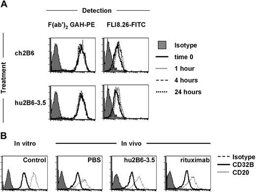
Antigen modulation following antibody treatment is a well-established mechanism for resistance to immunotherapeutics.24 Therefore, CD32B expression following exposure to the antibody was assessed both in vitro and in vivo using Daudi cells. Treatment of Daudi cells in vitro with a saturating concentration of ch2B6 or hu2B6-3.5 for 24 hours did not change CD32B expression levels (Figure 7A). The effect of antibody treatment on CD32B modulation was examined in vivo using the Daudi xenograft model. Following 8 weekly doses of hu2B6-3.5, rituximab, or PBS, residual tumors were allowed to grow in vivo for an additional week and subsequently isolated for analysis of CD32B and CD20 expression by flow cytometry. Administration of hu2B6-3.5 had no apparent impact on the expression of either CD20 or CD32B. Likewise, treatment with rituximab had no detectable effect on CD32B or CD20 expression in this model (Figure 7B).
The antitumor activity of 2B6 in B-lymphoma xenografts requires a functional Fc. Daudi cells (5 × 10⁶/mouse) were suspended in Matrigel and injected subcutaneously into the right flank of 7-week-old BALB/c nude female mice (N = number of mice per group). Intraperitoneal injections of antibodies (250 μg/mouse): ch2B6 (□), ch2B6-N297Q (•), rituximab (♦), or PBS (○), were performed weekly for 8 weeks, starting at day 0. Mean tumor volumes are plotted and error bars represent the SE of measurement.
The antitumor activity of 2B6 in B-lymphoma xenografts requires a functional Fc. Daudi cells (5 × 10⁶/mouse) were suspended in Matrigel and injected subcutaneously into the right flank of 7-week-old BALB/c nude female mice (N = number of mice per group). Intraperitoneal injections of antibodies (250 μg/mouse): ch2B6 (□), ch2B6-N297Q (•), rituximab (♦), or PBS (○), were performed weekly for 8 weeks, starting at day 0. Mean tumor volumes are plotted and error bars represent the SE of measurement.
Discussion
In this report, we show that the human low-affinity inhibitory Fc-γ receptor IIB, CD32B, is a target for immunotherapy of B-cell malignancies. A novel mouse mAb specific for CD32B was chimerized and humanized with excellent preservation of the affinity and selectivity of the original murine mAb. Both the chimeric and humanized versions of this mAb were capable of mediating ADCC in vitro against human Burkitt B-cell lymphoma lines and demonstrate potent antitumor activity in a mouse xenograft model of lymphoma.
CD32B has been shown to be expressed at high levels by lymphomas of B-cell origin. Overexpression of the FcγRIIB2 isoform of CD32B has been reported in certain FLs,25 the result of a common chromosomal translocation at 1q21.26 Furthermore, overexpression of the FcγRIIB1 isoform of CD32B has been shown to enhance the in vivo tumorigenic potential in xenograft experiments.27 An immunohistochemistry study performed by Camilleri-Broet et al, using a polyclonal antiserum directed against a unique CD32B intracellular epitope, showed that 54% (60 of 112) of all the NHLs surveyed were positive for CD32B expression.28 More significantly, within this series, all mantle cell lymphomas (MCLs; 5 of 5), splenic marginal zone (8 of 8), and small lymphocytic lymphomas (7 of 7) were positive for CD32B, whereas roughly half (13 of 24) of FLs examined had CD32B expression. Consistent with these results, in a small panel of samples we have tested we observed reactivity with 2B6 in only one of 3 FLs, whereas all MCL (4 of 4) and marginal zone lymphoma (3 of 3) samples reacted intensely for CD32B (data not shown). The recent finding of CD32B expression on tumor cells of extra-hematopoietic origin hints at the possibility that anti-CD32B mAbs may also have applications outside the range of B cell–related malignancies.29
In vivo, ch2B6 or hu2B6 were effective at reducing the rate of tumor growth in a xenograft mouse model of lymphoma. Several antibodies directed against B-cell markers demonstrate tumor-depleting activity in vivo. Cragg and Glennie found that the ability of the anti-CD20 mAbs, 1F5 and rituximab, to mediate complement-dependent cytotoxicity (CDC) activity in vitro correlated with the contribution of complement-mediated lysis to tumor prevention in vivo.30 They also demonstrate that another anti-CD20 antibody, B1, was capable of inducing apoptosis and did not depend on CDC or ADCC to reduce tumor burden in vivo. Antibodies against other B-cell antigens can exert ADCC or inhibit proliferation in vitro.31-33 These data indicate that the mechanism of action of anti–B-cell mAbs differs depending on the target and may differ within a given target with respect to the specific epitope recognized. The precise mechanism of 2B6-mediated antitumor activity observed in vivo remains unknown, but several pieces of evidence suggest a cell-dependent mechanism. First, induction of apoptosis by 2B6 in vitro was marginal even on secondary cross-linking (data not shown) and CDC was not detected with m2B6, ch2B6, or hu2B6-3.5 (data not shown). These data suggest that neither activity contributed to the antitumor effect observed in vivo. The lack of detectable CDC activity is noteworthy because several side effects of rituximab (chills, fever, cytokine release) could be attributed to the efficient complement binding and CDC induction observed with this mAb.34 Lastly, both in vitro and in vivo effects of 2B6 were eliminated by the introduction of a mutation (N297Q) capable of abolishing Fc function.20 Taken together, these data suggest that the antitumor effects of chimeric and humanized 2B6 were dependent on FcR engagement on effector cells, consistent with the ability of the mAb to induce cell-dependent cytotoxicity in vitro.
CD32B antigen modulation following antibody treatment in vitro and in vivo. (A) Daudi cells were cultured in the presence of ch2B6 (1 μg/mL) or hu2B6-3.5 (1 μg/mL) for 24 hours and analyzed for expression of CD32B or the presence of anti-CD32B antibodies, at the indicated time points. (B) Residual Daudi tumors, treated with 8 weekly doses of hu2B6-3.5 (25 μg/mouse), rituximab (25 μg/mouse), or PBS were allowed to grow in vivo for an additional week. Tumors were removed, dissociated, and incubated with the following antibodies (1-2 μg/mL): anti–CD32B-FITC (2B6, bold line), anti–CD20-FITC (L27, dotted line), or murine IgG1-FITC (solid area). The anti-CD32B (2B6) and anti-CD20 (L27) antibodies used do not cross-react with the corresponding mouse antigens.
CD32B antigen modulation following antibody treatment in vitro and in vivo. (A) Daudi cells were cultured in the presence of ch2B6 (1 μg/mL) or hu2B6-3.5 (1 μg/mL) for 24 hours and analyzed for expression of CD32B or the presence of anti-CD32B antibodies, at the indicated time points. (B) Residual Daudi tumors, treated with 8 weekly doses of hu2B6-3.5 (25 μg/mouse), rituximab (25 μg/mouse), or PBS were allowed to grow in vivo for an additional week. Tumors were removed, dissociated, and incubated with the following antibodies (1-2 μg/mL): anti–CD32B-FITC (2B6, bold line), anti–CD20-FITC (L27, dotted line), or murine IgG1-FITC (solid area). The anti-CD32B (2B6) and anti-CD20 (L27) antibodies used do not cross-react with the corresponding mouse antigens.
The selectivity of 2B6 for CD32B and its lack of reactivity with CD32A make this antibody unique among the anti-CD32 mAbs. Thus, 2B6 did not specifically react with neutrophils or platelets (M.-C.V., S.G., H.L., S.B., L.H., S.J., J.S., Jeffrey V. Ravetch, Kathryn E. Stein, E.B., and S.K., manuscript in preparation), cells known to express high levels of this activating FcR. This eliminates or greatly reduces the potential for triggering untoward activating effects (eg, cytokine release) or depletion of these critical hematopoietic components. A potential concern, however, is that CD32B targeting may adversely affect the mononuclear phagocyte effector cell population. Although peripheral blood monocytes only express minimal levels of CD32B and at minimal density (Figure 2 and data not shown), macrophages do express the inhibitory receptor (Figure 4) together with a full array of activating FcRs (CD16A, CD32A, CD64).8 We have shown here that 2B6 was capable of directing macrophage-dependent cell cytotoxicity against lymphoma cells in vitro, indicating that targeting CD32B on the macrophage surface was not detrimental to their cytotoxic activity. Furthermore, significant lysis of the macrophage effector cell population in the presence of anti-CD32B mAbs was not observed (data not shown). Therefore, a direct negative effect of 2B6 on mononuclear cells is unlikely.
In addition to ADCC, targeting CD32B in humans could lead to modulation of effector cell function through additional mechanisms. It has been proposed that the balance of activating and inhibitory FcR signaling can influence the tumor-depleting activity of therapeutic anticancer antibodies. In support of this hypothesis, genetically engineered mice deficient for the inhibitory FcγRIIB receptor showed increased response to antibody-based tumor immunotherapy.35,36 2B6 is capable of inhibiting immune complex binding to human CD32B.18,19 It is therefore possible that blockade of CD32B in the presence of 2B6 may further enhance immune cell effector function and improve the antitumor response. Because 2B6 does not cross-react with rodent FcγRIIB, the impact of in vivo blockade of CD32B cannot be modeled in wild-type mice. Transgenic models are being developed to perform additional mechanistic and toxicologic studies in response to targeting CD32B in the host.
Prepublished online as Blood First Edition Paper, June 6, 2006; DOI 10.1182/blood-2006-05-020602.
All of the authors are employed by a company (MacroGenics Inc) whose potential product was studied in the present work. Several of the authors (C.T.R., M.-C.V., S.G., L.H., N.T., S.J., J.S., S.K., E.B.) have filed patent applications related to the work that is described in the present study.
The online version of this article contains a data supplement.
The publication costs of this article were defrayed in part by page charge payment. Therefore, and solely to indicate this fact, this article is hereby marked “advertisement” in accordance with 18 U.S.C. section 1734.
The authors would like to thank Anmarie Boutrin, Valentina Ciccarone, Carolyn Dismond, Marco Goicochea, Yanira Manzanarez, Kalpana Shah, Tosan Tutse-Tonwe, and Weili Wang for exceptional technical assistance, and Dr Jeffrey L. Nordstrom, Kathryn E. Stein, and Sarah Kurz for critical review of the manuscript.

This feature is available to Subscribers Only
Sign In or Create an Account Close Modal