Abstract
Adoptive transfer of dendritic cells (DCs) transfected with in vitro–transcribed, RNA-encoding, tumor-associated antigens has recently entered clinical testing as a promising approach for cancer immunotherapy. However, pharmacokinetic exploration of RNA as a potential drug compound and a key aspect of clinical development is still pending. While investigating the impact of different structural modifications of RNA molecules on the kinetics of the encoded protein in DCs, we identified components located 3′ of the coding region that contributed to a higher transcript stability and translational efficiency. With the use of quantitative reverse transcription–polymerase chain reaction (RT-PCR) and eGFP variants to measure transcript amounts and protein yield, we showed that a poly(A) tail measuring 120 nucleotides compared with a shorter one, an unmasked poly(A) tail with a free 3′ end rather than one extended with unrelated nucleotides, and 2 sequential β-globin 3′ untranslated regions cloned head to tail between the coding region and the poly(A) tail each independently enhanced RNA stability and translational efficiency. Consecutively, the density of antigen-specific peptide/MHC complexes on the transfected cells and their potency to stimulate and expand antigen-specific CD4+ and CD8+ T cells were also increased. In summary, our data provide a strategy for optimizing RNA-transfected DC vaccines and a basis for defining release criteria for such vaccine preparations.
Introduction
Antigen-encoding RNA1,2 has the advantages of a genetic vaccine (delivery of all epitopes of the whole antigen, easy manufacturing, standardized purification) and the added value of a safe pharmaceutical characterized by transient expression and lack of integration into the genome of the treated host. The combination of this versatile antigen delivery molecule with dendritic cells (DCs) as the most potent antigen-presenting cells is regarded as an attractive approach to induce cellular and potentially therapeutic immune responses in patients with cancer. Reports demonstrated convincingly that the use of RNA results in efficient induction of antigen-specific immune responses in vitro and in animal models1,3-7 and paved the way for trials in humans. Early clinical trials showed feasibility, lack of toxicity, and promising efficacy based on immunologic and clinical read-outs.8-12 At this early phase of clinical development, antigen-specific RNA has the status of a drug compound requiring detailed exploration.
Basic pharmacologic issues that must be addressed in drug development include the pharmacokinetics of the compound of interest within the system of its physiological activity after administration. In the quoted clinical trials, this system would be represented by immature or mature DCs. A key objective of such investigations is better understanding of the impact of the structural features of the drug formulation on its pharmacologic properties. Neither of these questions has thus far been addressed for antigen-encoding RNA.
In vitro–transcribed RNA generated from plasmid templates and used in such studies typically consists of a 5′ cap structure followed by the sequence encoding the antigen of interest and a poly(A) tail 30 to 70 nucleotides in length.4,5 In some reports, the poly(A) tail is attached enzymatically by terminal polyadenylation instead of being encoded in the template.13 The gene of interest is used with its autochthonous noncoding regions12 or is flanked by the untranslated region (UTR) of the human or Xenopus laevis β-globin gene.14
The effects of each of these structural features on the intracellular bioavailability of the RNA molecule have not been systematically investigated. In fact, surprisingly little is known about the time-dose curve and the translational efficiency of in vitro–transcribed (IVT) antigen-encoding RNA after its electroporation into DCs. Similarly, a dose-response relationship between the levels of antigen-encoding RNA within the cell and the peptide/MHC molecules on the cell surface has not been established. Thus, the extent to which the density of peptide/MHC complexes on the cell surface can be influenced by augmenting RNA stability remains unclear. Our objective was to characterize and improve the pharmacokinetic properties of IVT RNA as a tool for antigen delivery to DCs.
To our knowledge, this is the first study to determine the structural modifications that increase the stability and translational efficiency of RNA molecules in DCs. We show that this optimization improves the surface presentation of epitopes and the induction of T-cell responses. Therefore, we expect that the implementation of modified RNAs into clinical vaccine constructs will contribute to an improved outcome.
Materials and methods
Cells and cell lines
OT-I CD8+ T cells transgenic for the T-cell receptor (TCR) and recognizing the Kb-specific peptide SIINFEKL from chicken OVA (OVA257-264) were kindly provided by H. Schild (Institute for Immunology, University of Mainz, Germany). All animal experiments complied with the guidelines set by the Johannes-Gutenberg University of Mainz.
Human monocytes were enriched with anti-CD14 microbeads (Miltenyi Biotec, Bergisch-Gladbach, Germany) from peripheral blood mononuclear cells (PBMCs) of healthy blood bank donors. Immature DCs (iDCs) were differentiated by culture in RPMI 1640 with 2 mM glutamine, 100 U/mL penicillin, 100 μg/mL streptomycin, 1 mM sodium pyruvate, nonessential amino acids, and 10% heat-inactivated human AB serum (all from Invitrogen, Karlsruhe, Germany) supplemented with 1000 U/mL GM-CSF (Essex, Lucerne, Switzerland) and 1000 U/mL IL-4 (Strathmann Biotech, Hamburg, Germany). Maturation of human DCs (mDCs) was induced by culturing for 2 days with 500 U/mL IL-4, 800 U/mL GM-CSF, 10 ng/mL IL-1β (PharMingen, Hamburg, Germany), 10 ng/mL TNF-α (Sigma, Taufkirchen, Germany), 1000 U/mL IL-6 (Strathmann Biotech), and 1 μg/mL PG-E2 (Sigma). Immature DCs were typically major histocompatibility complex (MHC) class IIpos, CD80low CD86low, CD83–, and CD14– and, after maturation, MHC class IIhigh, CD80high, CD86high, CD83+, and CD14–. Murine bone marrow–derived DCs (BMDCs) for in vitro or in vivo stimulation were generated as described by Lutz et al.15 Briefly, bulk cells obtained from bone cavities of C57BL/J6 mice were cultured for 7 days in medium supplemented with 200 U/mL rmGM-CSF (PeproTech/Tebu, Frankfurt, Germany). At day 7, cells were CD11c+, MHC class IIpos, CD80low, CD86low, DEC205neg and were used for quantification of peptide/MHC complexes. For in vivo expansion of T cells, BMDCs were matured after electroporation for 16 hours with poly(I:C) (50 μg/mL; Amersham Bioscience, Freiburg, Germany).
Vectors for in vitro transcription
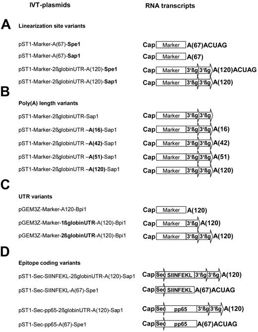
A list of all vectors is provided in Figure 1. A series of vectors for the IVT of polyadenylated RNA molecules under the control of the SP6 promoter were derived from a modified pGEM3Z-Vektor (Promega, Madison, WI) into which we had cloned a 120-bp poly(A) tail (pGEM3Z-A[120]). To engineer the pGEM3Z-β-globin UTR-A(120) series, the 3′ UTR of the β-globin molecule flanked by XhoI/SalI restriction enzyme sites was amplified from human bone marrow (3′β-globin UTR sense, 5′-tta ctc gag agc tcg ctt tct tgc tgt cca att tct-3′), 3′β-globin UTR antisense, 5′-tta gtc gac gca gca atg aaa ata aat gtt ttt tat tag gca-3′; and a single (pGEM3Z-1β-globin UTR-A[120]) or 2 serial fragments (pGEM3Z-2β-globin UTR-A[120]) were inserted in front of the poly(A) tail.
Vectors used for in vitro transcription and mRNAs derived from them. DNA templates coding for the marker proteins eGFP or d2eGFP but varying in (A) linearization site, (B) poly(A) tail length, (C) 3′ untranslated region (UTR), and (D) vectors encoding an ovalbumin-derived T-cell epitope and pp65 protein for investigation of antigen presentation and immune responses were transcribed in vitro in the presence of a cap analog to generate the mRNA species shown on the right.
Vectors used for in vitro transcription and mRNAs derived from them. DNA templates coding for the marker proteins eGFP or d2eGFP but varying in (A) linearization site, (B) poly(A) tail length, (C) 3′ untranslated region (UTR), and (D) vectors encoding an ovalbumin-derived T-cell epitope and pp65 protein for investigation of antigen presentation and immune responses were transcribed in vitro in the presence of a cap analog to generate the mRNA species shown on the right.
All vector backbones were equipped with the open reading frame (ORF) of the reporter molecule eGFP amplified from the peGFP-C1 vector (Becton Dickinson, Heidelberg, Germany) with the use of specific primers (eGFP sense, 5′-gga tcc acc atg gtg agc aag ggc gag gag-3′; eGFP-stop antisense, 5′-gga tcc tta ctt gta cag ctc gtc cat gcc g-3′). Vectors carrying the d2 variant of eGFP, which, because of a PEST domain, has a shorter half-life, were obtained by substituting the eGFP fragment in the respective construct with a BamHI/XhoI site-flanked d2eGFP fragment amplified from pTRE-d2GFP (BD Biosciences, Heidelberg, Germany) with specific primers (d2GFP-sense, 5′-gag gga tcc acc atg gtg agc aag ggc gag gag ctg-3′; d2GFP-stop antisense, 5′-gag ctc gag gaa ttc cta cac att gat cct agc aga -3′).
The pST1-2β-globin UTR-A(120) construct obtained by introduction of T7-promoter, 2-serial human 3′β-globin UTR poly(A) tails of different lengths and the neomycin-resistance gene into the pCMV-Script-Vector (Stratagene, La Jolla, CA) served as ancestor for a second series of vectors, allowing in vitro transcription of polyadenylated RNAs under the control of the T7-promoter.
To introduce sites for linearization with SapI (pST1-2β globin UTR-A[120]) and BpiI (pGEM3Z-A[120]), fragments encoding consensus sequences for these enzymes on the complementary strand were synthesized (Geneart, Regensburg, Germany) and inserted 3′ to the poly(A) tail. BpiI and SapI are type IIS restriction enzymes, which, in contrast to the broadly used type II endonucleases, cleave staggered to rather than within their specific recognition sites. In addition, eGFP and d2eGFP were inserted into pST1-A(120) and its variants.
To obtain the pST1-Sec-SIINFEKL vector, a BamHI site–flanked fragment representing base pairs 766 to 801 (aa 255-266) of the ovalbumin sequence encoding the immunodominant peptide epitope was synthesized by Geneart and cloned into the backbone. In addition, a 78-bp signal peptide derived from an MHC class I molecule (Sec) was amplified from activated PBMCs (primers: Sec sense, 5′-aag ctt agc ggc cgc acc atg cgg gtc acg gcg ccc cga acc-3′; Sec antisense, 5′-ctg cag gga gcc ggc cca ggt ctc ggt cag-3′) and inserted in front of the SIINFEKL sequence. The pST1-Sec-HCMV pp65 vector was generated accordingly by ligation of a PCR product obtained by amplification of the pp65 (UL83) ORF from a lysate of human cytomegalovirus (HCMV)–infected fibroblasts (BioWhittaker, Walkersville, MD) with specific oligonucleotides (pp65-sense, 5′-gga tcc acc atg gag tcg cgc ggt cgc cgt tgt ccc gaa atg-3′; pp65 Stop antisense, 5′-gga tcc tca acc tgg gcg tcg agg cga tgc-3′).
A cDNA fragment amplified from human testis mRNA coding for NY-ESO-I using NY-ESO sense (5′-gga tcc gcc acc atg cag gcc gaa ggc cgg ggc aca-3′) and antisense 5′-gga tcc gcg cct ctg ccc tga ggg agg-3′) oligonucleotides was cloned into the pST1 vector and used to generate control RNA.
Generation of IVT RNA
To generate templates for IVT, plasmids were linearized downstream of the poly(A) tract and purified by phenol/chloroform extraction and sodium acetate precipitation. Linearized vector DNAs were quantified spectrophotometrically and subjected to in vitro transcription using commercial kits according to the manufacturer's instructions.
In vitro transcription of pST1-A120–based plasmids was carried out with the mMessage mMachine T7 ultra kit (Ambion, Austin, TX), which attaches the anti–reverse cap analog (ARCA; 7-methyl(3′-O-methyl)GpppG) m7G(5′)ppp(5′)G in an ultra high–yield transcription reaction.
The pGEM3Z-A(120)–based plasmids were capped with 7-methylGpppG using the mMessage mMachine SP6 Kit (Ambion). For some experiments, vectors were linearized upstream of the plasmid encoded poly(A) tail. After in vitro transcription, RNA was enzymatically polyadenylated using the poly(A) tail kit (Ambion). Capped polyadenylated RNA was purified using MEGAclear Kit (Ambion).
Transfer of IVT RNA into cells
Cells were suspended in 250 μL X-VIVO 15 medium (Cambrex, Verviers, Belgium) and transferred to a 4-mm gap sterile electroporation cuvette (Bio-Rad, Hercules, CA). After addition of the appropriate amount of IVT RNA, cells were electroporated using a Gene-Pulser-II apparatus (Bio-Rad, Munich, Germany) applying the voltage and capacitance settings established for each cell type (K562, 200 V/300 μF; human iDC and mDC, 290 V/150 μF; murine BMDC, 276 V/150 μF; EL4, 300 V/150 μF). Cells were diluted in culture medium immediately after electroporation.
Quantification of eGFP transcript levels by real-time RT-PCR
Total cellular RNA was extracted with RNeasy Mini Kit (Qiagen, Hilden, Germany), reverse transcribed with hexamer primers using Superscript II (Invitrogen, Carlsbad, CA), and subjected to real-time quantitative analysis on ABI PRISM 7700 Sequence Detection System instrument and software (Applied Biosystems, Foster City, CA) with QuantiTect SYBR Green PCR Kit (Qiagen). Reactions were performed for 40 cycles in triplicate with specific primers amplifying eGFP and d2eGFP (eGFP-sense, 5′-cac atg aag cag cac gac ttc-3′; eGFP-antisense, 5′-cac ctt gat gcg gtt ctt ctg-3′; each 300 nM) with initial denaturation/activation for 15 minutes at 95°C, 30 seconds at 94°C, 30 seconds at 62°C, and 30 seconds at 72°C. Expression of eGFP transcripts was quantified relative to 18sRNA as internal standard to normalize for variances in the quality of RNA and the amount of input cDNA.
Flow cytometric analysis
For immunophenotyping of immature and mature DCs, antibodies were used with nonreactive isotype-matched controls (BD Biosciences, Heidelberg, Germany).
For quantification of eGFP and d2eGFP protein, cells were washed in PBS and incubated with propidium iodide (10 μg/mL) before flow cytometric analysis. For eGFP analysis in DC cultures, gating was performed on cells exhibiting large forward scatter (FSC) and side scatter (SSC) for exclusion of contaminating lymphocytes. Propidium iodide–negative cells were gated, and mean fluorescence intensity (MFI) expression was determined.
For quantification of the SIINFEKL peptide presented on the H-2 Kb molecule, mouse thymoma EL4 cells or mature BMDCs were harvested, washed 3 times with cold buffer (PBS-1% FCS), blocked with PBS-10% FCS, stained for 30 minutes at 4°C with 25.D1.16 antibody, which specifically detects OVA257-264 peptide SIINFEKL in conjunction with H-2 Kb molecules,16 and thereafter stained with goat anti–mouse-APC secondary antibody (Jackson ImmunoResearch, West Grove, PA). For generation of a standard curve for conversion of MFI values to equivalent molar concentrations of SIINFEKL peptide loaded on cells, the respective cells were pulsed for 2 hours at 37°C with titrated amounts of SIINFEKL peptide (Biosyntan, Berlin, Germany; 2 pM to 30 μM; 3-fold dilutions). Flow cytometric analysis was performed on a FACSCalibur analytical flow cytometer (BD Biosciences, Heidelberg, Germany) with CellQuest software (BD Biosciences).
In vivo expansion of murine T cells
For the assessment of antigen-specific in vivo expansion, splenocytes from TCR tg OT-I mice were prepared and adoptively transferred at day 0 into the tail veins of recipient C57BL/J6 mice. Cell number was adjusted to 1 × 10⁵ TCR tg CD8+ T cells. For immunization, BMDCs were electroporated with 50 pmol RNA and then activated by poly(I:C) for 16 hours. BMDCs (1 × 10⁶) were administered at day 1 by intraperitoneal injection. Retro-orbital blood samples were collected at day 4 and stained with anti-CD8 (Caltag Laboratories, Burlingame, CA) and SIINFEKL tetramer (H-2Kb/SIINFEKL 257-264; Beckman Coulter, Fullerton, CA).
In vitro expansion of human T cells
CD4+ and CD8+ T cells were isolated from PBMCs of HCMV-seropositive healthy donors by positive magnetic cell sorting with antibody-coated microbeads (Miltenyi Biotec, Bergisch-Gladbach, Germany). Autologous DCs (2 × 10⁵) were either electroporated with IVT RNA or pulsed with peptide pools, washed, and cocultured with 1 × 10⁶ CD4+ or CD8+ effector T cells in complete medium supplemented with 10% AB serum, 10 U/mL IL-2 (R&D Systems, Minneapolis, MN), and 5 ng/mL IL-7 (R&D Systems) in 24-well plates (BD Biosciences, Heidelberg, Germany). Enzyme-linked immunospot (ELISpot) assays were performed 7 days after the initiation of stimulation.
Enzyme-linked immunospot assay
Microtiter plates (96-well; Millipore, Bedford, MA) were coated overnight at 4°C with 0.1 μg/mL anti-IFN antibody (1-D1K; Mabtech, Stockholm, Sweden), washed 4 times with PBS, and blocked with 2% human albumin. Autologous DCs used as stimulator cells were plated in triplicate (2 × 10⁴ target cells/well) after loading with 1.75 μg/mL pp65 or NY-ESO-I peptide pools. Peptides (15 mer) with 11 amino acid overlaps covering the whole HCMV pp65 sequence and an analogous peptide pool derived from the NY-ESO-I antigen (both from Jerini, Berlin, Germany) as negative control were used. CD4+ or CD8+ responder cells from the T-cell expansion culture were added at a concentration of 1 to 3 × 10⁴ cells/well. Plates were incubated overnight (37°C, 5% CO2), washed with PBS 0.05% Tween 20, and incubated for 2 hours with anti-IFN (biotinylated mAb 7-B6-1; Mabtech) at a final concentration of 1 μg/mL at 37°C. After washing in PBS containing 0.05% Tween 20, avidin-bound horseradish peroxidase H (Vectastain Elite Kit; Vector Laboratories, Burlingame, CA) was added and incubated for 1 hour at room temperature. Plates were developed after a final wash with PBS containing 0.05% Tween 20 using 3-amino-9-ethyl carbazole (Sigma). Spots were counted using a computer-based evaluation system and KS-ELISpot software version 4.4.35 (Carl Zeiss Vision, Eching, Germany).
Results
Density of peptide/MHC complexes on the cell surface correlates with the dose of antigen-encoding RNA electroporated into cells
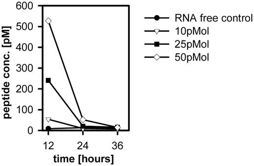
To investigate the dose-response relationship between the levels of antigen-encoding RNA transferred into the cell and the peptide/MHC molecules on the cell surface, we took advantage of an antibody allowing flow cytometric quantification of antigen-specific peptide/MHC complexes (H2-Kb-OVA257-264).17 Increasing amounts of RNA transcribed in vitro from the linearized Sec-SIINFEKL-A67-Spe1 vector and encoding the OVA257-264 peptide SIINFEKL were electroporated into H2-Kb–expressing EL4 tumor cells and analyzed for surface expression kinetics of specific peptide/MHC complexes. Even after electroporation of RNA amounts as low as 10 pmol, surface expression of peptide/MHC molecules was easily detected 12 hours after transfection, declined over time, and was below the detection level 24 hours after RNA transfer (Figure 2). Density and persistence of peptide/MHC complexes correlated with the amount of RNA transferred into the cells.
Enzymatic polyadenylation of RNA results in a mixture of transcripts varying in length
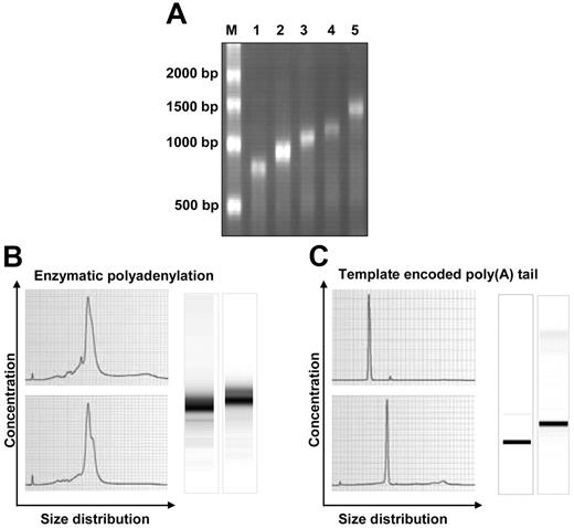
Poly(A) tailing of RNA can be accomplished either by enzymatic polyadenylation after IVT of the plasmid template,13 or by cloning a poly(A) stretch into the template vector. We observed that the length of an enzymatically synthesized poly(A) tail highly depended on the reaction conditions. Variations in the quantity of poly(A) polymerase (Figure 3A) and in the incubation time (data not shown) led to profound differences in poly(A) length. Because many parameters influenced poly(A) tail length during the polyadenylation reaction, the number of attached ATP nucleotides could not be precisely controlled even if the same reaction conditions were applied. Accordingly, enzymatically polyadenylated RNA samples generated in independent production runs were not directly comparable. In addition to this variability between different preparations, each individual preparation appeared to contain a mixture of RNA species differing in poly(A) tail length. This was documented by the fuzzy signal observed in Bioanalyzer (Agilent, Santa Clara, CA) electropherograms and gel images of enzymatically tailed RNA (Figure 3B). In contrast, IVT RNA with a template encoded poly(A) tail appeared as a steep peak and showed a sharp band in the gel image (Figure 3C). In summary, template-encoded poly(A) tails were clearly superior because they reproducibly provided a well-defined product of sufficient purity.
Dose-response relationship between the levels of peptide-encoding RNA within the cell and peptide/MHC molecules on the cell surface. EL4 cells were electroporated (settings 300 V/150 μF) with different amounts of Sec-SIINFEKL-A(67)ACUAG RNA. Transfected cells harvested at different time points were stained with 25D1.16 antibody to determine surface SIINFEKL/Kb-complexes. Concentrations of SIINFEKL peptide were calculated from the mean fluorescence values of viable cells using a peptide titration as standard curve.
Dose-response relationship between the levels of peptide-encoding RNA within the cell and peptide/MHC molecules on the cell surface. EL4 cells were electroporated (settings 300 V/150 μF) with different amounts of Sec-SIINFEKL-A(67)ACUAG RNA. Transfected cells harvested at different time points were stained with 25D1.16 antibody to determine surface SIINFEKL/Kb-complexes. Concentrations of SIINFEKL peptide were calculated from the mean fluorescence values of viable cells using a peptide titration as standard curve.
Comparison of enzymatically polyadenylated RNA with tailed IVT RNA transcribed from a template encoding the poly(A) tract. (A) Template IVT RNA before polyadenylation (lane 1) compared with IVT RNA products obtained in independent polyadenylation reactions with increasing concentrations of poly(A) polymerase (lanes 2-5), resulting in increasing sizes of poly(A) tail lengths. (B) IVT RNA with an enzymatically attached poly(A) tail was purified and analyzed. The electropherograms (left) and gel images (right) disclose a mixture of RNA species differing in length. (C) Electropherograms (left) and gel images (right) of purified IVT RNA obtained from a plasmid template encoding the poly(A) tail show a well-defined homogenous RNA species.
Comparison of enzymatically polyadenylated RNA with tailed IVT RNA transcribed from a template encoding the poly(A) tract. (A) Template IVT RNA before polyadenylation (lane 1) compared with IVT RNA products obtained in independent polyadenylation reactions with increasing concentrations of poly(A) polymerase (lanes 2-5), resulting in increasing sizes of poly(A) tail lengths. (B) IVT RNA with an enzymatically attached poly(A) tail was purified and analyzed. The electropherograms (left) and gel images (right) disclose a mixture of RNA species differing in length. (C) Electropherograms (left) and gel images (right) of purified IVT RNA obtained from a plasmid template encoding the poly(A) tail show a well-defined homogenous RNA species.
Overhang at the 3′ end of the poly(A) tail hampers translational efficiency of IVT RNA
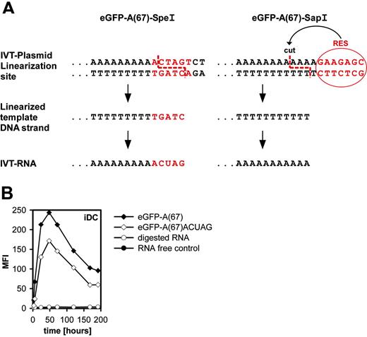
Vectors encoding the antigen of interest together with a poly(A) tail had to be linearized downstream to the poly(A) stretch before in vitro transcription. This is typically achieved by using a type II restriction enzyme that cuts the vector backbone 3′ of the poly(A) tract.4,12,18 Part of the consensus sequence of the restriction enzyme remained as overhang extending the poly(A) tail at the 3′ end (Figure 4A, left). To investigate the impact of this on protein yield, we generated IVT RNA with a free-ending poly(A) tail taking advantage of type IIS restriction enzymes such as Sap1 and Bpi1. Whereas type II restriction endonucleases digest within their recognition site, type IIS enzymes cut several base pairs aside it (Figure 4A). We replaced the Spe1 restriction enzyme site in the original vector encoding eGFP with a poly(A)67 tail by introducing a Sap1 site on the complementary strand. Thus, the poly(A) tails of the 2 eGFP-encoding RNA species we obtained either had (eGFP-A(67)ACUAG) or did not have (eGFP-A(67)) an additional 3′ extension.
For assessment of RNA stability, we used the standard technique of monitoring decay kinetics of the translation product in transiently transfected cells, plotting the amount of the specific protein as a function of time.19 Protein decay kinetics is regarded as a state-of-the-art tool favored over RNA kinetics because it allows integrated recording of all relevant translational characteristics in the function-oriented manner required for the objective of our investigations.20
Human immature DCs were electroporated with 50 pmol of both eGFP IVT RNA species, and the kinetics were determined by flow cytometric quantification of eGFP protein. Linearization with Sap1 improved expression significantly compared with restriction with Spe1 because the RNA with free-ending poly(A) tail resulted in 1.5-fold higher maximum protein levels and prolonged detectability of the protein. Interestingly, visual inspection of the curves showed that the message stability (eg, the length of time over which protein continued to accumulate) of both eGFP-A(67) and eGFP-A(67)ACUAG RNAs appeared to be comparable (Figure 4B). In addition, functional RNA half-life (eg, time needed to complete a 50% decay in the capacity of an RNA to synthesize protein,21 which is a more accurate parameter than physical half-life because it also considers translational competence of an RNA species) is comparable for eGFP-A(67) and eGFP-A(67)ACUAG RNAs. Apparently, the difference in protein yield can be accounted for by the higher translational efficiency (eg, the slope of the curve) of RNA with a free poly(A) tail compared with one with a 3′ extension.
Role of a free-ending poly(A) tail on translation efficiency. (A) Rationale for using a type IIS restriction enzyme (eg, Sap1) instead of a type II restriction enzyme (eg, Spe1) for linearization of the plasmid template before in vitro transcription. Type IIS restriction enzymes cut adjacent to rather than within their recognition site and prevent an overhang of nucleotides derived from the vector backbone and remaining as 3′ attachment at the poly(A) tail. (B) Impact of an unmasked free poly(A) tail on translational efficiency in dendritic cells. Immature dendritic cells were electroporated with eGFP-A(67) RNA, which contains an unmasked poly(A) tail or with eGFP-A(67)ACUAG, in which 4 additional nucleotides are attached 3′ to the poly(A) tail. Electroporation without RNA or with RNase-digested RNA served as controls. Cells were harvested at different time points (3 hours, 6 hours, 24 hours, 48 hours, 72 hours, 120 hours, 168 hours, 192 hours), and the eGFP fluorescence of viable cells was measured by flow cytometry.
Role of a free-ending poly(A) tail on translation efficiency. (A) Rationale for using a type IIS restriction enzyme (eg, Sap1) instead of a type II restriction enzyme (eg, Spe1) for linearization of the plasmid template before in vitro transcription. Type IIS restriction enzymes cut adjacent to rather than within their recognition site and prevent an overhang of nucleotides derived from the vector backbone and remaining as 3′ attachment at the poly(A) tail. (B) Impact of an unmasked free poly(A) tail on translational efficiency in dendritic cells. Immature dendritic cells were electroporated with eGFP-A(67) RNA, which contains an unmasked poly(A) tail or with eGFP-A(67)ACUAG, in which 4 additional nucleotides are attached 3′ to the poly(A) tail. Electroporation without RNA or with RNase-digested RNA served as controls. Cells were harvested at different time points (3 hours, 6 hours, 24 hours, 48 hours, 72 hours, 120 hours, 168 hours, 192 hours), and the eGFP fluorescence of viable cells was measured by flow cytometry.
Poly(A) tail length has an impact on translational efficiency
To assess the impact of the poly(A) tail on protein yield, we used a series of green fluorescence protein variant–encoding IVT RNAs that differed merely in their poly(A) tail lengths and, through electroporation, inserted equimolar amounts of them into different cell types.
eGFP is a protein with a relatively long physical half-life of 17.3 hours. We sought to determine whether the stabilizing effect of the modification we introduced still applied for proteins with much shorter half-lives and therefore included a recently described destabilized eGFP variant, d2eGFP,22 in several of the experiments. This variant has a half-life of 2 hours because of a C-terminal PEST region that promotes protein degradation.
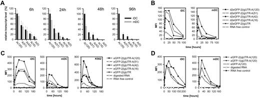
As exemplified for immature and mature DCs by real-time RT PCR analysis, intracellular IVT RNA levels of d2eGFP variants at different time points after transfection were highest for poly(A)120-tailed RNA, suggesting that RNAs with shorter poly(A) tails undergo faster degradation (Figure 5A). Flow cytometric time-course analysis of d2eGFP protein in the same batch of cells showed that this contributed to increased, prolonged protein expression (Figure 5B). Interestingly, the level and stability of d2eGFP RNA and the protein amounts were higher in immature than in mature DCs. Moreover, in immature DCs, the decay rate at the onset of degradation appeared to be protracted (Figure 5B).
Impact of regulatory components located 3′ of the coding region on transcript stability and protein yield in dendritic cells and cell lines. (A) Influence of poly(A) tail length on transcript stability in iDCs and mDCs. Cells were electroporated with equal amounts of d2eGFP-encoding IVT RNA species, which differed in the lengths of their poly(A) tails. Cells were harvested after 6 hours, 24 hours, 48 hours, and 96 hours, and eGFP transcript levels were quantified by real-time RT-PCR. Cells electroporated without RNA served as controls. For each time point, the transcript levels (+SEM) are shown relative to expression levels obtained for d2eGFP-2β-globinUTR-A(120) in iDCs. (B) Flow cytometric analysis of protein levels in the same cells used in the experiment described in panel A, which were transfected with the short-lived d2eGFP variant as reporter molecule. (C) Cells were transfected with eGFP-encoding IVT RNA variants differing in the lengths of their poly(A) tails. RNase-digested RNA and untailed RNA served as controls. Cells were harvested after 6 hours, 24 hours, 48 hours, 72 hours, 96 hours, 144 hours, and 192 hours, and eGFP fluorescence of viable cells was measured by flow cytometry. (D) Influence of the 3′ untranslated region (UTR) on translational efficiency. Immature and mature dendritic cells were electroporated with eGFP RNA variants differing in their 3′ UTR. Cells were harvested after 6 hours, 24 hours, 48 hours, 72 hours, 96 hours, 144 hours, 168 hours, or 218 hours, and the eGFP fluorescence of viable cells was measured by flow cytometry.
Impact of regulatory components located 3′ of the coding region on transcript stability and protein yield in dendritic cells and cell lines. (A) Influence of poly(A) tail length on transcript stability in iDCs and mDCs. Cells were electroporated with equal amounts of d2eGFP-encoding IVT RNA species, which differed in the lengths of their poly(A) tails. Cells were harvested after 6 hours, 24 hours, 48 hours, and 96 hours, and eGFP transcript levels were quantified by real-time RT-PCR. Cells electroporated without RNA served as controls. For each time point, the transcript levels (+SEM) are shown relative to expression levels obtained for d2eGFP-2β-globinUTR-A(120) in iDCs. (B) Flow cytometric analysis of protein levels in the same cells used in the experiment described in panel A, which were transfected with the short-lived d2eGFP variant as reporter molecule. (C) Cells were transfected with eGFP-encoding IVT RNA variants differing in the lengths of their poly(A) tails. RNase-digested RNA and untailed RNA served as controls. Cells were harvested after 6 hours, 24 hours, 48 hours, 72 hours, 96 hours, 144 hours, and 192 hours, and eGFP fluorescence of viable cells was measured by flow cytometry. (D) Influence of the 3′ untranslated region (UTR) on translational efficiency. Immature and mature dendritic cells were electroporated with eGFP RNA variants differing in their 3′ UTR. Cells were harvested after 6 hours, 24 hours, 48 hours, 72 hours, 96 hours, 144 hours, 168 hours, or 218 hours, and the eGFP fluorescence of viable cells was measured by flow cytometry.
As expected, protein yield was higher for eGFP IVT RNA variants than for d2eGFP RNA species (Figure 5C). Importantly, however, higher protein levels resulted in poly(A)120-tailed RNA than in shorter RNA. Again, protein levels were higher and decay was protracted in immature DCs compared with mature ones, with maximum protein levels sustained in steady state until 120 hours after electroporation (Figure 5C). Poly(A) tails longer than 120 bases did not have a significant effect on eGFP or d2eGFP expression (data not shown).
Two 3′ UTRs of the human β-globin gene cloned in tandem improve protein level
In several preclinical studies, the antigen of interest is flanked by the 3′ UTR of the human or Xenopus laevis β-globin gene.5,14,23 The impact of this modification has never been systematically investigated. We compared poly(A)120-tailed eGFP RNA without any UTR with RNA incorporating the human β-globin 3′ UTR. As a cloning artifact, we also generated a plasmid template that incorporated 2 sequential β-globin 3′ UTRs fused head to tail. Interestingly, increased protein yields, particularly in immature DCs, were not as pronounced as expected when comparing RNA with one β-globin 3′ UTR to the tailed control RNA lacking a 3′ UTR (Figure 5D). However, 2 reiterated β-globin 3′ UTRs between the coding sequence and the poly(A) tail resulted in significantly higher maximum protein levels and prolonged persistence of the protein. In addition, for this structural modification, the translational characteristics influenced in immature DCs (primarily message stability and functional half-life) differed from those in mature DCs (primarily translation efficiency). Notably, inclusion of the human β-globin 5′ UTR did not have an additional beneficial effect on protein yield (data not shown).
The combination of the optimal poly(A) length, a free-ending poly(A) tail, and an optimized 3′ UTR act synergistically on RNA stability and translational efficiency
In summary, our data showed that a poly(A) tail of 120 nucleotides compared with a shorter one, a poly(A) tail with a free 3′ end rather than one with an extension, and 2 tandemly reiterated β-globin 3′ UTRs each improved the translational characteristics of RNA and resulted in higher and sustained protein expression. We combined these features in one RNA molecule (eGFP-2β-globinUTR-A(120)) and compared it with a standard IVT RNA containing a masked poly(A) tail of 67 bp and lacking regulatory UTR (eGFP-A(67)ACUAG). Quantification of the transcript in immature DCs by real-time RT-PCR 48 hours after RNA transfer disclosed that combining these modifications significantly improved RNA stability by approximately 2 orders of magnitude (Figure 6A). Fluorescence microscopy of transfected, mature DCs after 24 hours confirmed this differential stability on protein level (Figure 6B).
In flow cytometric time kinetics with the short-lived d2eGFP variant, we observed that the combination of the respective structural features resulted not only in higher protein levels but was sustained over a longer time and that this applied to molecules with short half-lives (Figure 6C). Again, prolonged stability was particularly prominent in immature DCs (Figure 6C) in which we observed significant MFIs, even approximately 80 hours after electroporation. Flow cytometric data presented in Figure 6C feature results of 3 independent experiments conducted in parallel, showing nearly no SD and proving the high reproducibility of this procedure.
Stabilized IVT RNA increases the number of antigen-specific peptide/MHC complexes on the cell surface and improves the expansion of antigen-specific T cells in vivo
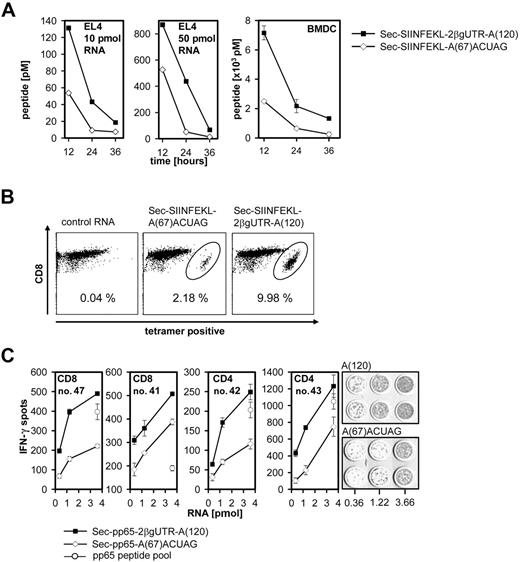
Next we investigated whether improved RNA stability and consecutively higher protein levels in antigen-presenting cells translate into more efficient T-cell stimulation. We first quantified the effect on cell-surface expression of peptide/MHC complexes. The SIINFEKL peptide was cloned into the vector representing all optimizations (pST1-Sec-SIINFEKL-2β-globinUTR-A(120)-Sap1) and into a vector with standard features (pST1-Sec-SIINFEKL-A(67)-Spe1). IVT RNAs derived from both vectors were electroporated into EL4 cells and BMDCs. We detected significantly higher numbers of OVA peptide/Kb complexes on the cell surface sustained over a longer time course upon electroporation of the genetically improved RNA Sec-SIINFEKL-2β-globinUTR-A(120) (Figure 7A).
Improvement of RNA stability by combination of the optimized structural features. (A) Immature dendritic cells were transfected with different eGFP variants featuring combinations of the improved structural characteristics. Cells were harvested 48 hours after transfection, and the eGFP transcript level was assessed by real time RT-PCR. Cells electroporated with buffer or with RNase-digested RNA were used as controls and reference. Transcript levels were shown relative to expression levels obtained for eGFP-2β-globinUTR-A(120). (B) Fluorescence microscopy of mature dendritic cells 24 hours after transfection with standard eGFP-A(67)ACUAG and optimized eGFP-2β-globinUTR-A(120) IVT RNA. To allow comparison, images were obtained using equal acquisition parameters. Images were taken with an Olympus-IX71 inverted microscope (Hamburg, Germany) with a 20×/0.4 NA objective lens. TILLvisION software (TILL Photonics, Gräfeling, Germany) was used for image acquisition. (C) Immature and mature dendritic cells were transfected with different IVT RNA constructs encoding for the short-lived d2eGFP variant. Mean fluorescence intensities of viable cells were determined at different time points after transfection in 3 independent experiments. Data for both cell types are shown as the mean ± SEM of 3 experiments.
Improvement of RNA stability by combination of the optimized structural features. (A) Immature dendritic cells were transfected with different eGFP variants featuring combinations of the improved structural characteristics. Cells were harvested 48 hours after transfection, and the eGFP transcript level was assessed by real time RT-PCR. Cells electroporated with buffer or with RNase-digested RNA were used as controls and reference. Transcript levels were shown relative to expression levels obtained for eGFP-2β-globinUTR-A(120). (B) Fluorescence microscopy of mature dendritic cells 24 hours after transfection with standard eGFP-A(67)ACUAG and optimized eGFP-2β-globinUTR-A(120) IVT RNA. To allow comparison, images were obtained using equal acquisition parameters. Images were taken with an Olympus-IX71 inverted microscope (Hamburg, Germany) with a 20×/0.4 NA objective lens. TILLvisION software (TILL Photonics, Gräfeling, Germany) was used for image acquisition. (C) Immature and mature dendritic cells were transfected with different IVT RNA constructs encoding for the short-lived d2eGFP variant. Mean fluorescence intensities of viable cells were determined at different time points after transfection in 3 independent experiments. Data for both cell types are shown as the mean ± SEM of 3 experiments.
To assess the impact on stimulatory capacity, we resorted to the OT-I TCR, which has been used extensively on the C57BL/J6 (B6) background to detect MHC class I presentation of the SIINFEKL peptide.24,25 At day 0, animals were adoptively transferred with OT-I CD8+ T cells. The next day, BMDCs of C57BL/J6 mice electroporated with SIINFEKL encoding RNA construct variants and matured for 16 hours were administered intraperitoneally into mice. At day 4, OT-I CD8+ T cells were measured in peripheral blood with tetramer technology. We found in vivo expansion of antigen-specific TCR transgenic CD8+ T cells to be significantly superior using Sec-SIINFEKL-2β-globinUTR-A(120) RNA for antigen delivery compared with Sec-SIINFEKL-A(67)ACUAG RNA (Figure 7B).
To assess whether stabilized IVT RNA constructs for antigen delivery improved antigen-specific stimulation of human T cells, we resorted to HCMV pp65, the immunodominant antigen of HCMV that is frequently used for the validation of autologous stimulation of polyepitopic T-cell responses. CD4+ and CD8+ T cells purified from HCMV seropositive healthy donors were cocultured with autologous DCs electroporated with the respective IVT RNA variants encoding pp65. Expansion of T cells measured on day 7 in an IFN-γ ELISpot using autologous DCs pulsed with a pool of overlapping peptides covering either the entire pp65 protein sequence or a control protein showed superiority of Sec-pp65-2β-globinUTR-A(120), with effects most prominent for the expansion of CD4+ T cells.
Discussion
The use of DCs transfected with antigen-encoding IVT RNA in clinical applications provides an opportunity to address several of the shortcomings of current vaccination strategies at once. The success and the ultimate clinical usefulness of this approach will depend on the optimization of parameters contributing to the induction and efficient expansion of T-cell responses. Our aim was to refine translational characteristics of transiently expressed RNA in DCs and thus improve the pharmacokinetics and consecutively the T-cell stimulatory capacity of these engineered antigen-presenting cells.
Impact of stabilized IVT RNA constructs on T-cell stimulation in vivo and in vitro. (A) Increase of antigen-specific peptide/MHC complexes by using stabilized IVT RNA constructs. Cells were electroporated with Sec-SIINFEKL-A(67)ACUAG RNA or Sec-SIINFEKL-2β-globinUTR-A(120) RNA (EL4 cells, 10 pmol, 50 pmol; C57Bl/J6 immature BMDCs in triplicate, 150 pmol). Electroporation with buffer only was used as control. Cells were stained for SIINFEKL/Kb-complexes with 25D1.16 antibody. Concentrations of SIINFEKL peptide were calculated from the mean fluorescence values of viable cells using a peptide titration as standard curve. Data for BMDCs are shown as the mean ± SEM of 3 experiments. (B) Improved in vivo T-cell expansion with stabilized IVT RNA constructs. TCR transgenic CD8+ OT-I cells (1 × 10⁵) were adoptively transferred to C57Bl/J6 mice. BMDCs of C57Bl/J6 mice were transfected with 50 pmol RNA (Sec-SIINFEKL-A(67)ACUAG, Sec-SIINFEKL-2β-globinUTR-A(120) or control RNA), matured for 16 hours with poly(I:C) (50 μg/mL), and injected intraperitoneally 1 day after T-cell transfer (n = 3). Peripheral blood was taken at day 4 and stained for SIINFEKL tetramer–positive CD8+ T cells. Dot plots show CD8+ T cells, and the numbers given represent the percentages of tetramer-positive CD8+ T cells. (C) Improved in vitro expansion of human T cells with stabilized IVT RNA constructs. CD8+ and CD4+ lymphocytes from HCMV-seropositive healthy donors were cocultivated with autologous DCs transfected with Sec-pp65-A(67)ACUAG RNA or Sec-pp65-2β-globinUTR-A(120) RNA, pp65 peptide pool (1.75 μg/mL) as positive control, or control RNA (data not shown). After expansion for 7 days, each effector cell population (4 × 10⁴/well) was tested in IFN-γ ELISpot on autologous DCs (3 × 10⁴/well) loaded with pp65 peptide pool or an irrelevant peptide pool (1.75 μg/mL). Graphs represent the mean ± SEM spot number of triplicates.
Impact of stabilized IVT RNA constructs on T-cell stimulation in vivo and in vitro. (A) Increase of antigen-specific peptide/MHC complexes by using stabilized IVT RNA constructs. Cells were electroporated with Sec-SIINFEKL-A(67)ACUAG RNA or Sec-SIINFEKL-2β-globinUTR-A(120) RNA (EL4 cells, 10 pmol, 50 pmol; C57Bl/J6 immature BMDCs in triplicate, 150 pmol). Electroporation with buffer only was used as control. Cells were stained for SIINFEKL/Kb-complexes with 25D1.16 antibody. Concentrations of SIINFEKL peptide were calculated from the mean fluorescence values of viable cells using a peptide titration as standard curve. Data for BMDCs are shown as the mean ± SEM of 3 experiments. (B) Improved in vivo T-cell expansion with stabilized IVT RNA constructs. TCR transgenic CD8+ OT-I cells (1 × 10⁵) were adoptively transferred to C57Bl/J6 mice. BMDCs of C57Bl/J6 mice were transfected with 50 pmol RNA (Sec-SIINFEKL-A(67)ACUAG, Sec-SIINFEKL-2β-globinUTR-A(120) or control RNA), matured for 16 hours with poly(I:C) (50 μg/mL), and injected intraperitoneally 1 day after T-cell transfer (n = 3). Peripheral blood was taken at day 4 and stained for SIINFEKL tetramer–positive CD8+ T cells. Dot plots show CD8+ T cells, and the numbers given represent the percentages of tetramer-positive CD8+ T cells. (C) Improved in vitro expansion of human T cells with stabilized IVT RNA constructs. CD8+ and CD4+ lymphocytes from HCMV-seropositive healthy donors were cocultivated with autologous DCs transfected with Sec-pp65-A(67)ACUAG RNA or Sec-pp65-2β-globinUTR-A(120) RNA, pp65 peptide pool (1.75 μg/mL) as positive control, or control RNA (data not shown). After expansion for 7 days, each effector cell population (4 × 10⁴/well) was tested in IFN-γ ELISpot on autologous DCs (3 × 10⁴/well) loaded with pp65 peptide pool or an irrelevant peptide pool (1.75 μg/mL). Graphs represent the mean ± SEM spot number of triplicates.
The mechanisms underlying the steady state levels of naturally expressed mRNAs in the cytoplasm have received considerable attention recently because the regulation of mRNA turnover plays a major role in the overall control of gene expression.26,27
mRNA stability and translation are controlled by a complex network of RNA/protein interactions and depend on the primary and secondary structures of mRNAs, their rates of translation, and the initiation of one of the various degradation mechanisms.28-31 Sequence elements within the 3′ and 5′ noncoding regions of the RNA molecule have been characterized as important factors in regulating the stability of a transcript in ways that are not completely understood.32,33 However, most data stem from studies in yeast cell lysates or prokaryotic cells,34-36 whereas data in eukaryotic cell lines are less conclusive and seem to depend on the cell type used.37 For human DCs, this is the only study systematically investigating the influence of several 3′ elements on translational characteristics of transiently expressed RNA.
The poly(A) tail regulates the fate of mRNA by synergy with the 5′ cap, with the internal ribosomal entry site, and with various RNA stability determinants.31,38-41 Antigen-encoding RNA constructs used currently for preclinical studies have poly(A) tails measuring 30 to 70 nucleotides in length,4,5 whereas those administered in clinical trials measure 64 nucleotides in length.8,12 According to our data, the pharmacokinetic properties of RNA in DCs can be further improved by a poly(A) tail 120 bp long. This impact of poly(A) tail lengths on the pharmacologic properties of IVT RNA implicates the need for pure preparations with defined and uniform tail lengths of IVT RNA. As we show, this can be achieved by encoding the poly(A) tract in the template vector rather than attaching it by enzymatic polyadenylation. For linearization of such vectors downstream of the poly(A) stretch before in vitro transcription, we propose type IIS endonucleases instead of the usually used type II restriction enzymes.4,8,12,14,42 This offers the advantage of cutting within the poly(A) stretch preventing an overhang extending the poly(A) tail, which may compromise efficient protein synthesis of the encoded antigen. Vectors used currently for preclinical and clinical studies resort either to UTRs of β-globin5,14,23 or to autochthonous UTRs of the antigen they want to deliver,12 which are not further characterized in terms of their impact on RNA stability. According to our data, 2 serial 3′ UTRs of the β-globin gene upstream of the poly(A) tail are better than one at improving the translational characteristics of RNA. As previous reports show, the length of the 3′ UTR as such has an effect on the stability and translational efficiency of poly(A)-negative RNA but not of poly(A)-positive RNA, for which a 27-bp 3′ UTR appears to be optimal.20 Therefore, we do not expect this effect to be mediated simply by the increase of the 3′ noncoding region. Systematic screening approaches for 3′ UTRs conferring even better translational characteristics may be worthwhile for further optimizing RNA vaccines. By combining these structural components, we achieved not only a more than 5-fold increase of maximum protein concentration but also persistence of protein expression over a longer time.
In principle, the improvement of RNA kinetics by these modifications is not restricted to DCs but is similar in K562 cells and may also be of interest for other cellular settings, such as T cells,43 B cells,44 and muscle cells,45 which are attractive host cells for the expression of IVT RNA–encoded proteins. Interestingly, we observed that stability and decay kinetics of the RNAs differ in immature and mature human DCs, most likely because of differences in the complex network of RNA/protein interactions in both cell types. Notably, by revisiting our database of genomewide expression profiles of immature and mature human DCs obtained with microarrays,46 we found that factors involved in RNA stability are induced in the course of maturation (data not shown).
The mode of action of such a vaccine and its capacity to induce efficient T-cell responses depends on prolonged surface presentation of sufficient amounts of MHC-bound peptide epitopes derived from the RNA-encoded antigen. Previous studies reported that the density of MHC epitopes presented on the cell surface correlates with the amount of intracellular protein detected at a defined time point.47 However, it has also been reported that the production of MHC-associated epitopes and priming of cytotoxic T-lymphocytes is achieved even if the full-length protein encoded by the transferred RNA cannot be detected in DCs.42,48 These observations are not contradictory but may be explained by the fact that in addition to steady state protein levels, defective ribosomal products (DriPs) such as unfinished or misfolded proteins are quickly processed into peptides and contribute significantly to MHC-associated epitopes.49 The latter will obviously increase with the rate of de novo translation, which again is a function of stability and translational efficiency of the RNA molecule. In line with this, with a SIINFEKL-encoding RNA combining all structural improvements we described, more than 56 pmol SIINFEKL/MHC complexes (having been reported as the amount required for induction of robust IFN-γ secretion of specific T cells in this particular system17 ) are expressed on the antigen-presenting cell up to 36 hours after electroporation. This is particularly interesting in light of migration kinetics of DCs from skin to regional lymph nodes. Mouse DCs start to enter the regional lymph node 18 hours after inoculation in the skin, reaching a maximum within 72 hours.50,51 Similar time kinetics have been reported for intradermally injected DCs in melanoma patients.52,53 Thus, prolonging the duration of antigen presentation may have a significant effect on the stimulation of immune effectors in the lymph node.
Our findings also apply for the expansion of human antigen-specific T cells, as exemplified by stimulation of sorted T-cell populations from CMV-seropositive donors with pp65 antigen. Antigen titration shows a higher impact of stabilized IVT RNA constructs on CD4+ compared with CD8+ immune responses, most likely because of the preexistence of high-affinity CD8+ T cells against immunodominant epitopes in the respective donor repertoires.
In summary, we confirm our starting assumption that the optimization of structural features of antigen-encoding RNA increases the density and stability of peptide/MHC complexes on the cell surface and translates into improved stimulation of CD4+ and CD8+ T cells in murine and human systems. Moreover, our report underscores that one of the many advantages of formulating the antigen as IVT RNA is the added value provided by recombinant DNA technology of easily engineering elements into vaccine constructs that enhance their potency.
We expect that strategies to manipulate RNA stability in genetically engineered cells for control of heterologous protein expression will emerge into a promising future direction in pharmaceutical applications.
Authorship
Contribution: S.H. and S.K. designed, performed, and analyzed experiments; A.S., P.S., and M.K. performed and analyzed experiments; C.H. advised on research; Ö.T. designed research and wrote the paper; and U.S. designed research, analyzed data, and wrote the paper.
Conflict-of-interest disclosure: U.S., Ö.T., S.H., and S.K. hold a patent related to the work described in the present study. A.S., P.S., M.K., and C.H. declare no competing financial interests.
S.H. and S.K. contributed equally to this work. Ö.T. and U.S. contributed equally to this work.
Prepublished online as Blood First Edition Paper, August 29, 2006; DOI 10.1182/blood-2006-04-015024.
The publication costs of this article were defrayed in part by page charge payment. Therefore, and solely to indicate this fact, this article is hereby marked “advertisement” in accordance with 18 USC section 1734.
This work was supported by the Combined Project Grant SFB432, the Heisenberg scholarship TU 115/2-1 of the Deutsche Forschungsgemeinschaft, and the Immunology Cluster of Excellence at the University of Mainz.


This feature is available to Subscribers Only
Sign In or Create an Account Close Modal