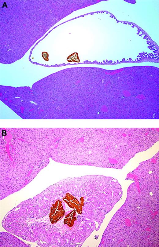
Pigment gallstones in “Berkeley” transgenic sickle cell mice. H&E. (A) Stones in a dilated gallbladder; original magnification, × 12.5. (B) Stones in a hyperplastic gallbladder; original magnification, × 40.
Pigment gallstones in “Berkeley” transgenic sickle cell mice. H&E. (A) Stones in a dilated gallbladder; original magnification, × 12.5. (B) Stones in a hyperplastic gallbladder; original magnification, × 40.
We appreciate the histopathologic survey of “Berkeley” transgenic sickle-cell mice by Manci and colleagues,1 and we have similar results in a larger range of ages (2 to 23 months) in another subcolony of these mice originally from Drs Chris Pastzy and Mohandas Narla. We also concur that the hemizygote mice are poor models for human sickle trait.2 However, we would like to add 2 additional similarities between these mice and human sickle cell disease that may have relevance to pathophysiology of sickle cell disease.
While Manci et al found no gallstones, we consistently see a 20% to 30% incidence of pigmented gallstones in examining nearly 100 of these mice. Photomicrographs (see figure) were visualized using an Olympus BX51 microscope (Olympus, Melville, NY) equipped with UPlan Apop 4 ×/0.16 and PlanApo 1.25 × 0.04 objective lenses. Images were captured with an Olympus DP70 camera, and were formatted using Adobe Photoshop software (Adobe Systems, San Jose, CA). Affected mice typically have a dilated gallbladder containing a single large stone and several smaller ones. Some gallbladders with gallstones are hyperplastic (Figure 1B), but none have histologic signs of cholecystitis or biliary duct obstruction, and we conclude that these gallstones are asymptomatic. The youngest mouse with pigmented gallstones was 6 weeks old. All mice exclusively expressing human sickle hemoglobin have elevated serum total bilirubin (13 ± 1 μM/L; mean and SEM, n = 16). This is another manifestation of human sickle-cell disease in the mouse model, and it correlates with the high-grade hemolysis in these mice. The only other mice with pigmented gallstones are those with spherocytosis due to the nb/nb mutation, manifesting gallstones after 6 months of age.3,4 Dogs, prairie dogs, and guinea pigs also form pigmented stones.5
We have also observed priapism in the Berkeley transgenic sickle-cell mice, as well as the same biochemical phenotype of nitric oxide resistance and phosphodiesterase 5A dysregulation as in priapic mice with knockout of nitric oxide synthases.6 These functional abnormalities of vasoregulation are present despite very subtle histologic changes in the penis.
Both gallstones and priapism contribute significantly to the complications of sickle-cell disease. These 2 functional manifestations in this transgenic mouse model of severe sickle-cell disease highlight the possible links between hemolysis and low nitric oxide bioavailability. Some disease similarities may accumulate as the mice age. While we acknowledge that there are differences in the physiology of mice compared with humans, the animal model has potential utility for functional studies relevant to sickle-cell disease pathophysiology.


This feature is available to Subscribers Only
Sign In or Create an Account Close Modal