Abstract
Store-operated Ca2+ entry (SOCE) is a major mechanism for Ca2+ influx in platelets and other cells. De novo conformational coupling between elements in the plasma membrane and Ca2+ stores, where the actin cytoskeleton plays an important regulatory role, has been proposed as the most likely mechanism to activate SOCE in platelets. Here we have examined for the first time changes in platelet F-actin levels on a subsecond time scale. Using stopped-flow fluorimetry and a quenched-flow approach, we provide evidence for the involvement of cofilin in actin filament reorganization and SOCE in platelets. Thrombin (0.1 U/mL) evoked an initial decrease in F-actin that commenced within 0.1 second and reached a minimum 0.9 second after stimulation, prior to the activation of SOCE. F-actin then increased, exceeding basal levels approximately 2.5 seconds after stimulation. Thrombin also induced cofilin dephosphorylation and activation, which paralleled the changes observed in F-actin, and rapid Btk activation. Inhibition of cofilin dephosphorylation by LFM-A13 resulted in the loss of net actin depolymerization and an increased delay in SOCE initiation. These results suggest that cofilin is important for the rapid actin remodeling necessary for the activation of SOCE in platelets through de novo conformational coupling.
Introduction
In nonexcitable cells such as platelets, store-operated calcium entry (SOCE), regulated by the filling state of the intracellular Ca2+ stores, is a major mechanism for calcium influx.1-5 Different models that have been presented to account for the activation of SOCE in distinct cell types can be grouped into 2 main categories that propose either indirect or direct coupling between the Ca2+ stores and the plasma membrane (PM). Indirect coupling models assume the generation of a small diffusible molecule that operates as a Ca2+ influx factor (CIF), so gating Ca2+ channels in the PM.6-8 On the other hand, direct or constitutive conformational coupling models propose a physical interaction between Ca2+ channels in the PM and IP3 receptors in the membrane of the intracellular Ca2+ stores.5,9 It also has been suggested that SOCE might be activated by the insertion of preformed channels into the PM by vesicle fusion in a secretion-like coupling model.10,11
A modification of the constitutive conformational coupling model proposes a dynamic and reversible conformational coupling based on the transport of portions of the endoplasmic reticulum (ER) containing IP3 receptors to the PM to facilitate de novo protein coupling.9,12-14 De novo conformational coupling requires a mechanical support provided by the actin cytoskeleton.14 In addition, the cortical actin cytoskeleton plays a regulatory role by acting as a negative clamp that prevents constitutive coupling and activation of SOCE.4,5,14,15 The de novo conformational coupling model requires that the cortical actin barrier be rapidly disrupted following cell stimulation to allow the coupling to occur.9,12,16 Earlier determinations of agonist-evoked changes in platelet F-actin have been made on a time scale of seconds or minutes17-19 ; however, we are not aware of determinations of F-actin content on the same time scale as agonist-evoked Ca2+ entry, which can commence within a second of stimulation.16,20
Proteins involved in actin filament remodeling include a large number of actin-binding proteins (ABPs), which control actin reorganization by direct contact with F-actin or with G-actin monomers. Some ABPs, like tropomodulin or Cap Z, bind to the barbed ends of F-actin, preventing the addition of more G-actin subunits.21-24 Twinfilin, verprolin/WIP, DNase I, and thymosin β4 inhibit actin polymerization by sequestering G-actin units.21,24-27 The role of other ABPs on actin remodeling, such as profilin and Srv2, is more subtle. Profilin-bound actin monomers cannot add to the pointed end but add to the barbed end, and Srv2 appears to be a shuttle, transferring actin monomers from cofilin to profilin and allowing nucleotide exchange on the monomers.28,29 Other ABPs, such as gelsolin, actively reduce the length of actin filaments by promoting the cleavage of the filaments.21,30,31 Cofilin accelerates depolymerization, though the mechanism of this effect is not completely understood.21
ABPs can be regulated by several intracellular mechanisms. A major pathway involves the activation of phosphatidylinositol 3-kinase (PI3K) by a LIM kinase-Rho-protein kinase C (PKC)-Ca2+/calmodulin-dependent pathway.32-35 Recent studies have presented an alternative pathway for actin reorganization based on the equilibrium between Ser/thr kinases and phosphatases.33-37 Especially relevant is the regulation of cofilin by the Ser/thr phosphatase, slingshot, which dephosphorylates the residue Ser3, allowing cofilin to bind to the actin cytoskeleton and reduce the length of the actin filament by removing G-actin monomers from the barbed end.32-39
Here we have investigated the temporal relationship between Ca2+ entry and actin filament reorganization on a subsecond time scale. The results suggest that actin depolymerization precedes release of Ca2+ from intracellular stores and the activation of Ca2+ entry, the former indicating that it is independent of rises in cytosolic free Ca2+ concentration ([Ca2+]c). Rapid actin depolymerization requires cofilin dephosphorylation and is compatible with membrane trafficking underlying the de novo conformational coupling of the type 2 inositol 1,4,5-trisphosphate receptor (IP3RII) to the store-operated Ca2+ entry channel hTRPC1 with subsequent activation of SOCE.12,16
Materials and methods
Materials
Fura-2 acetoxymethyl ester (fura-2/AM) was from Molecular Probes (Leiden, The Netherlands). Apyrase (grade 7), aspirin, bovine serum albumin, dithiothreitol, and fluorescein isothiocyanate-conjugated (FITC) phalloidin and thrombin were from Sigma (Poole, Dorset, United Kingdom). LFM-A13 was from Calbiochem (Nottingham, United Kingdom). Anti-phospho-Btk (Y-223) antibody and anti-Btk antibody were from Cell Signaling Technology (Beverly, MA). Anti-phosphoSer3-cofilin antibody, anticofilin antibody (C-20), horseradish peroxidase-conjugated donkey anti-goat IgG antibody, and horseradish peroxidase-conjugated goat anti-rabbit IgG antibody were from Santa Cruz Biotechnology (Santa Cruz, CA). All other reagents were of analytical grade.
Platelet preparation
Fura-2-loaded platelets were prepared as described previously.14 Briefly, blood was obtained from drug-free healthy volunteers. Approval was obtained from the University of Cambridge institutional review board for these studies. Informed consent was provided according to the Declaration of Helsinki. Blood was mixed with one-sixth volume of acid/citrate dextrose anticoagulant containing (in mM) 85 sodium citrate, 78 citric acid, and 111 D-glucose. Platelet-rich plasma then was prepared by centrifugation for 5 minutes at 700 g and aspirin (100 μM) and apyrase (40 μg/mL) added. Platelet-rich plasma was incubated at 37°C with 2 μM fura-2/AM for 45 minutes. Cells were then collected by centrifugation at 350 g for 20 minutes and resuspended in HEPES-buffered saline (HBS) containing (in mM) 145 NaCl, 10 HEPES, 10 D-glucose, 5 KCl, 1 MgSO4, pH 7.45, and supplemented with 0.1% wt/vol bovine serum albumin and 40 μg/mL apyrase.
Stopped-flow kinetic measurements
The kinetics of fluorescence change from fura-2-loaded platelets was investigated by stopped-flow fluorimetry at 37°C using a Hi-Tech Scientific SF-61SX2 Single-Mixing Stopped-Flow System (Hi-Tech Ltd, Salisbury, Wiltshire, United Kingdom) with an excitation wavelength of 340 or 360 nm and emission at 500 nm. Dye-loaded cells (100 μL; 4 × 10⁸ cells/mL) and an agonist solution (100 μL) were introduced into the sample flow circuit via separate reservoirs at the top of the sample-handling unit.16 Mn2+ influx was monitored as a quenching of fura-2 fluorescence at the iso-emissive wavelength of 360 nm, presented on an arbitrary linear scale.40 Results were corrected for the effects of quenching extracellular fura-2 and basal leak of Mn2+ into the cells by subtraction of agonist-free control runs.40 To reduce leakage of Mn2+ into the cells before the experiment, 50 μM MnCl2 was added to the cell suspension, and 350 μM MnCl2 was added to the agonist solution, giving a final concentration of 200 μM extracellular Mn2+ after mixing.
F-actin kinetic measurements
The kinetics of F-actin reorganization were determined in samples stimulated for various times before fixing using a rapid quench flow system16 (Hi-Tech Ltd). Briefly, the cell suspension (75 μL) and an agonist solution (75 μL) were introduced into the sample flow circuit via separate reservoirs at the top of the sample-handling unit and then mixed after the times indicated with 75 μL 3% (wt/vol) formaldehyde in phosphate-buffered saline (PBS) before storing on ice for 10 minutes. Fixed platelets were permeabilized by incubation for 10 minutes with 0.025% (vol/vol) Nonidet P-40 detergent dissolved in PBS. Platelets then were incubated for 30 minutes with fluorescein isothiocyanate-labeled phalloidin (1 μM) in PBS supplemented with 0.5% (wt/vol) bovine serum albumin. After incubation the platelets were collected by centrifugation for 60 seconds at 3000 g and resuspended in PBS. Staining of cells was measured using a Perkin-Elmer fluorescence spectrofluorimeter (Perkin-Elmer, Norwalk, CT). Samples were excited at 496 nm, and emission was at 516 nm.
Western blotting
Platelets were stimulated for various times at 37°C using a Hi-Tech Scientific RQF-63 Rapid Quench-Flow System16 as above for F-actin determinations, except that the stimulation was terminated by mixing with 75 μL of 2 × Laemmli buffer41 with 10% dithiothreitol, followed by heating for 5 minutes at 95°C. One-dimensional sodium dodecyl sulfate (SDS)-electrophoresis was performed on 12.5% sodium dodecyl sulfate-polyacrylamide gels, and separated proteins were electrophoretically transferred for 2 hours at 0.8 mA/cm2 in a semidry blotter (Hoefer Scientific, Newcastle, Staffordshire, United Kingdom) onto nitrocellulose for subsequent probing. Blots were incubated overnight with 10% (wt/vol) BSA in Tris-buffered saline with 0.1% Tween 20 (TBST) to block residual protein binding sites. Blocked membranes were then incubated with the anti-phospho-Btk (Y-223) antibody or the anti-Btk antibody diluted 1:1000 in TBST or with the anti-phosphoSer3-cofilin antibody diluted 1:250 in TBST or with anticofilin antibody diluted 1:100 in TBST for 1 hour. The primary antibody was removed and blots washed 6 times for 5 minutes each with TBST. To detect the primary antibody, blots were incubated with horseradish peroxidase-conjugated goat anti-rabbit IgG antibody or horseradish peroxidase-conjugated donkey anti-goat IgG antibody, diluted 1:2000 and 1:2500 in TBST, respectively; washed 6 times in TBST, and exposed to enhanced chemiluminescence reagents for 1 minute. Blots were then exposed to photographic films, and the optical density was estimated using scanning densitometry.
Statistical analysis
Analysis of statistical significance was performed using Student unpaired t test. For multiple comparisons, one-way analysis of variance combined with the Dunnet test was used.
Results
Latencies of Ca2+ release from intracellular stores and of Mn2+ entry stimulated by the physiologic agonist thrombin
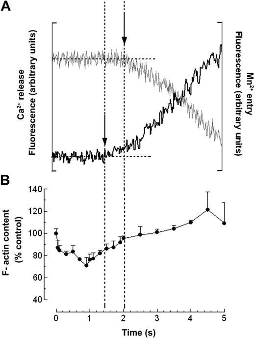
Human platelets were stimulated with 0.1 U/mL thrombin, a concentration that activates SOCE but not noncapacitative Ca2+ entry,16,42 and the latencies of thrombin-evoked release of Ca2+ from intracellular stores and of thrombin-evoked Mn2+ entry were determined by stopped-flow fluorimetry. Fura-2-loaded platelets were rapidly mixed with thrombin at a final concentration of 0.1 U/mL in the presence of 100 μM EGTA and 200 μM MnCl2. Recording fura-2 fluorescence at an excitation wavelength of 340 nm indicated that the delay in onset of thrombin-induced Ca2+ release from the intracellular stores was 1.64 ± 0.09 seconds (mean ± SE; Figure 1; n = 20). At the iso-emissive wavelength of 360 nm, Mn2+ quench of fura-2 fluorescence was first detected with latencies of 2.14 ± 0.09 seconds (Figure 1; n = 20). As shown previously,16 mixing cells with agonist-free HBS containing 100 μM EGTA and 200 μM MnCl2 did not modify fura-2 fluorescence at either excitation wavelength (not shown), confirming that the mixing procedure per se did not activate the cells.
Comparison of the latency and time course of thrombin-evoked Ca2+ release, Mn2+ entry, and actin reorganization. (A) Fura-2-loaded human platelets were rapidly mixed with thrombin (at 0 seconds) at a final concentration of 0.1 U/mL in the presence of 100 μM of EGTA and 200 μM MnCl2. Fura-2 fluorescence was recorded at excitation wavelengths of 340 nm (black trace, left axis) and 360 nm (gray trace, right axis). Traces are representative of 20 runs made on cell preparations from 10 donors. (B) Human platelets were rapidly mixed with 0.1 U/mL thrombin (•) or with HBS (dashed line) and incubated at 37°C for various time periods (1-5 seconds) before mixing with formaldehyde (3% in PBS). Actin filament content was determined as described in “Materials and methods.” Results shown are presented as percentage of the F-actin content in resting cells and expressed as mean ± SE of 21 runs made on cell preparations from 12 donors. Vertical dashed lines represent the starting times for Ca2+ release and Mn2+ entry.
Comparison of the latency and time course of thrombin-evoked Ca2+ release, Mn2+ entry, and actin reorganization. (A) Fura-2-loaded human platelets were rapidly mixed with thrombin (at 0 seconds) at a final concentration of 0.1 U/mL in the presence of 100 μM of EGTA and 200 μM MnCl2. Fura-2 fluorescence was recorded at excitation wavelengths of 340 nm (black trace, left axis) and 360 nm (gray trace, right axis). Traces are representative of 20 runs made on cell preparations from 10 donors. (B) Human platelets were rapidly mixed with 0.1 U/mL thrombin (•) or with HBS (dashed line) and incubated at 37°C for various time periods (1-5 seconds) before mixing with formaldehyde (3% in PBS). Actin filament content was determined as described in “Materials and methods.” Results shown are presented as percentage of the F-actin content in resting cells and expressed as mean ± SE of 21 runs made on cell preparations from 12 donors. Vertical dashed lines represent the starting times for Ca2+ release and Mn2+ entry.
Time course of thrombin-evoked actin filament reorganization
It has been reported that actin filament reorganization is necessary for the activation of SOCE in a number of cells, including smooth muscle cells,43 corneal endothelial cells,7 pancreatic acinar cells,5 glioma C6 cells,44 and platelets.14 To investigate the time course of actin filament remodeling, platelet samples were prepared by quenched flow for subsequent staining with FITC-phalloidin. The delay times between the rapid mixing of cells with 0.1 U/mL thrombin and the subsequent fixation with 3% formaldehyde in PBS (see “Materials and methods”) were set at 100- to 200-ms intervals for the first 2 seconds and at 500 ms for subsequent time points, commencing 100 ms after the mixing of cells with thrombin. Thrombin (0.1 U/mL) evoked an initial decrease in platelet F-actin that commenced within 100 ms and reached a minimum 0.9 second after stimulation at 70.7% ± 4.4% of the resting level (Figure 1; P < .05; n = 21). The F-actin content then increased, exceeding basal levels again approximately 2.5 seconds after stimulation. As reported above for Ca2+ mobilization, mixing cells with agonist-free HBS did not evoke significant changes in F-actin content.
Time course of cofilin phosphorylation evoked by thrombin
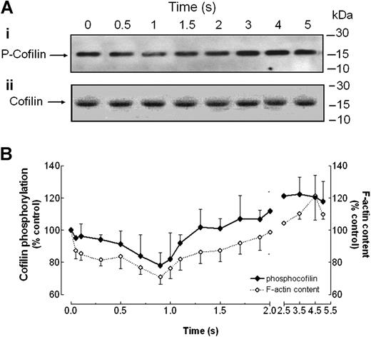
The actin depolymerizing factor cofilin is an ABP involved in actin reorganization that is negatively regulated by phosphorylation at Ser3 and reactivated by dephosphorylation.34,36 In view of the initial net actin depolymerization induced by thrombin, we have investigated the time course of changes in cofilin phosphorylation after treatment with 0.1 U/mL thrombin. To do this, platelet samples were prepared by quenched flow for subsequent sodium dodecyl sulfate-polyacrylamide gel electrophoresis (SDS-PAGE) and Western blot analysis. The delay times between the rapid mixing of cells with thrombin and the subsequent mixing with Laemmli buffer (“Materials and methods”) were set as described above for determination of F-actin content. Thrombin (0.1 U/mL) evoked an initial dephosphorylation of cofilin that commenced within 100 ms and reached a minimum 0.9 second after stimulation at 78.1% ± 7.7% of the resting level, as detected by Western blotting with an anti-phosphoSer3-cofilin antibody (Figure 2, top panel and graph; P < .05; n = 15). Cofilin phosphorylation at Ser3 then increased, exceeding resting levels again approximately 1.5 seconds after stimulation. Western blotting with an anticofilin antibody revealed that a similar amount of the protein was loaded in all lanes (Figure 2A, lower panel).
Comparison of the latency and time course of thrombin-evoked cofilin phosphorylation and actin reorganization. Platelets were rapidly mixed with thrombin at a final concentration of 0.1 U/mL or with agonist-free HBS solution (control at 0 seconds) and incubated at 37°C for various time periods (1-5 seconds) before mixing with lysis buffer (for cofilin phosphorylation) or with formaldehyde (3% in PBS; for F-actin measurement) using a quenched-flow system. For cofilin phosphorylation, proteins were separated by SDS-PAGE followed by Western blotting with either anti-phosphoSer3-cofilin antibody (Ai) or anticofilin antibody (Aii) as described in “Materials and methods.” Bands were revealed using chemiluminescence and were quantified using scanning densitometry. Positions of molecular-mass markers are shown on the right. Actin filament content was determined as described in “Materials and methods.” (B) Graph represents the quantification of cofilin phosphorylation (filled symbols) and F-actin content (open symbols). Values are mean ± SE of 15 runs made on cell preparations from 11 donors expressed as the percentage of cofilin phosphorylation or F-actin content in resting cells.
Comparison of the latency and time course of thrombin-evoked cofilin phosphorylation and actin reorganization. Platelets were rapidly mixed with thrombin at a final concentration of 0.1 U/mL or with agonist-free HBS solution (control at 0 seconds) and incubated at 37°C for various time periods (1-5 seconds) before mixing with lysis buffer (for cofilin phosphorylation) or with formaldehyde (3% in PBS; for F-actin measurement) using a quenched-flow system. For cofilin phosphorylation, proteins were separated by SDS-PAGE followed by Western blotting with either anti-phosphoSer3-cofilin antibody (Ai) or anticofilin antibody (Aii) as described in “Materials and methods.” Bands were revealed using chemiluminescence and were quantified using scanning densitometry. Positions of molecular-mass markers are shown on the right. Actin filament content was determined as described in “Materials and methods.” (B) Graph represents the quantification of cofilin phosphorylation (filled symbols) and F-actin content (open symbols). Values are mean ± SE of 15 runs made on cell preparations from 11 donors expressed as the percentage of cofilin phosphorylation or F-actin content in resting cells.
Time course of actin filament reorganization and cofilin phosphorylation evoked by thrombin in the presence of LFM-A13
We recently have demonstrated the involvement of Bruton tyrosine kinase (Btk) in actin remodeling and SOCE in thapsigargin-treated human platelets.45 Hence, we have investigated whether the role of Btk in SOCE might be mediated by cofilin and the regulation of actin reorganization. The activation of Btk was analyzed by Western blotting using a rabbit monoclonal phosphospecific anti-Btk antibody that only detects Btk autophosphorylated at the tyrosine residue 223, which has been shown to be the full activated form of Btk.46,47 Treatment of human platelets with thrombin (0.1 U/mL) evoked a rapid activation of Btk that was detectable within 100 ms and reached a maximum 2.5 seconds after stimulation at 385% ± 27% of the resting level (Figure 3A, top panel, and Figure 3B; n = 6). Western blotting with an anti-Btk antibody revealed that a similar amount of protein was loaded in all lanes (Figure 3A, lower panel).
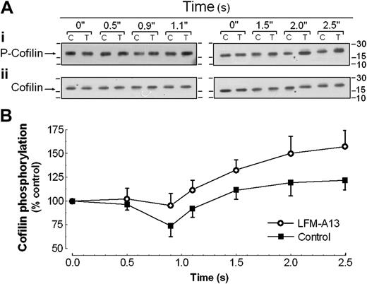
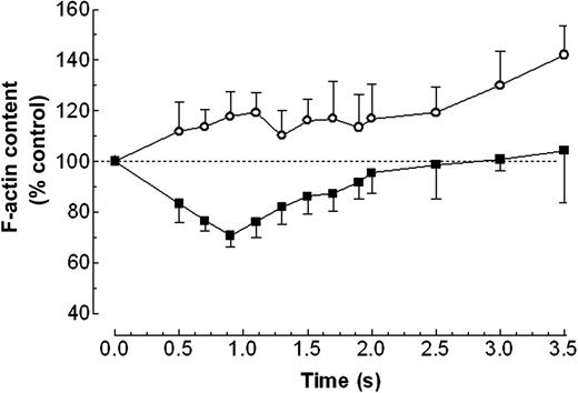
Pretreatment of platelets for 10 minutes with 10 μM LFM-A13, which abolishes Btk activation,45 inhibited the thrombin-evoked dephosphorylation of cofilin detected in the first 1.5 seconds after stimulation with the agonist and enhanced thrombin-evoked cofilin phosphorylation as detected by Western blotting with an anti-phosphoSer3-cofilin antibody (Figure 4; n = 6). Consistent with this, treatment of the cells for 10 minutes with 10 μM LFM-A13 abolished thrombin-evoked net actin depolymerization (Figure 5). After treatment with LFM-A13 the F-actin content 0.9 second after treatment with thrombin (0.1 U/mL) was 110% of basal, compared with 70% of basal in controls (Table 1; P < .001).
Effect of Btk inhibition on thrombin-evoked actin reorganization and Ca2+ mobilization
. | Control . | LFM-A13 . |
|---|---|---|
| F-actin content, % control | 70.7 ± 9.8 | 110.3 ± 4.36* |
| Ca2+ release, latency, s | 1.64 ± 0.09 | 1.85 ± 0.14 |
| Ca2+ entry, latency, s | 2.14 ± 0.09 | 2.60 ± 0.17† |
. | Control . | LFM-A13 . |
|---|---|---|
| F-actin content, % control | 70.7 ± 9.8 | 110.3 ± 4.36* |
| Ca2+ release, latency, s | 1.64 ± 0.09 | 1.85 ± 0.14 |
| Ca2+ entry, latency, s | 2.14 ± 0.09 | 2.60 ± 0.17† |
Human platelets were preincubated for 10 minutes with 10 μM LFM-A13 and then were rapidly mixed with 0.1 U/mL thrombin. F-actin content was determined 0.9 second after thrombin stimulation as described in “Materials and methods.” Results shown are presented as percentage of the F-actin content in resting cells and expressed as mean ± SE of 6 independent experiments.
P < .001. Fura-2 fluorescence was recorded at excitation wavelengths of 340 nm (for Ca2+ release) and 360 nm (for Ca2+ entry). Values are expressed as mean ± SE of at least 6 runs made on cell preparations from 6 donors and 7 to 10 separate experiments
P < .05
Treatment of platelets with LFM-A13 did not significantly increase the latency of thrombin-evoked Ca2+ release. The thrombin-evoked increase in [Ca2+]c was first detected after a delay of 1.85 ± 0.14 seconds after a 10-minute pretreatment with 10 μM LFM-A13 (n = 6), compared with 1.64 ± 0.09 seconds in controls (Table 1; P = .20). In contrast, the latency of thrombin-evoked Mn2+ entry was increased by the Btk inhibitor (Table 1). Mn2+ entry was first detected 2.60 ± 0.17 seconds (n = 10) after agonist addition, following a 10-minute pretreatment with 10 μM LFM-A13, compared with 2.14 ± 0.09 seconds in controls (P < .05). We have previously shown that pretreatment of human platelets for 10 minutes with 10 μM LFM-A13 decreased thrombin-evoked Ca2+ entry by about 30% without having any effect on thrombin-evoked release of Ca2+ from the intracellular stores.45
Thrombin-evoked rapid Btk phosphorylation and activation in human platelets. Platelets were rapidly mixed with thrombin at a final concentration of 0.1 U/mL, or with agonist-free HBS solution (control at 0 seconds) and incubated at 37°C for various time periods (0.1-2.5 seconds) before mixing with lysis buffer using a quenched-flow system. Proteins were separated by SDS-PAGE followed by Western blotting with either anti-phospho-Btk (Y-223) antibody (Ai) or anti-Btk antibody (Aii) as described in “Materials and methods.” Bands were revealed using chemiluminescence and were quantified using scanning densitometry. Positions of molecular-mass markers are shown on the right. (B) Graph represents the quantification of Btk phosphorylation. Values are mean ± SE of 6 runs made on cell preparations from 6 donors expressed as the percentage of Btk phosphorylation in resting cells.
Thrombin-evoked rapid Btk phosphorylation and activation in human platelets. Platelets were rapidly mixed with thrombin at a final concentration of 0.1 U/mL, or with agonist-free HBS solution (control at 0 seconds) and incubated at 37°C for various time periods (0.1-2.5 seconds) before mixing with lysis buffer using a quenched-flow system. Proteins were separated by SDS-PAGE followed by Western blotting with either anti-phospho-Btk (Y-223) antibody (Ai) or anti-Btk antibody (Aii) as described in “Materials and methods.” Bands were revealed using chemiluminescence and were quantified using scanning densitometry. Positions of molecular-mass markers are shown on the right. (B) Graph represents the quantification of Btk phosphorylation. Values are mean ± SE of 6 runs made on cell preparations from 6 donors expressed as the percentage of Btk phosphorylation in resting cells.
Effect of Btk inhibition on the latency and time course of thrombin-evoked cofilin phosphorylation. Human platelets were preincubated for 10 minutes in the presence of 10 μM LFM-A13 (○) or the vehicle (▪) and then rapidly mixed with thrombin (0.1 U/mL) or with agonist-free HBS solution (control at 0 seconds) and incubated at 37°C for various time periods before mixing with lysis buffer using a quenched-flow system. Proteins were separated by SDS-PAGE followed by Western blotting with either anti-phosphoSer3-cofilin antibody (Ai) or anticofilin antibody (Aii) as described in “Material and methods.” C indicates control; T, LFM-A13-treated cells. Bands were revealed using chemiluminescence and were quantified using scanning densitometry. Positions of molecular-mass markers are shown on the right. (B) Graph represents the quantification of cofilin phosphorylation. Values are mean ± SE of 6 runs made on cell preparations from 6 donors expressed as the percentage of cofilin phosphorylation in resting cells.
Effect of Btk inhibition on the latency and time course of thrombin-evoked cofilin phosphorylation. Human platelets were preincubated for 10 minutes in the presence of 10 μM LFM-A13 (○) or the vehicle (▪) and then rapidly mixed with thrombin (0.1 U/mL) or with agonist-free HBS solution (control at 0 seconds) and incubated at 37°C for various time periods before mixing with lysis buffer using a quenched-flow system. Proteins were separated by SDS-PAGE followed by Western blotting with either anti-phosphoSer3-cofilin antibody (Ai) or anticofilin antibody (Aii) as described in “Material and methods.” C indicates control; T, LFM-A13-treated cells. Bands were revealed using chemiluminescence and were quantified using scanning densitometry. Positions of molecular-mass markers are shown on the right. (B) Graph represents the quantification of cofilin phosphorylation. Values are mean ± SE of 6 runs made on cell preparations from 6 donors expressed as the percentage of cofilin phosphorylation in resting cells.
Effect of LFM-A13 on the latency and time course of thrombin-evoked actin reorganization. Human platelets were preincubated for 10 minutes in the presence of 10 μM LFM-A13 (○) or the vehicle (▪) and then rapidly mixed with 0.1 U/mL thrombin and incubated at 37°C for various time periods before mixing with formaldehyde (3% in PBS). Actin filament content was determined as described in “Material and methods.” Results are presented as the percentage of the F-actin content of nonstimulated cells (indicated by the dashed line) and expressed as mean ± SE of 6 runs made on cell preparations from 6 donors.
Effect of LFM-A13 on the latency and time course of thrombin-evoked actin reorganization. Human platelets were preincubated for 10 minutes in the presence of 10 μM LFM-A13 (○) or the vehicle (▪) and then rapidly mixed with 0.1 U/mL thrombin and incubated at 37°C for various time periods before mixing with formaldehyde (3% in PBS). Actin filament content was determined as described in “Material and methods.” Results are presented as the percentage of the F-actin content of nonstimulated cells (indicated by the dashed line) and expressed as mean ± SE of 6 runs made on cell preparations from 6 donors.
Discussion
We previously have proposed that SOCE in human platelets may be activated by a de novo conformational coupling model in which Ca2+ store depletion leads to trafficking of portions of the ER toward the PM to allow coupling between IP3RII in the ER membrane and hTRPC1 in the PM.12-16 In support of this model, we have shown that SOCE is reduced by inhibitors of actin polymerization such as cytochalasin D or latrunculin A, which impair reorganization of the cytosolic actin network that provides support to the transport of the ER toward the PM or by stabilization of the membrane cytoskeleton using jasplakinolide, which interferes with the depolymerization of the membrane cytoskeleton and which might thus prevent the approach of the ER and PM through the dense cortical F-actin layer present in platelets.14 We also have shown in co-immunoprecipitation experiments that Ca2+ store depletion following treatment with thapsigargin (TG) or the physiologic agonist thrombin results in de novo coupling of IP3RII and hTRPC1.12,15,16 This coupling is inhibited by agents that interfere with remodeling of the actin cytoskeleton15 and is reversed if the Ca2+ stores are allowed to refill.48 The coupling between IP3RII and hTRPC1 is closely temporally correlated with the activation of Ca2+ release from the ER and the activation of Ca2+ entry in thrombin-stimulated platelets,16 supporting the hypothesis that this coupling event may underlie the activation of SOCE.
Rapid coupling of IP3RII in the ER membrane to hTRPC1 in the PM requires an equally rapid reorganization of the actin cytoskeleton to allow the approach of the ER and PM, which are normally separated by a dense cortical layer of F-actin.14 Here we have monitored for the first time thrombin-stimulated changes in platelet F-actin content on a subsecond time scale. Using a quenched-flow approach to fix platelet samples at varying times after stimulation with 0.1 U/mL thrombin, we have shown that there is an initial decrease in F-actin content that commenced within 100 ms of stimulation. The F-actin content of the cells reached a minimum around 70% of the resting level 0.9 second after stimulation and then began to increase again, exceeding the resting level about 2.5 seconds after stimulation. The early decrease in platelet F-actin content preceded the release of Ca2+ from intracellular stores, which was determined by stopped-flow fluorimetry in parallel experiments on the same platelet preparations to occur about 1.6 seconds after stimulation with thrombin. This early decrease and subsequent rise in platelet F-actin content may play an important role in the trafficking of portions of the ER toward the PM. The fact that the actin remodeling preceded Ca2+ store release might explain the close temporal correlation between Ca2+ release and the coupling of IP3RII to hTRPC1 that we have previously reported.16
Since remodeling of the actin cytoskeleton occurred prior to the release of Ca2+ from intracellular stores, it cannot be dependent on either Ca2+ store depletion or a rise in [Ca2+]c. To investigate the mechanism of early thrombin-evoked changes in platelet F-actin, we focused on the possible role of cofilin, an actin binding protein that promotes actin depolymerization by removing G actin subunits from the barbed ends of the filaments.21,25-38 Cofilin has been shown to be activated by the Ser/Thr phosphatase, slingshot, which dephosphorylates the residue Ser3.33-39 Cofilin dephosphorylation has been shown to be involved in transient association with the actin cytoskeleton,49 decreasing cofilin affinity for actin after phosphorylation at Ser3.50 Western blotting using platelet samples prepared by quenched flow from the same cell preparations used for F-actin measurements revealed that the platelet content of cofilin phosphorylated on Ser3 changed with a similar time course to the content of F-actin. Thrombin (0.1 U/mL) evoked an initial dephosphorylation of cofilin that commenced within 100 ms, reached a minimum 0.9 second after stimulation at around 78% of the resting level, and then cofilin phosphorylation at Ser3 increased again exceeding resting levels about 1.5 seconds after stimulation. The close temporal correlation between the changes in platelet phosphocofilin and F-actin contents are compatible with cofilin playing a role in the observed early actin depolymerization.
A role for cofilin in the early thrombin-evoked actin depolymerization is supported by our observations with the Btk inhibitor, LFM-A13. Pretreatment of platelets for 10 minutes with 10 μM LFM-A13, which we have previously shown abolishes Btk activation,45 inhibited the early cofilin dephosphorylation and the actin depolymerization evoked by thrombin. Our results demonstrate that thrombin induced rapid Btk phosphorylation and activation, which is consistent with a role for Btk in the activation of cofilin and actin filament reorganization stimulated by thrombin. We previously have shown that Btk inhibition reduced Ca2+ entry evoked following thrombin-evoked Ca2+ store depletion.45 Here we found that Btk inhibition increased the latency of thrombin-evoked Ca2+ entry as assessed by Mn2+ quench of fura-2 fluorescence. These alterations in the activation of Ca2+ entry following Btk inhibition might be explained by a failure of SOCE activation by de novo conformational coupling when actin depolymerization is inhibited. The residual Ca2+ entry observed following inhibition of actin depolymerization may be accounted for by the cytoskeleton-independent SOCE pathway activated via extracellular signal-related kinase 1/2 (ERK1/2) that we have previously described.51
Another mechanism of regulating cofilin activity is through its binding to phosphotidylinositol (4,5)-bisphosphate (PIP2), which inhibits the actin-binding ability of cofilin.52-54 This mechanism has been shown to be important for the spatial regulation of cofilin activity. The PIP2- and actin-binding sites have been localized in residues W104-M115 of the cofilin primary sequence.55 In addition to the described mechanism of cofilin activation through the activation of Btk, thrombin also might regulate cofilin activity by the activation of phospholipase C, which might co-operate to remodel the actin cytoskeleton through PIP2 hydrolysis, leading to activation of cofilin.53
In summary, we have shown that platelet activation by thrombin is associated with rapid depolymerization of F-actin prior to the release of Ca2+ from intracellular stores and the activation of Ca2+ entry. This early actin depolymerization and later polymerization may be important in allowing trafficking of portions of the ER toward the PM to allow the de novo coupling of IP3RII to hTRPC1, which we have suggested may underlie the activation of SOCE in human platelets.12-16,48 The activation of cofilin by its dephosphorylation at Ser3 parallels the early thrombin-evoked actin depolymerization and appears to be essential for this to occur. In NIH3T3 cells, cofilin dephosphorylation has been shown to lie downstream of Ras activation and to involve 2 Ras effector pathways, Raf-MEK and PI3K.56 We previously have demonstrated that inhibition of Ras,57 MEK,51 or PI3K58 results in inhibition of SOCE in human platelets. These earlier data are compatible with an important role for cofilin activity in the activation of SOCE by a reversible de novo coupling model in human platelets.
Supported by the Wellcome Trust (064070).
Prepublished online as Blood First Edition Paper, October 18, 2005; DOI 10.1182/blood-2005-05-2015.
The publication costs of this article were defrayed in part by page charge payment. Therefore, and solely to indicate this fact, this article is hereby marked “advertisement” in accordance with 18 U.S.C. section 1734.
M.T.H. held a British Heart Foundation Studentship. P.C.R. was supported by a DGESIC fellowship (BFI2001-0624) and held a fellowship from Junta de Extremadura.


This feature is available to Subscribers Only
Sign In or Create an Account Close Modal