Abstract
Chemokine receptor signaling is critical for lymphocyte trafficking across high endothelial venules (HEVs), but the exact mode of action of individual chemokines expressed in the HEVs is unclear. Here we report that CXCL13, expressed in a substantial proportion of HEVs in both lymph nodes (LNs) and Peyer patches (PPs), serves as an arrest chemokine for B cells. Whole-mount analysis of mesenteric LNs (MLNs) showed that, unlike T cells, B cellsa dhere poorly to the HEVs of CXCL13–/– mice and that B-cell adhesion is substantially restored in CXCL13–/– HEVs when CXCL13 is added to the MLNs by superfusion, as we have previously observed in PP HEVs by intravital microscopy. In vitro, CXCL13 activated the small guanosine triphosphatase (GTPase) Rap1 in B cells, and corroborating this observation, a deficiency of RAPL, the Rap1 effector molecule, caused a significant reduction in shear-resistant B-cell adhesion to intercellular adhesion molecule 1 (ICAM-1). In addition, CXCL13 induced B-cell adhesion to mucosal addressin cell adhesion molecule 1 (MAdCAM-1) by activating α4 integrin. These data identify CXCL13 as an arrest chemokine for B cells in HEVs and show that CXCL13 plays an important role in B-cell entry into not only PPs but also MLNs.
Introduction
Certain chemokines that are produced in or localized to the venular wall induce the rapid integrin-mediated arrest of leukocytes and their adhesion to the venular endothelium, essential steps for the recruitment of these cells to the secondary lymphoid tissues or sites of inflammation.1,2 For instance, CCL21 plays an indispensable role in the migration of naive T cells to lymph nodes (LNs) and Peyer patches (PPs).3,4 CCL21 is expressed on the luminal surface of high endothelial venules3 (HEVs), and desensitization of its receptor, CCR7, results in severe impairment of leukocyte function–associated antigen 1 (LFA-1)–mediated T-cell arrest at HEVs.3,4 Plt/plt mice congenitally lack expression of CCL21 and CCL195 and show severely impaired T-cell arrest at HEVs; arrest is restored by the reconstitution of CCL21 expression.3 Therefore, CCL21 fulfills the criteria proposed by Ley6 for an arrest chemokine in that (1) it is luminally expressed, (2) its arrest function is prevented by blocking the function of CCL21 or the function of its receptor, and (3) its arrest function is restored when its endothelial expression is reconstituted. CCL21 appears to trigger signal transduction pathways involving small guanosine triphosphatase (GTPase) Rap1 and a Rap1 effector molecule RAPL;,7,8 and RAPL-deficient mice indeed showed impaired trafficking of T cells, B cells, and dendritic cells.9
In contrast, an arrest chemokine for B cells at HEVs has not been identified. Okada et al10 reported that chemokine signals through CXCR4 and CCR7 are important for B-cell entry into LNs, whereas signals through CXCR4, CCR7, and CXCR5 are important for B-cell entry into PPs. However, how the ligands of these receptors contribute to B-cell migration to these lymphoid tissues has not been characterized in detail.
The CXCR5 ligand is the chemokine CXCL13, which selectively attracts mature B cells11 and a small subset of T cells.12 Mice deficient in CXCL13 show developmental defects in peripheral LNs (PLNs) and PPs but not mesenteric LNs (MLNs).13,14 They also show impaired B-cell trafficking in MLNs.13 Cupedo et al15 recently reported that the differential requirement of CXCL13 in genesis of MLNs and PLNs may be explained by differences in subpopulations of LN organizer cells in developing LN anlagen.
We reported previously that (1) CXCL13 is luminally expressed in about 50% of the HEVs in LNs and PPs, (2) CXCL13–/– PP HEVs show much-reduced levels of B-cell adhesion compared with T-cell adhesion, and (3) reconstitution of CXCL13 expression by the superfusion of CXCL13–/– PPs with CXCL13 significantly restores B-cell arrest in PP HEVs,13 indicating that CXCL13 plays a critical role in B-cell arrest/adhesion at the HEVs of PPs. The exact mode of action of the HEV-associated CXCL13 for B cells, however, has not been determined. In addition, significance of CXCL13 expressed in LN HEVs in B-cell arrest/adhesion has not been directly evaluated although Okada et al previously reported that the entry of CXCR5-deficient B cells into LNs was hardly affected.10 Here we examined the mode of action of CXCL13 on B cells in MLN HEVs by the use of CXCL13-deficient mice and report that CXCL13 is an arrest chemokine for B cells at HEVs. CXCL13 activates signal transduction pathways involving a small GTPase Rap1 and its effector molecule, RAPL, which results in integrin activation in B cells.
Materials and methods
Animals
C57BL/6 mice (Japan SLC, Hamamatsu, Japan) and green fluorescent protein (GFP)–transgenic mice,16 kindly provided by Dr M. Okabe of Osaka University, were housed under specific pathogen-free conditions. CXCL13–/– mice and RAPL–/– mice were established and used as described previously.9,13 Our experimental protocol was approved by the Ethics Review Committee for Animal Experimentation of Osaka University Medical School.
Lymphocyte isolation and antibodies
B cells and T cells were isolated by incubating spleen cells with a mixture of anti-CD3 and anti-CD11b monoclonal antibodies (mAbs) or anti-B220 and anti-CD11b mAbs, respectively, followed by AutoMACS separation (Miltenyi Biotec, Bergisch Gladbach, Germany), as described previously.13 The purity of the B and T cells was more than 90% on average, as determined by flow cytometry. The MECA-89 mAb17 was purified from ascites and labeled with Alexa Fluor 594 or Alexa Fluor 647 (Molecular Probes, Eugene, OR) in our laboratory. Unless otherwise noted, the origin of the antibodies used in this study was as described previously.13
Whole-mount microscopy
Purified B cells and T cells from GFP-transgenic mice were resuspended in warmed saline and injected intravenously into wild-type (WT) and CXCL13–/– mice (n = 3; 1 × 10⁷ cells/mouse). Ten minutes after the lymphocyte injection, Alexa Fluor 594– or Alexa Fluor 647–labeled MECA-89 mAb was injected intravenously to label the mucosal addressin cell adhesion molecule 1-positive (MAdCAM-1+) HEVs in situ. Mice were humanely killed 15 minutes after the lymphocyte injection and perfused with saline and 4% paraformaldehyde in phosphate-buffered saline (PBS). The MLNs were collected, fixed with 4% paraformaldehyde, and treated with increasing concentrations of saccharose (10%, 20%, and 30%). Cell adhesion to MAdCAM-1+ MLN HEVs was analyzed by confocal microscopy (LSM510META; Carl Zeiss, Jena, Germany, or Randiance 2100; Bio-Rad, Hercules, CA). Digital images were processed using Photoshop 6.0 software (Adobe Systems, San Jose, CA). For quantification of the lymphocyte adhesion to HEVs, the number of lymphocytes that adhered to each HEV segment and the length of the HEV segments were evaluated for 80 HEV segments/mouse. Superfusion with mouse CXCL13 (Dako, Glostrup, Denmark; 25 μg/mL in PBS for 90 minutes) and immunohistochemical analyses were performed as described previously.13 In some experiments, purified B cells were preincubated with pertussis toxin (PTX; 100 ng/mL; Calbiochem, San Diego, CA) at 37°C for 2 hours, washed, and injected intravenously into mice.
Static adhesion assay
Multiwell (4-mm diameter) glass slides were coated with rat intercellular adhesion molecule 1 (ICAM-1)/IgG18 (a gift from Dr Y. Iigo, Daiichi Pharmaceutical, Tokyo, Japan), rat MAdCAM-1/IgG,19 or human IgG (50 ng/well) overnight at 4°C and blocked with fetal calf serum. B cells (1 × 10⁵) were then added and allowed to settle for 15 minutes at 37°C. Subsequently, mouse CXCL13 (Dako) was added to the wells and incubated for 5 minutes at 37°C to induce cell adhesion. Phorbol myristate acetate (PMA; final concentration of 25 ng/mL; Sigma-Aldrich, St Louis, MO) was used as a positive control. After the unbound cells were washed off, the bound cells were counted. For antibody blocking, the cells or immobilized ligands were preincubated with an mAb or control antibody for 30 minutes at 37°C; anti–mouse CD11a (KBA),20 anti–rat CD54 (1A29),21 and anti–rat MAdCAM-1 (OST2)19 were used at 20 μg/mL and anti-CD49d (PS/2)22 was used as a hybridoma culture supernatant. In some experiments, cells were preincubated with PTX (100 ng/mL) at 37°C for 15 minutes, washed, and used in binding studies.
Detachment assay
ICAM-1/IgG (10 μg/mL) and CXCL13 (2 μM) were immobilized on the inside walls of an approximately 15-μL glass capillary tube (ϕ: 0.69 mm; Drummond, Broomal, PA) as described previously,23 so that the upstream half of each tube was coated with ICAM-1/IgG alone and the downstream half with ICAM-1/IgG plus CXCL13. B cells (1 × 10⁶ cells/mL) were injected into the capillary tubes at 0.25 dynes/cm2 for 5 minutes at 37°C. Shear stress was generated with an automated Harvard syringe pump (PHD2000; Instech Laboratories, Plymouth Meeting, PA). The flow was then increased in 2-fold increments every 20 seconds. The experiments were videotaped for analysis, and the number of B cells that remained bound was determined by counting, at each interval. In some experiments, cells were treated with PTX or KBA (see “Static adhesion assay”). For the functional comparison of wild-type and RAPL-deficient B cells, shear-resistant B-cell adhesion to immobilized ICAM-1/IgG (0.5 μg/mL) was assessed in a parallel flow chamber as described.7
Flow adhesion assay
MAdCAM-1/IgG or CXCL13 was immobilized on glass capillary tubes as described. B cells (1 × 10⁶ cells/mL) were then injected into the capillary tubes at 1.0 dyne/cm2 at 37°C, and cell images were recorded with a cell-viewing system (SRM-100; Nikon, Tokyo, Japan) and video recorder (BR-S600; Victor, Yokohama, Japan), using a Nikon Plan Fluor DL 10 ×/0.3 numeric aperture objective. Digital images were prepared using Quick Time 6.5.2 (Apple, Cupertino, CA) and Adobe Photoshop 6.0. Three categories of B-cell tethers under continuous flow were determined as described previously24 ; tethers were defined as “transient” if the cells attached briefly (< 3 seconds) to the substrate, as “rolling” if cells rolled on the substrate at least 3 seconds, and as “arrested” if cells were arrested on the substrate and remained adherent and stationary for at least 20 seconds. All cellular interactions were determined by manually tracking individual cells in a 0.39-mm2 area for 1 minute. For the blocking of MAdCAM-1, the tubes were pretreated with OST2 (20 μg/mL) for 30 minutes at room temperature. In some experiments, B cells were preincubated with PS/2 or PTX (see “static adhesion assay”).
Detachment assay with cultured endothelial cells
The BC1 mouse endothelial cells were derived from the cortical bone of BALB/c mice25 and used in detachment assays under flow as previously described.7 A monolayer of BC1 cells was grown on a gelatin-coated plastic dish and placed in a flow chamber (Glycotech, Gaithersburg, MD). The endothelial cell monolayer was preincubated with 1 μM CXCL13 for 5 minutes, and unbound CXCL13 was removed by gentle washing. B cells (5 × 10⁶/mL) were injected into the chamber at 0.1 dyne/cm2 for 8 minutes. Next, a higher shear stress (2 dynes/cm2) was applied for 10 minutes.7 Experiments were videotaped for analysis, and the number of adherent B cells was counted. In blocking experiments, B cells were pretreated with PTX, KBA, or PS/2 as described.
Rap1 activation
Purified B cells were stimulated with 30 nM mouse CXCL13 at 37°C for the indicated time period. The cells (1 × 10⁷) were lysed in ice-cold lysis buffer (50 mM Tris [tris(hydroxymethyl)aminomethane]–HCl, pH 7.4, 500 mM NaCl, 1% NP-40, 2.5 mM MgCl2, 10% glycerol, 10 μg/mL aprotinin, and 10 μg/mL leupeptin) and subjected to a pull-down assay using a glutathione S-transferase (GST)–tagged Ral guanosine diphosphate dissociation stimulator (GDS)–Ras–binding domain (RBD) fusion protein coupled to glutathione-agarose beads as previously described.26 Rap1 protein was detected by Western blotting using an anti-Rap1 antibody (BD PharMingen, San Diego, CA).
Results
B-cell adhesion to MLN HEVs is significantly impaired in CXCL13–/– mice and is restored by the reconstitution of CXCL13 expression
We first sought to quantify B-cell adhesion in the MLN HEVs of CXCL13–/– and WT mice. For this purpose, GFP-transgenic B and T cells were injected into WT and CXCL13–/– mice, and their behavior in HEVs was analyzed 15 minutes after the lymphocyte injection by whole-mount microscopy of the MLNs. As shown in Figure 1A, in WT mice, a few B cells were detected on the wall of MLN HEVs, whereas in CXCL13–/– mice, the number of B cells adhering to the HEVs was reduced, decreasing to about 40% of that seen in WT mice (Figure 1B). When the frequency distribution of adherent B cells per HEV segment was compared between the CXCL13–/– and WT mice, the distribution curve of the CXCL13–/– mice was shifted to the left compared with that of WT mice (Figure 1C), confirming that the B-cell adhesion to the MLN HEVs was significantly impaired in the absence of CXCL13 expression. On the other hand, the number of T cells that adhered to the MLN HEVs, which was greater than that of B cells, was comparable between the WT and CXCL13–/– mice (Figure 1A-B), indicating that the reduction in B-cell adhesion was not due to secondary effects, such as a difference in the blood flow rate in the HEVs of these mice. It should be noted, however, that there was a low but a significant level of B-cell adhesion in the CXCL13–/– HEVs (Figure 1A-B), which was almost completely abrogated by the treatment of B cells with a Gαi-specific inhibitor, PTX (data not shown), indicating that CXCL13 is not the only chemokine that regulates B-cell arrest/adhesion in LN HEVs.
B-cell adhesion to MLN HEVs is impaired in CXCL13-deficient mice and is restored by CXCL13 expression. (A) Whole-mount microscopy of MLNs from CXCL13–/– and WT mice. GFP-transgenic B or T cells (green) were injected intravenously into WT and CXCL13–/– mice. Subsequently, Alexa Fluor 594–conjugated MECA89 mAb (red) was injected to label the HEVs in situ. Images were obtained using a Zeiss LSM510 META microscope (Carl Zeiss, Jena, Germany) with a 20×/0.5 numeric aperture (NA) objective. Zeiss Plan NeoFluar. (B) B- and T-cell adhesion to CXCL13–/– and WT MLN HEV segments. HEVs were divided into segments as shown in panel A, and the number of cells bound to each segment was determined. In each experiment, 80 HEV segments/mouse were examined. Data represent the mean ± SD of the number of bound cells per HEV segment in 3 mice. Note that the B-cell adhesion to CXCL13–/– MLN HEVs was about 40% of that seen to WT MLN HEVs (*P < .01). (C) The frequency distribution of adherent B cells per HEV segment. (D) Restoring CXCL13 to the MLN HEVs of CXCL13–/– mice restored B-cell adhesion. After superfusion of MLNs with PBS or CXCL13, GFP-transgenic B cells and Alexa Fluor 647–conjugated MECA89 mAbs (blue) were injected and analyzed as in panel A (right panels). Frozen sections of the MLNs of these mice were stained with an anti-CXCL13 antibody (red; left panels, arrowheads). Images were obtained using a Bio-Rad Radience 2100 microscope (Bio-Rad, Hercules, CA) with a Nikon Plan Fluor 20×/0.45 NA objective.
B-cell adhesion to MLN HEVs is impaired in CXCL13-deficient mice and is restored by CXCL13 expression. (A) Whole-mount microscopy of MLNs from CXCL13–/– and WT mice. GFP-transgenic B or T cells (green) were injected intravenously into WT and CXCL13–/– mice. Subsequently, Alexa Fluor 594–conjugated MECA89 mAb (red) was injected to label the HEVs in situ. Images were obtained using a Zeiss LSM510 META microscope (Carl Zeiss, Jena, Germany) with a 20×/0.5 numeric aperture (NA) objective. Zeiss Plan NeoFluar. (B) B- and T-cell adhesion to CXCL13–/– and WT MLN HEV segments. HEVs were divided into segments as shown in panel A, and the number of cells bound to each segment was determined. In each experiment, 80 HEV segments/mouse were examined. Data represent the mean ± SD of the number of bound cells per HEV segment in 3 mice. Note that the B-cell adhesion to CXCL13–/– MLN HEVs was about 40% of that seen to WT MLN HEVs (*P < .01). (C) The frequency distribution of adherent B cells per HEV segment. (D) Restoring CXCL13 to the MLN HEVs of CXCL13–/– mice restored B-cell adhesion. After superfusion of MLNs with PBS or CXCL13, GFP-transgenic B cells and Alexa Fluor 647–conjugated MECA89 mAbs (blue) were injected and analyzed as in panel A (right panels). Frozen sections of the MLNs of these mice were stained with an anti-CXCL13 antibody (red; left panels, arrowheads). Images were obtained using a Bio-Rad Radience 2100 microscope (Bio-Rad, Hercules, CA) with a Nikon Plan Fluor 20×/0.45 NA objective.
We next applied CXCL13 topically to the MLNs of CXCL13–/– mice. This maneuver restored the CXCL13 expression and increased the B-cell adhesion/arrest in CXCL13–/– MLN HEVs significantly (Figure 1D), indicating that the forced expression of CXCL13 restored B-cell adhesion to the MLN HEVs in CXCL13–/– mice. These in vivo observations altogether indicate that CXCL13 plays a critical role in B-cell adhesion to MLN HEVs.
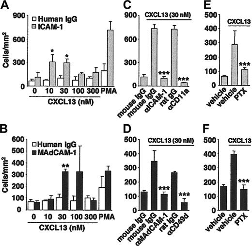
CXCL13 induces B-cell adhesion to ICAM-1 and MAdCAM-1 under static conditions. B cells were stimulated with CXCL13 and allowed to bind to immobilized ICAM-1 (A,C,E), MAdCAM-1 (B,D,F), or control IgG. PMA was used as a positive control. After the unbound B cells were washed off, the bound B cells were counted. The binding of B cells to ICAM-1 (A) was abrogated by mAbs to ICAM-1 or CD11a (C) or by PTX (E). Similarly, the CXCL13-induced B-cell binding to MAdCAM-1 (B) was inhibited by mAbs to CD49d or MAdCAM-1 (D) or by PTX (F). Data represent the mean ± SD of the number of bound B cells in triplicate fields. *P < .05 compared with untreated B cells. **P < .01 compared with untreated B cells. ***P < .01 compared with control IgG- or vehicle-treated B cells.
CXCL13 induces B-cell adhesion to ICAM-1 and MAdCAM-1 under static conditions. B cells were stimulated with CXCL13 and allowed to bind to immobilized ICAM-1 (A,C,E), MAdCAM-1 (B,D,F), or control IgG. PMA was used as a positive control. After the unbound B cells were washed off, the bound B cells were counted. The binding of B cells to ICAM-1 (A) was abrogated by mAbs to ICAM-1 or CD11a (C) or by PTX (E). Similarly, the CXCL13-induced B-cell binding to MAdCAM-1 (B) was inhibited by mAbs to CD49d or MAdCAM-1 (D) or by PTX (F). Data represent the mean ± SD of the number of bound B cells in triplicate fields. *P < .05 compared with untreated B cells. **P < .01 compared with untreated B cells. ***P < .01 compared with control IgG- or vehicle-treated B cells.
CXCL13 induces B-cell adhesion to immobilized ICAM-1 and MAdCAM-1 under static as well as flow conditions
To elucidate the mode of action of CXCL13 on B cells, we examined whether CXCL13 could induce integrin-mediated B-cell adhesion to the HEV-associated endothelial ligands, ICAM-1 and MAdCAM-1. Under static conditions, CXCL13 significantly promoted B-cell adhesion to ICAM-1 (Figure 2A) and MAdCAM-1 (Figure 2B), yielding bell-shaped dose-response curves that are typical for chemotactic responses. The CXCL13-induced B-cell adhesion to ICAM-1 was specifically inhibited by an anti-CD11a or anti–ICAM-1 mAb (Figure 2C), and the induced cell adhesion to MAdCAM-1 was also specifically inhibited by an anti-CD49d or anti–MAdCAM-1 mAb (Figure 2D). A Gαi-specific inhibitor, PTX, inhibited the CXCL13-induced B-cell adhesion to ICAM-1 (Figure 2E) and MAdCAM-1 (Figure 2F), reducing it to basal levels. Taken together, these results indicate that CXCL13 regulates B-cell adhesion mainly via Gαi-dependent, integrin-dependent signal-transduction pathways.
Next, we examined whether CXCL13 promotes shear-resistant B-cell adhesion to ICAM-1. For this purpose, B cells were first allowed to interact with immobilized ICAM-1 at a low shear stress, and then they were subjected to progressively increasing shear stress. Whereas most of the B cells that initially adhered to the immobilized ICAM-1 detached with increments of shear stress, when CXCL13 was coimmobilized with ICAM-1, the majority of B cells remained adherent even at the highest shear stress examined (16 dyne/cm2), indicating that CXCL13 effectively induced shear-resistant B-cell adhesion (Figure 3A). The augmented B-cell adhesion was inhibited by PTX to the basal level (Figure 3B), and also by anti-CD11a mAb (data not shown), indicating that CXCL13 activated LFA-1 on B cells via PTX-sensitive signal transduction to induce shear-resistant cell adhesion to ICAM-1.
In addition, immobilized CXCL13 induced B-cell arrest on MAdCAM-1 in a dose-dependent manner under physiologic flow conditions (1 dyne/cm2), although it did not significantly affect the total number of B cells interacting with the immobilized MAdCAM-1 (Figure 4A). In the absence of CXCL13, almost no B cells were arrested on MAdCAM-1, whereas in the presence of CXCL13, they adhered to it (Figure 4B). The CXCL13-promoted cell adhesion was almost completely inhibited by treating the capillary tube with an anti–MAdCAM-1 mAb or the B cells with an anti-CD49 mAb (Figure 4C) or PTX (Figure 4D), but not by treatment with the control IgG, indicating that CXCL13 enables the α4 integrin on B cells to bind to MAdCAM-1 to induce cell arrest under physiologic flow conditions.
CXCL13 induces shear-resistant B-cell adhesion to an endothelial cell monolayer
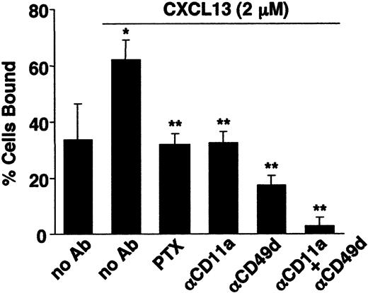
Next, we examined the effects of CXCL13 on shear-resistant B-cell adhesion to a BC1 endothelial cell monolayer. When the BC1 monolayer was pretreated with CXCL13, more than 60% of the input B cells remained adherent under high-shear stress flow conditions, whereas when the monolayer was pretreated with culture medium alone, only about 30% of the input B cells were adherent under the same conditions (Figure 5). PTX, anti-CD11a mAb, and anti-CD49d mAb all inhibited the shear-resistant B-cell adhesion to basal levels, and the combination of anti-CD11a and anti-CD49d almost completely inhibited the CXCL13-induced shear-resistant adhesion, indicating that CXCL13 induced LFA-1– and α4 integrin–dependent adhesion of B cells to the BC1 monolayer. Corroborating this, the BC1 cells expressed ICAM-1 and vascular cell adhesion molecule 1 (VCAM-1) and showed a readily detectable level of CXCL13 binding, as evidenced by flow cytometry (data not shown). Collectively, these results demonstrate that CXCL13 can activate both LFA-1 and α4 integrins on B cells, which results in the induction of shear-resistant B-cell adhesion to endothelial cells.
CXCL13 induces shear-resistant B-cell adhesion to ICAM-1 under flow conditions. (A) Shear-resistant B-cell adhesion to ICAM-1 alone or to ICAM-1 with CXCL13. The B cells that remained adherent at each shear-stress level were counted and expressed as a percentage of the initially bound B cells. (B) Pretreatment of B cells with PTX. The shear-resistant B-cell adhesion induced by CXCL13 (16 dyne/cm2) was abrogated by PTX. Data represent the mean ± SD of 3 independent experiments. *P < .01 compared with vehicle-treated B cells.
CXCL13 induces shear-resistant B-cell adhesion to ICAM-1 under flow conditions. (A) Shear-resistant B-cell adhesion to ICAM-1 alone or to ICAM-1 with CXCL13. The B cells that remained adherent at each shear-stress level were counted and expressed as a percentage of the initially bound B cells. (B) Pretreatment of B cells with PTX. The shear-resistant B-cell adhesion induced by CXCL13 (16 dyne/cm2) was abrogated by PTX. Data represent the mean ± SD of 3 independent experiments. *P < .01 compared with vehicle-treated B cells.
CXCL13 induces B-cell arrest on MAdCAM-1 under flow conditions. (A) B cells were injected at 1 dyne/cm2 into capillary tubes coated with MAdCAM-1 with or without various concentrations of CXCL13. The B-cell interactions with MAdCAM-1 were classified into 3 categories as described in “Materials and methods.” (B) B-cell arrest (dotted squares) was seen only in the CXCL13 coimmobilized area. Images were acquired as described in “Flow adhesion assays.” (C) The adhesive interaction of B cells with MAdCAM-1 was abrogated by mAbs to CD49d or MAdCAM-1. (D) The CXCL13-induced B-cell arrest was inhibited by pretreatment of the B cells with PTX. Data represent the mean ± SD for 3 separate experiments. *P < .05 compared with vehicle-treated B cells.
CXCL13 induces B-cell arrest on MAdCAM-1 under flow conditions. (A) B cells were injected at 1 dyne/cm2 into capillary tubes coated with MAdCAM-1 with or without various concentrations of CXCL13. The B-cell interactions with MAdCAM-1 were classified into 3 categories as described in “Materials and methods.” (B) B-cell arrest (dotted squares) was seen only in the CXCL13 coimmobilized area. Images were acquired as described in “Flow adhesion assays.” (C) The adhesive interaction of B cells with MAdCAM-1 was abrogated by mAbs to CD49d or MAdCAM-1. (D) The CXCL13-induced B-cell arrest was inhibited by pretreatment of the B cells with PTX. Data represent the mean ± SD for 3 separate experiments. *P < .05 compared with vehicle-treated B cells.
CXCL13 induces integrin-dependent B-cell adhesion to BC1 endothelial cells. B cells were perfused at 0.1 dyne/cm2 on BC1 cell monolayers that were untreated or had been treated with CXCL13. Shear stress was applied at 2 dyne/cm2 for 10 minutes and the remaining adherent B cells were counted. Results are expressed as the percentage of input cells remaining bound. For inhibition studies, B cells were treated with PTX or mAbs to CD11a or CD49d. Data represent the mean ± SD for 3 separate experiments. *P < .01 compared with untreated BC1 cells. **P < .01 compared with vehicle-treated or control IgG-treated B cells.
CXCL13 induces integrin-dependent B-cell adhesion to BC1 endothelial cells. B cells were perfused at 0.1 dyne/cm2 on BC1 cell monolayers that were untreated or had been treated with CXCL13. Shear stress was applied at 2 dyne/cm2 for 10 minutes and the remaining adherent B cells were counted. Results are expressed as the percentage of input cells remaining bound. For inhibition studies, B cells were treated with PTX or mAbs to CD11a or CD49d. Data represent the mean ± SD for 3 separate experiments. *P < .01 compared with untreated BC1 cells. **P < .01 compared with vehicle-treated or control IgG-treated B cells.
CXCL13 rapidly activates Rap1 and requires RAPL for integrin activation in B cells
Next we investigated CXCL13 signal-transduction pathways that lead to integrin activation in B cells. Because the activation of small GTPase Rap1 has been implicated in the chemokine-induced integrin activation in lymphocytes,7 we examined its involvement using a pull-down assay with a GST-RalGDS fusion protein. As shown in Figure 6A, CXCL13 stimulation activated Rap1 in B cells at 30 seconds; it was then down-regulated to basal levels within 5 minutes. To evaluate the functional relevance of the Rap1 activation in B-cell adhesion to integrin ligands, we then investigated the contribution of RAPL,8,9 an effector molecule of Rap1, in CXCL13-induced B-cell adhesion. As shown in Figure 6B, B cells lacking RAPL showed much-reduced levels of shear-resistant adhesion to ICAM-1 compared with that observed with WT B cells. RAPL deficiency, however, did not completely abrogate the CXCL13-induced B-cell adhesion, indicating that RAPL-independent mechanisms also contribute to B-cell binding. The surface expression of LFA-1 in RAPL-deficient B cells was comparable to that of WT B cells (data not shown). These observations indicate that CXCL13 activates signal-transduction pathways involving Rap1 and its effector molecule RAPL, resulting in integrin activation in B cells.
Discussion
The present study demonstrates for the first time that CXCL13, which is expressed by substantial portions of the HEVs in LNs and PPs,13 functions as an arrest chemokine for B cells in MLNs. We found that B-cell adhesion was selectively impaired in CXCL13-deficient MLN HEVs and was substantially restored by the reconstitution of CXCL13 expression. Furthermore, CXCL13 induced integrin-dependent shear-resistant adhesion of purified B cells in vitro, and the CXCL13-induced integrin activation of B cells was dependent on Gαi and involved Rap1/RAPL-mediated signaling. These results, together with our previous intravital observations in PP HEVs,13 clearly show that CXCL13 plays a critical role in B-cell arrest in both MLN HEVs and PP HEVs.
In our study, B-cell, but not T-cell, adhesion was significantly reduced in CXCL13–/– MLN HEVs compared with their adhesion in WT mice. Although a CXCL13 deficiency severely affects the development of secondary lymphoid tissues,14 it has relatively little effect on the MLNs, where the HEVs are normally found, and the CXCL13–/– HEVs express CCL21, CXCL12, and MAdCAM-1 at levels comparable to those seen in WT mice (Y.E. and T. Tanaka, unpublished observation, June 2003). When we quantified the B-cell interactions with HEVs of comparable sizes and lengths in CXCL13–/– and WT mice (the cumulative lengths of the HEVs examined were 9396 ± 710.0 μm for CXCL13–/– mice and 9510 ± 696.8 μm for WT mice; the average lengths of the HEV segments used for analysis were 117.5 ± 46.7 μm for CXCL13–/– mice and 118.9 ± 46.7 μm for WT mice; and the mean diameters of HEVs used for analysis were 24.4 ± 4.3 μm for CXCL13–/– mice and 28.8 ± 1.8 μm for WT mice), we found that B-cell adhesion to HEVs was significantly lower in the CXCL13–/– mice, suggesting that the reduction was likely due to the absence of CXCL13 expression in HEVs but not to secondary effects induced by the CXCL13 deficiency (such as changes in HEV structure and/or blood flow rate).
CXCL13 activates signal transduction pathways involving Rap1 and RAPL. (A) Activation of Rap1 in CXCL13-stimulated B cells. B cells were stimulated with CXCL13 (30 nM) for the indicated times and subjected to a pull-down assay with Ral GDS-RBD. The amounts of GTP-Rap1 (top) and total Rap1 protein (bottom) were examined by Western blotting with an anti-Rap1 antibody. (B) Shear-resistant adhesion of wild-type (WT) and RAPL-deficient B cells to ICAM-1. Cells were stimulated with CXCL13 (100 nM) and allowed to bind ICAM-1. Shear-resistant B-cell adhesion (2 dyne/cm2) was examined by parallel flow chamber assay.
CXCL13 activates signal transduction pathways involving Rap1 and RAPL. (A) Activation of Rap1 in CXCL13-stimulated B cells. B cells were stimulated with CXCL13 (30 nM) for the indicated times and subjected to a pull-down assay with Ral GDS-RBD. The amounts of GTP-Rap1 (top) and total Rap1 protein (bottom) were examined by Western blotting with an anti-Rap1 antibody. (B) Shear-resistant adhesion of wild-type (WT) and RAPL-deficient B cells to ICAM-1. Cells were stimulated with CXCL13 (100 nM) and allowed to bind ICAM-1. Shear-resistant B-cell adhesion (2 dyne/cm2) was examined by parallel flow chamber assay.
The defective B-cell adhesion in CXCL13–/– MLN HEVs found in this study is consistent with our previous observations that B cells, but not T cells, migrated into the MLNs at reduced levels in CXCL13-deficient mice in a short-term in vivo lymphocyte migration assay.13 However, this finding is at variance with previous observations made by Okada et al,10 who reported that CXCL13 is required for B-cell trafficking into PPs but not LNs. Of note in this regard is that we examined the trafficking of WT B cells in CXCL13–/– mice, whereas Okada et al10 investigated the trafficking of B cells lacking CXCR5, a specific receptor for CXCL13, in WT mice. Although it should be verified experimentally, one possible explanation for the apparently contradictory observations is that the CXCR5 deficiency resulted in compensatory changes in B-cell responses to chemokines such as CCL21 and CXCL12, which might have masked the functional significance of CXCL13 in LN HEVs. Further investigation is clearly needed to resolve this issue.
It should be noted that CXCL13 is not the sole chemokine that triggers firm B-cell adhesion to HEVs, because there is a low but readily detectable level of B-cell adhesion in the CXCL13–/– MLN HEVs. Indeed, a study by Okada et al10 demonstrated the involvement of multiple chemokines in the B-cell entry into LNs and PPs via HEVs. In accordance with their observations, we detected CCL21, CCL19, and CXCL12, in addition to CXCL13, in substantial portions of the HEVs in LNs and PPs (Ebisuno et al13 ; Y.E., unpublished observation, June 2003). It will thus be important to determine how these chemokine signals influence each other and regulate B-cell adhesion to and transmigration across HEVs.
Although our preliminary experiments indicated that B-cell adhesion to cervical LN HEVs is also impaired in CXCL13–/– mice (Y.E., unpublished observation, July 2004; among peripheral PLNs, cervical LNs are less affected in CXCL13-deficient mice14 ), severe and generalized developmental defects in the PLNs prevented us from a further examination of B-cell trafficking to the PLN HEVs of these mice. It seems likely, however, that CXCL13 functions as an arrest chemokine for B cells also in the PLN HEVs because readily detectable levels of CXCL13 can be observed in a majority (> 80%) of the PLN HEVs in WT mice.13
With regard to the signaling pathways elicited by CXCL13, Rap1 activation was rapidly induced in B cells on stimulation with CXCL13. Together with the report that Rap1 activation was also observed in CXCL12-stimulated B cells,27 these results indicate a critical role for Rap1-dependent signal transduction pathways in the chemokine-induced integrin activation in B cells, as reported in T cells.7 Supporting this idea, B cells lacking RAPL, an effector molecule of Rap1, showed remarkably reduced levels of shear-resistant adhesion to ICAM-1, and RAPL-deficient B cells showed severely impaired trafficking to LNs.9 RAPL deficiency, however, did not completely abrogate the CXCL13-induced shear-resistant binding of B cells, suggesting the presence of an alternative mechanism for the CXCL13-induced integrin activation in B cells. Recently, Nombela-Arrieta et al28 showed that DOCK2, which functions upstream of Rac, is also indispensable for the CXCL13-induced B-cell adhesion to integrin ligands. Elucidation of the molecular mechanisms by which CXCL13 regulates integrin functions in B cells awaits further investigation.
Our results established that CXCL13 stimulation rapidly activates multiple integrins in B cells, including LFA-1 and α4 integrin, and this activation is dependent on Gαi. The activation of multiple integrins in lymphocytes by signaling through a common chemokine receptor was also reported previously.24 The stimulation of CXCR4 or CCR7 can activate both LFA-1 and α4 integrins on the same cell, which is thought to occur at different lipid microdomains via distinct Gαi protein–associated machineries.24 Rapid activation of multiple integrins by a single chemokine in distinct cell-surface microdomains might be beneficial in inducing robust lymphocyte adhesion in the presence of continuous blood flow in vivo.
An additional implication of our finding is that CXCL13 may contribute to the selective recruitment into LNs of circulating T cells expressing CXCR5. Although the great majority of T cells do not express CXCR5, a small fraction of activated or memory T cells (follicular helper T cells) do express it.12,29 CXCL13 expressed by HEVs may promote the trafficking of such T cells into LNs so they can interact with B cells within the secondary lymphoid tissues. Similar speculation has been made regarding human tonsils, where CXCL13 is expressed by a fraction of the HEVs.12 Experimental verification of this issue is now needed.
In summary, we showed that CXCL13, which is expressed in considerable portions of HEVs in both the LNs and PPs, serves as an arrest chemokine for B cells. Our results also indicate that CXCL13 is an important target molecule for the regulation of B-cell trafficking to secondary lymphoid tissues, such as LNs and PPs, and possibly to tertiary lymphoid tissues that bear HEV-like venules, which are often found in rheumatoid synovia and atopic skin.
Prepublished online as Blood First Edition Paper, June 21, 2005; DOI 10.1182/blood-2005-01-0133.
Supported by a Grant-in-Aid of the Ministry of Education, Culture, Sports, Science and Technology of Japan and a grant for Advanced Research on Cancer from the Ministry of Education, Culture, Sports, Science and Technology of Japan. N.K., Y.E., T. Tanaka, and M.M. designed the research; N.K., Y.E., T. Tanaka, K.O., H.H., K.K., and T. Kinashi performed the research; T. Kaisho, S.A., N.F., and T. Tsuruo contributed vital new reagents or analytical tools; N.K., Y.E., T. Tanaka, K.O., H.H., K.K., T. Kinashi, and M.M. analyzed data; and N.K., Y.E., T. Tanaka, and M.M. wrote the paper.
N.K., Y.E., and T. Tanaka contributed equally to this work.
The publication costs of this article were defrayed in part by page charge payment. Therefore, and solely to indicate this fact, this article is hereby marked “advertisement” in accordance with 18 U.S.C. section 1734.
We thank Dr M. Okabe of the Research Institute for Microbial Diseases, Osaka University for the GFP-transgenic mice, Dr Y. Iigo of Daiichi Pharmaceutical Co for the rat ICAM-1/IgG, and Drs T. Hirata and M. H. Jang for critically reading the manuscript. We also thank Ms S. Yamashita and M. Komine for secretarial assistance and Ms T. Kondo for technical help.


This feature is available to Subscribers Only
Sign In or Create an Account Close Modal