Comment on Bacchiocchi et al, page 2175
In this issue of Blood, Bacchiocchi and colleagues seek new intracellular transducers operative in ALK-positive hematopoietic malignancies and demonstrate that α-DGK is constitutively activated in the ALCL-derived cell line, Karpas 299.
Anaplastic large-cell lymphoma (ALCL) is most frequent in the first 3 decades of life, the anaplastic lymphoma kinase (ALK)–positive form accounting for 10% to 30% of childhood lymphomas. The characteristic t(2;5)(p23;q35) is found in most patients with clonal T-cell receptor (TCR) gene rearrangements. This results in fusion between the ALK gene on chromosome 2 and the nucleophosmin (NPM) gene on chromosome 5, with the production of a chimeric protein where the N-terminal portion of NPM combines with the intracytoplasmic portion of ALK.
ALK positivity has been associated with a favorable prognosis in ALCL, which, in general, has a better prognosis than other T-cell non-Hodgkin lymphomas (T-NHLs), with its high-grade histologic appearance belying its generally favorable clinical course and outcome. ALK is a receptor tyrosine kinase, the main role of which remains to be completely elucidated. Recent cDNA microarray studies have demonstrated that suppressor of cytokine signaling 3 (SOCS3) is highly expressed in ALK-positive ALCL,1 while signal transducer and activator of transcription 3 (Stat3) activity has been shown to be pathogenetically important by down-regulating the expression of multiple target proteins that are involved in the control of apoptosis and cell cycle progression.2 Survivin is a target of Stat3 signaling, and there is a strong association between survivin expression and Stat3 activation, with survivin-negative cases having significantly better 5-year failure-free survivals.3 Additionally, Stat3 activation leads to induction of genes such as Mcl-1, Bcl-2, and Bcl-X(L) and contributes to high tissue inhibitor of metalloproteinase-1 (TIMP-1) expression in ALCL.4 The retinoblastoma protein is frequently absent or phosphorylated in ALCL, and absence is associated with better clinical outcome.5 FIG1
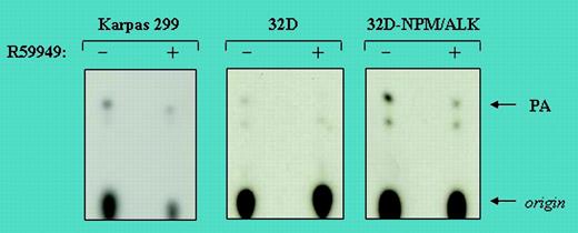
α-DGK activity in NPM/ALK-positive total cell lysates in the presence (+) or absence (-) of the DGK inhibitor R59949. See the complete figure in the article beginning on page 2175.
α-DGK activity in NPM/ALK-positive total cell lysates in the presence (+) or absence (-) of the DGK inhibitor R59949. See the complete figure in the article beginning on page 2175.
The work of Bacchiocchi et al reported in this issue provides further insight into this complex field. The authors sought to identify new intracellular transducers operative in ALK-positive malignancies and investigated the potential involvement of diacylglycerol kinase (DGK). There are 9 isoenzymes of DGK and they have a role in the regulation of signal transduction processes. The lipid second messenger, diaglycerol (DAG), is phosphorylated to phosphatidic acid (PA) by DGKs. In an elegant series of experiments, Bacchiocchi et al demonstrated initially, using the human ALK-positive ALCL-derived cell line Karpas 299, high constitutive DGK activity. Similar results were observed in the murine hematopoietic 32D cell line infected with a retrovirus expressing a green fluorescent protein (GFP)–NPM/ALK fusion protein. In contrast, significantly lower levels were found in the parental 32D cells infected with the empty vector, thus linking the DGK activity to the expression of NPM/ALK. Further experiments identified α-DGK as the main isoform involved. Bacchiocchi et al's experiments also indicated that the ALK-induced activation of α-DGK takes place through a p60src tyrosine kinase–dependent mechanism, although it remains unclear whether this is dependent or independent of p60src activation. Intriguingly, the authors also provide evidence that pharmacologic inhibition of α-DGK by R59949 significantly impaired the responsiveness of genetically engineered NIH 3T3 cells, overexpressing the chimeric epidermal growth factor receptor (EGFR)–ALK receptor (NIH-EGFR/ALK), to epidermal growth factor (EGF), as well as the growth rate of NPM/ALK cells (see figure). Although it is premature to identify α-DGK as a possible therapeutic target, the work of Bacchiocchi et al provides a tantalizing glimpse of how better understanding of the disordered molecular and cellular mechanisms in ALCL, along with recent developments in immunotherapy using the human monoclonal anti-CD30 antibody 5F11, may lead to more specific, less toxic treatment and further improvement in outcomes for patients with ALK-positive ALCL. ▪


This feature is available to Subscribers Only
Sign In or Create an Account Close Modal