Aspergillus and cytomegalovirus are major causes of morbidity/mortality after haploidentical hematopoietic transplantation. The high degree of mismatching makes cell immunotherapy impossible as it would result in lethal graft-versus-host disease (GvHD). We generated large numbers of donor T-cell clones specific for Aspergillus or cytomegalovirus antigens. We identified clones potentially responsible for causing GvHD by screening them for cross-reactivity against recipient mononuclear cells. Nonrecipient reactive, pathogen-specific clones were infused soon after transplantation. They were CD4+ and produced high levels of interferon-γ and low levels of interleukin-10. In 46 control transplant recipients who did not receive adoptive therapy, spontaneous pathogen-specific T cells occurred in low frequency 9 to 12 months after transplantation and displayed a nonprotective low interferon-γ/high interleukin-10 production phenotype. In the 35 recipients who received adoptive therapy, one single infusion of donor alloantigen-deleted, pathogen-specific clones in the dose range of 105 to 106 cells/kg body weight did not cause GvHD and induced high-frequency T-cell responses to pathogens, which exhibited a protective high interferon-γ/low interleukin-10 production phenotype within 3 weeks of infusion. Frequencies of pathogen-specific T cells remained stable over time, and were associated with control of Aspergillus and cytomegalovirus antigenemia and infectious mortality. This study opens new perspectives for reducing infectious mortality after haploidentical transplantations.
Introduction
Transplantation of very high doses of extensively T-cell-depleted peripheral blood hematopoietic stem cells from full human leukocyte antigen (HLA) haplotype-mismatched (“haploidentical”) family members is an option for patients with high-risk acute leukemia who urgently need a graft and do not have matched sibling or unrelated donors.1-3
Despite antifungal or antiviral prophylaxis and preemptive therapy,4,5 Aspergillus fumigatus and cytomegalovirus (CMV) are still major clinical problems in haploidentical transplant recipients.4,5
Aspergillosis, one of the most common causes of infection-related mortality, does not respond satisfactorily to antifungal agents. Specific defects in adaptive immune effector mechanisms play a role in susceptibility to Aspergillus infection. Indeed, “late-onset” infections are common even after neutrophil counts have recovered and their incidence is increased by graft-versus-host disease (GvHD) and its immune-suppressive treatment.6,7 Conversely, patients surviving aspergillosis show significant antigen-specific proliferation of interferon-γ (IFN-γ)-producing T cells.8
CMV is the other major cause of infection-related mortality, and indeed CMV positivity is an independent risk factor for mortality even in recipients of matched sibling or unrelated donor transplants. Until CMV-specific T-cell responses develop several months after transplantation, CMV causes fatal pneumonia, encephalitis, and enteritis. CMV is itself immune suppressive as it impairs dendritic-cell function.9 Furthermore, anti-CMV agents cause leukopenia and T-cell immune suppression.10
Immune reconstitution after haploidentical transplantation could theoretically be hastened by transferring immune T lymphocytes. Although no data are available in adults, several strategies have been attempted in children with inborn errors or hematologic malignancies. Ex-vivo induction of anergy to alloantigens in T-cell-replete bone marrow transplants was associated, despite immunoprophylaxis, with acute GvHD in 3 of 11 patients and significant infection-related mortality.11 After T-cell-depleted transplantation, infusion of T cells depleted of antirecipient alloreactivity by anti-CD25 immunotoxin appeared to improve immunity to infection but was associated with more than grade I acute GvHD in 2 of 9 patients in one report and 2 of 12 in another.12,13
However alloreactivity is attenuated, T-cell-based adoptive therapy is problematic in the adult haploidentical transplant setting as infusion of grafts containing only a few more T cells than the threshold dose of 104/kg may cause lethal GvHD.1-3 To date, transferring donor immunity after haploidentical transplantation has been prevented by (a) the HLA barrier; (b) the need to transfer donor T cells early when patients are most vulnerable to GvHD; (c) lack of posttransplantation GvHD prophylaxis because immune suppression would protect against the GvHD potential of infused T cells; and (d) the increased susceptibility of adults to GvHD.
The present study was designed to (1) set up experimental conditions to reliably and positively identify alloreactive clones in the donor pathogen (Aspergillus and CMV)-specific repertoire so as to delete them; and (2) adoptively transfer allodeleted clones to prevent/control Aspergillus and CMV infections without causing GvHD.
Patients, materials, and methods
Transplant protocol
Patients with hematologic malignancies who received haploidentical transplants were included in this study. Approval was obtained from the Umbria Region Ethics Committee and from the Perugia University institutional review board. Indications to haploidentical transplantation were failure to find a matched donor or ineligibility for an unrelated donor search and an urgently needed transplantation. Donor selection criteria were as previously described.1-3,14
Cell collection, graft processing, and transplantation procedures
Before granulocyte-colony-stimulating factor (G-CSF) administration for stem-cell mobilization,1-3,15 200 mL blood was drawn from donors to collect peripheral blood mononuclear cells (PBMCs) and plasma to generate pathogen-specific T-cell clones. Blood (100 mL) was drawn from transplant candidates to collect PBMCs to serve as stimulators in mixed lymphocyte reactions using donor pathogen-specific T-cell clones as responders. Conditioning included total body irradiation (TBI) in 79 patients and melphalan in 12 patients, followed by thiotepa, fludarabine, and rabbit anti-thymocyte globulin (ATG) (8 patients in the control cohorts and 7 in the immunotherapy groups received 8 mg/kg total dose of Sangstat ATG [Genzyme Transplant, Cambridge, MA]. Forty-six control patients and 30 who received immunotherapy were given 25 mg/kg total dose of Fresenius ATG [Fresenius Biotech, Graefelfing, Germany]). No immunosuppression was administered as GvHD prophylaxis after transplantation. All transplant recipients received antifungal prophylaxis with liposomal amphotericin (1 mg/kg daily) from day -5 until the neutrophil count reached 1 × 10⁹/L. Patients with invasive aspergillosis received liposomal amphotericin (6 mg/kg daily). Anti-CMV prophylaxis for all patients was gancyclovir (10 mg/kg daily) from day -9 to day -2 and foscarnet (90 mg/kg daily) from day +4 to day +20. Patients with CMV reactivation received gancyclovir (10 mg/kg daily). Engraftment and GvHD were assessed by consensus criteria.16 Blood and marrow chimerism was assessed by polymerase chain reaction (PCR) amplification of variable-number tandem repeat regions with different DNA polymorphism patterns (DNA microsatellites).2,3
Preparation of nonrecipient-reactive, pathogen-specific donor clones
Origin, characteristics, culture conditions, and heat inactivation of the A fumigatus strain have already been described.17,18 Briefly, A fumigatus strain, from a fatal case of pulmonary aspergillosis, was grown on Sabouraud dextrose agar slants (Difco Laboratories, Detroit, MI) for 4 days at room temperature. Conidia were resuspended in phosphate-buffered saline (PBS) containing 0.025% (wt/vol) Tween 20 and filtered twice through a 40-μ pore nylon cell strainer. Conidia were heat-killed at 121°C for 15 minutes in an autoclave. All preparations were devoid of microbial growth on culturing on appropriate culture media and tested endotoxin-free by the limulus amebocyte lysate assay (BioWhittaker, Walkersville, MD). As heat-inactivated conidia elicit anti-Aspergillus human T-cell responses in vitro, they were used to induce Aspergillus-specific T-cell clones.19
CMV antigens were obtained from Microbix Biosystems (Toronto, ON). MRC-5 cell monolayers were infected, ensuring the full host of viral proteins was present upon harvest. Cells were disrupted to release CMV antigen; cellular debris was removed by centrifugation. The CMV antigen was inactivated using gamma radiation. This preparation contains CMV antigens from all parts of the virus cycle of replication including early and late antigens, nuclear antigen, cytoplasmic antigen, and structural and nonstructural proteins. The antigen is in glycine-buffered saline without detergent or preservative. Before use, preparations were screened for microbial contamination by culturing samples on appropriate culture media and testing for endotoxins by the limulus amebocyte lysate assay (BioWhittaker).
Before G-CSF stem-cell mobilization, 200 mL blood was drawn from donors to collect PBMCs and plasma for adoptive therapy. Donor PBMCs, to serve as Aspergillus or CMV antigen-presenting cells (APCs), were incubated with Aspergillus or CMV antigens for 4 hours at a concentration of 10 × 106 cells/mL and irradiated with 20 Gy. Cells were resuspended in RPMI-1640 medium containing 10% autologous serum. Cells (2 × 10⁵/well) were plated in U-bottomed 96-well plates. Nonirradiated PBMCs from the same donor were used as responder cells and were added to the plates (96 wells for each cell dose) in limiting dilution concentrations of 12 000, 6000, 3000, and 1500 cells/well for generating Aspergillus-specific clones, and 500, 250, 125, and 62 cells/well for generating CMV-specific clones. These ranges of cell/well concentrations were chosen because preliminary experiments on PBMCs from healthy donors had demonstrated growth of microcultures that were clonal in origin. Microcultures were supplemented with 105 irradiated donor PBMCs on day 6 of culture. As preliminary experiments (not shown) had demonstrated that adding exogenous interleukin-2 (IL-2, Proleukin; Chiron, Amsterdam, The Netherlands) before day 14 promotes growth of non-pathogen-specific cells, IL-2 (125 U/mL final concentration) was added only from day 14 onward to ensure specificity. Cultures were inspected daily by inverted microscopy for cell growth up to day 18. All clones obtained under these culture conditions exhibited a CD4+ phenotype (“Results”). According to the Poisson formulas, only microcultures growing in one out of four or fewer wells were clonal in origin and are referred to as clones throughout the article20,21 ; only these microcultures were selected for the following steps.
Clonal cultures were split and supplemented with IL-2 as necessary. After approximately another week's culture, 3 to 4 or more wells (containing about 2 × 10⁵ cells/well) were obtained for each clone. After clones had rested in IL-2-free medium for 24 hours, pathogen specificity and donor HLA restriction of pathogen-specific responses were checked by restimulation with pathogen-pulsed (Aspergillus or CMV antigen) irradiated donor PBMCs for 3 days. DNA synthesis was measured by uptake of H3-thymidine, which was added during the last 6 hours of culture (Amersham Biosciences, Little Chalfont, United Kingdom). Recipient HLA restriction of Aspergillus and CMV responses was tested by stimulating clones with recipient Aspergillus- or CMV-antigen-pulsed irradiated PBMCs. Alloreactivity was assessed by culturing clonal cells with irradiated recipient PBMCs at a ratio of 1:1 for 3 days and measuring DNA synthesis. Negative controls were obtained by stimulating clonal cells with irradiated autologous (donor) PBMCs. Clones that displayed higher thymidine incorporation in response to recipient PBMCs than to autologous (donor) PBMCs were considered alloreactive.
Clones were assessed for CD3, CD4, CD8, α-β, and γ-δ T-cell receptor phenotype by indirect immunofluorescence using primary monoclonal antibodies (BD Pharmingen, San Diego, CA) plus secondary fluorochrome-conjugated goat anti-mouse immunoglobulin antibodies (Southern Biotechnology Associates, Birmingham, AL) and flow cytometry (FACSCalibur; Becton Dickinson, San Jose, CA). T-cell clonal supernatants were assessed for IFN-γ and IL-10 content by enzyme-linked immunosorbent assays (ELISA; Euroclone, Paignton-Devon, United Kingdom).
Non-recipient-reactive clones were pooled, expanded, and infused as adoptive therapy. The release criteria were as follows: pathogen-specificity, lack of reactivity to recipient alloantigens, and no bacterial, fungal or mycoplasma contamination. Viral contamination was minimized by donor selection criteria (eg, no evidence of any ongoing viral infection, and no HIV and hepatitis seropositivity)1-3 and by sterile laboratory procedures.
Adoptive therapy protocol
Patients and donors provided informed consent, according to the Declaration of Helsinki. Consecutive patients were randomly assigned to the immunotherapy or control groups. Immunotherapy and control cohorts were matched for the following general inclusion criteria: full donor-type engraftment, no GvHD, no steroid treatment.
Only transplant recipients with evidence of invasive aspergillosis (eg, Aspergillus pneumonia)22,23 were recruited to the Aspergillus adoptive therapy trial (Table 1). Regardless of assignment to the immunotherapy (n = 10) or control group (n = 13), all patients with evidence of invasive aspergillosis received liposomal amphotericin (6 mg/kg per day).
Characteristics of patients of the study and controls
. | Aspergillus . | . | CMV . | . | ||
|---|---|---|---|---|---|---|
. | Infused patients . | Controls . | Infused patients* . | Controls† . | ||
| No. of patients | 10 | 13 | 27 | 41 | ||
| Median age (range) | 30 (16-48) | 26 (17-62) | 33 (16-58) | 35 (16-63) | ||
| Diagnosis | ||||||
| AML | 4 | 8 | 14 | 26 | ||
| ALL | 2 | 3 | 6 | 6 | ||
| CML | 1 | 2 | 2 | 1 | ||
| MM | 0 | 0 | 3 | 0 | ||
| NHL | 2 | 0 | 2 | 0 | ||
| Disease status | ||||||
| I to II CR | 5 | 6 | 13 | 21 | ||
| More than II CR | 2 | 1 | 2 | 2 | ||
| Refractory disease | 2 | 4 | 10 | 17 | ||
| CML-CP | 0 | 0 | 1 | 1 | ||
| CML-AP | 1 | 1 | 1 | 0 | ||
| CML-BC | 0 | 1 | 0 | 0 | ||
| Conditioning regimen | ||||||
| TBI-based | 9 | 9 | 23 | 38 | ||
| Melphalan-based | 1 | 4 | 4 | 3 | ||
| Graft content | ||||||
| CD34, × 106/kg | 9.7 ± 1.2 | 12.5 ± 4.2 | 11.6 ± 2.6 | 12.0 ± 1.4 | ||
| CD3, × 104/kg | 0.8 ± 0.6 | 0.8 ± 0.6 | 1.0 ± 0.6 | 1.4 ± 0.1 | ||
AML indicates acute myeloid leukemia; ALL, acute lymphoblastic leukemia; CML, chronic myeloid leukemia; MM, multiple myeloma; NHL, non-Hodgkin lymphoma; CR, complete remission; CP, chronic phase; AP, accelerated phase; and BC, blastic crisis.
Twenty-five of 27 patients received donor type CMV-specific T-cell clones; 2 of 27 patients received host type CMV-specific T-cell clones
Thirty-three CMV-positive recipients who received transplants from CMV-positive donors; 8 CMV-positive recipients who received transplants from CMV-negative donors
Because of high risk of CMV reactivation, the CMV adoptive therapy trial recruited haploidentical recipients satisfying general inclusion criteria to the immunotherapy (n = 25) or control (n = 33) group.2,3 CMV serostatus of donor/recipient pairs for the immunotherapy group was positive/positive in 23 and positive/negative in 2; for the control group, serostatus was positive/positive in 32 and positive/negative in 1. Regardless of group assignment, CMV reactivation (ie, positive antigenemia) was treated with gancyclovir (10 mg/kg per day).
Between days 13 and 37 after transplantation (median = 21 days), one infusion of clones was given to at least 3 recipients per cell dose. Doses started at 105 cells/kg and were escalated to 3 × 10⁶ cells/kg. Patients were closely monitored for adverse side effects.
Weekly for the first 3 weeks and then monthly, CD3+/CD4+ and CD3+/CD8+ T-cell subset reconstitution was monitored by flow cytometry. Frequencies of Aspergillus- or CMV-specific T cells were monitored by limiting dilution analyses of proliferating cells in response to Aspergillus- or CMV-antigen stimulation. Posttransplantation PBMCs were incubated with Aspergillus or CMV antigens, irradiated, resuspended in RPMI-1640 medium containing 10% autologous serum, and plated in U-bottomed wells (2 × 10⁵ cells/well). Nonirradiated PBMCs from the same patients who underwent transplantation, used as responder cells, were added to plates (48 wells for each cell dose) in limiting dilution concentrations of 100 000, 50 000, 25 000, and 12 500 cells/well for detecting Aspergillus- and CMV-specific clone frequencies. These ranges of cell/well concentrations were chosen because preliminary experiments on PBMCs from patients who underwent transplantation had demonstrated growth of microcultures that were clonal in origin (above). Under our culture conditions, all clones obtained from patients after immunotherapy, such as donor clones, exhibited a CD4+ phenotype. Screening, maintenance, expansion, phenotyping, pathogen-specificity assessment, and donor and recipient HLA restriction of posttransplantation clones were as described in “Preparation of nonrecipient-reactive, pathogen-specific donor clones.”
T-cell clonal supernatants were assessed for IFN-γ and IL-10 content by ELISA (Euroclone).
Posttransplantation CMV-specific CD8+ T-cell responses were monitored using immunomagnetically purified (MiniMACS; Miltenyi Biotec, Bergish Gladbach, Germany) CD8+ T cells cultured overnight with CMV-antigen-pulsed irradiated PBMCs. CD8+ cells were then assayed for IFN-γ production by enzyme-linked immunosorbent spot (ELISPOT) assay (Amplimedical, Buttigliera Alta, Italy).
Before and after immunotherapy and in the control cohort, serum Aspergillus galactomannan antigen levels were measured weekly by sandwich immunocapture ELISA (Platelia Aspergillus; Bio-Rad, Milan, Italy) as recommended by the manufacturer. Results were expressed as Aspergillus galactomannan content (ng/mL), compared with a standard containing 1 ng galactomannan. Values more than 1 ng/mL were considered indicative of invasive aspergillosis. Invasive aspergillosis was defined according to consensus criteria.23
CMV antigenemia was monitored weekly by standard immunofluorescence staining of peripheral leukocytes with an antibody pool against pp65 and expressed as positive cell number per 200 000 total cells. When indicated, biologic specimens from the bronchoalveolar lavage, gastrointestinal biopsy, lumbar puncture, and urine were analyzed for CMV by immunofluorescence, molecular, or cultural techniques. CMV disease was diagnosed according to standard criteria.24
In 2 other transplantations, from CMV-negative donors into CMV-positive recipients, we used pretransplantation recipient cells to obtain CMV-specific clones and infused nondonor reactive clones. Eight CMV-negative donor/CMV-positive recipient transplant pairs served as controls.
Statistical analysis
Differences between data sets were analyzed using a paired, 2-tailed Student t test. All data are presented as mean ± standard deviation. Cumulative incidence estimates25 were used for infectious mortality.
Results
Identifying and deleting donor pathogen-specific T-cell clones cross-reacting with recipient alloantigens
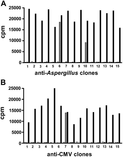
No donor culture failed to generate either CMV- (from CMV-seropositive donors) or Aspergillus-specific T-cell clones. Moreover, 100% of clones were specific for the pathogen against which they had been raised (ie, they proliferated when rechallenged with the relevant pathogen). On average, each clone expanded to a total of 8 × 10⁵ to 1 × 10⁶ cells within 21 to 28 days. For the purpose of the present immunotherapy trial, we established more than 1200 Aspergillus-specific clones from 10 donors and more than 3000 CMV-specific clones from 25 donors. All clones were CD4+. From 1% to 10% pathogen-specific clones displayed cross-reactivity to recipient PBMCs (Figure 1) (ie, they could potentially cause GvHD) and were discarded. Non-recipient-reactive clones were pooled and infused as adoptive therapy. All donor pathogen-specific clones responded to donor pathogen-pulsed PBMCs (Figure 1). Approximately 50% also responded to recipient pathogen-pulsed PBMCs (not shown).
Adoptive therapy
None of the controls developed acute or chronic GvHD at any time point in the study. None of the 34 patients who received anti-CMV or anti-Aspergillus immunotherapy in doses escalating from 1 × 10⁵ to 1 × 10⁶ cells/kg developed acute or chronic GvHD, or any other infusion-related toxicity. Infusion of 3 × 10⁶ (anti-CMV) cells/kg in one patient caused grade II acute GvHD that resolved with steroids. No other infusion-related adverse effect was observed in any patient at any dose of anti-CMV or anti-Aspergillus immunotherapy at any time point in the follow-up. Therefore, the safe dose emerged as 1 × 10⁶ cells or less/kg.
Tables 2, 3, 4, 5 provide details of the time course of adoptive transfer, pathogen-specific T-cell reconstitution, antigenemias, and clinical data in individual control patients (Table 2 for invasive aspergillosis; Table 4 for CMV) and recipients of adoptive therapy (Table 3 for invasive aspergillosis; Table 5 for CMV).
Invasive aspergillosis in 13 patients who did not receive Aspergillus-specific T-cell therapy
Patient no. . | Invasive aspergillosis . | Galactomannan antigenemia at diagnosis, ng/mL* . | Galactomannan antigenemia under amphotericin therapy,†ng/mL . | Outcome (time after diagnosis, wk) . |
|---|---|---|---|---|
| 1 | P: mult/bilat | 4.5 ± 0.7 | 3.1 ± 0.3 | Death (5) |
| 2 | P: monolat | 3.5 ± 0.6 | 3.6 ± 0.6 | Death (7) |
| 3 | P: mult/bilat | 1.9 ± 0.1 | 1.4 ± 0.1 | Death (4) |
| 4 | P: mult/bilat + pl eff | 4.1 ± 0.2 | 3.3 ± 0.5 | Resolution (17) |
| 5 | P: mult/bilat | 3.1 ± 0.1 | 1.5 ± 0.06 | Resolution (4) |
| 6 | P: mult/bilat | 1.8 ± 0.4 | 2.6 ± 0.6 | Resolution (4) |
| 7 | P: mult/bilat | 4.7 ± 0.4 | 2.7 ± 1.3 | Death (5) |
| 8 | P: mult/bilat | 2.8 ± 0.3 | 2.3 ± 0.8 | Death (4) |
| 9 | P: monolat | 2.9 ± 0.3 | 3.0 ± 0.15 | Resolution (4) |
| 10 | P: monolat + pl eff | 3.9 ± 0.3 | 3.3 ± 0.7 | Resolution (5) |
| 11 | P: monolat + pl eff | 1.8 ± 0.4 | 2.9 ± 0.5 | Death (4) |
| 12 | P: mult/bilat | 2.9 ± 0.2 | 3.3 ± 0.6 | Resolution (5) |
| 13 | P: monolat + pl eff | 3.2 ± 0.2 | 2.8 ± 0.3 | Resolution (6) |
P indicates pneumonia; mult/bilat, multiple, bilateral lung lesions; monolat, monolateral lung lesions; and pl eff, pleural effusion.
Mean ± SD of at least 3 assessments of serum Aspergillus galactomannan antigen levels by sandwich immunocapture ELISA. Values more than 1 ng/mL were considered indicative of invasive aspergillosis
Mean ± SD of weekly assessments for up to 15 weeks after transplantation
Aspergillus-specific immune reconstitution and outcome of invasive aspergillosis in 10 patients who received Aspergillus-specific T-cell therapy
Patient no. . | Invasive aspergillosis before cell therapy . | Galactomannan antigenemia before T-cell therapy,*ng/mL . | Cell therapy, day after transplantation . | Aspergillus-specific CD4+ reconstitution, day after cell therapy . | Galactomannan antigenemia after cell therapy,†ng/mL . | Outcome (time after diagnosis, wk) . |
|---|---|---|---|---|---|---|
| 1 | P: mult/bilat + pl eff + CNS | 3.4 ± 0.6 | 37 | 21 | 0.8 ± 0.1 | Death (4) |
| 2 | P: mult/bilat + sin | 4.2 ± 0.4 | 23 | 21 | 0.3 ± 0.1 | Resolution (4) |
| 3 | P: mult/bilat + pl eff | 1.6 ± 0.5 | 32 | 21 | 0.4 ± 0.1 | Resolution (5) |
| 4 | P: mult/bilat + pl eff | 2.0 ± 0.6 | 32 | 21 | 0.2 ± 0.0 | Resolution (12) |
| 5 | P: monolat + sin | 1.9 ± 0.7 | 25 | 21 | 0.4 ± 0.1 | Resolution (14) |
| 6 | P: mult/bilat | 2.8 ± 0.3 | 17 | 21 | 0.6 ± 0.5 | Resolution (5) |
| 7 | P: mult bilat + pl eff + sin | 2.1 ± 0.5 | 17 | 21 | 0.8 ± 0.1 | Resolution (10) |
| 8 | P: mult/bilat | 2.2 ± 0.1 | 21 | 21 | 0.7 ± 0.2 | Resolution (6) |
| 9 | P: monolat | 3.1 ± 0.2 | 21 | 21 | 0.6 ± 0.2 | Resolution (7) |
| 10 | P: mult/bilat | 3.1 ± 0.3 | 21 | 21 | 0.4 ± 0.3 | Resolution (8) |
P indicates pneumonia; mult/bilat, multiple, bilateral lung lesions; pl eff, pleural effusion; CNS, brain lesion; sin, maxillary and etmoidal sinusitis; and monolat, monolateral lung lesions.
Mean ± SD of at least 3 assessments of serum Aspergillus galactomannan antigen levels by sandwich immunocapture ELISA. Values more than 1 ng/mL were considered indicative of invasive aspergillosis
Mean ± SD of weekly assessments from 6 to 12 weeks after T-cell therapy. All patients also received liposomal amphotericin therapy
CMV reactivation/disease in 33 patients who did not receive CMV-specific T-cell therapy
Patient no. . | pp65 antigenemia* . | CMV disease (onset after transplantation, wk) . | Outcome (time after diagnosis, wk) . |
|---|---|---|---|
| 1 | 29 ± 22 | — | — |
| 2 | 20/5 | — | — |
| 3 | 33 | — | — |
| 4 | 8 ± 3 | — | — |
| 5 | 12 ± 10 | — | — |
| 6 | 2/10 | — | — |
| 7 | 20 | — | — |
| 8 | 13 | — | — |
| 9 | 5 ± 2 | Hepatitis + enteritis (7) | Resolution (2) |
| 10 | 7 ± 4 | IP (12) | Death (2) |
| 11 | 15 ± 14 | — | — |
| 12 | 2/2 | — | — |
| 13 | 14 ± 8 | — | — |
| 14 | 23 ± 20 | — | — |
| 15 | 43 ± 35 | IP (11) | Death (3) |
| 16 | 109 ± 53 | IP + CNS (15) | Death (2) |
| 17 | 22 ± 16 | — | — |
| 18 | 95 | — | — |
| 19 | 7 ± 6 | — | — |
| 20 | 128 ± 70 | Hepatitis (13) | Death (2) |
| 21 | 21 ± 16 | — | — |
| 22 | 39 ± 20 | IP (10) | Death (4) |
| 23 | 8 ± 2 | — | — |
| 24 | 6 ± 4 | IP + CNS (12) | Death (4) |
| 25 | 38 ± 27 | IP (14) | Death (4) |
| 26 | 77 ± 52 | IP (15) | Death (4) |
| 27 | 19 ± 16 | — | — |
| 28 | 34 | Cystitis (8) | Resolution (6) |
| 29 | 10 ± 3 | — | — |
| 30 | 13 ± 4 | — | — |
| 31 | 21 ± 11 | IP (11) | Resolution (5) |
| 32 | — | — | — |
| 33 | 9 ± 4 | IP (12) | Death (4) |
— indicates no manifestations of CMV disease and negative antigenemia; IP, interstitial pneumonia; CNS, central nervous system involvement.
Positive antigenemias (no. of positive cells/200 000 cells) detected weekly for up to 12 weeks after withdrawing antiviral foscarnet prophylaxis, at 20 days after transplantation. Single number indicates one episode of reactivation. Two numbers indicate 2 episodes of reactivation. Mean ± SD indicates 3 or more episodes of reactivation
CMV-specific immune reconstitution and outcome of CMV reactivation/disease in 25 patients who received CMV-specific T-cell therapy
Patient no. . | Cell therapy, day after transplantation . | CMV specific CD4+ reconstitution, day after cell therapy . | CMV-specific CD8+ reconstitution, day after cell therapy . | pp65 antigenemia after cell therapy* . | CMV disease (onset after transplantation, wk) . | Outcome (time after diagnosis, wk) . |
|---|---|---|---|---|---|---|
| 1 | 16 | 21 | 21 | — | — | — |
| 2 | 23 | 21 | 21 | — | — | — |
| 3 | 14 | 21 | 21 | — | — | — |
| 4 | 21 | 21 | 21 | — | — | — |
| 5 | 13 | 21 | 21 | 10 | IP/CNS (2) | Resolution (3) |
| 6 | 32 | 21 | 21 | — | — | — |
| 7 | 37 | 21 | 21 | 7 ± 6 | CNS (12) | Death (4) |
| 8 | 16 | 21 | 21 | 13 ± 7 | Enteritis (15) | Death (2) |
| 9 | 37 | 21 | 21 | — | — | — |
| 10 | 26 | 21 | 21 | — | — | — |
| 11 | 15 | 21 | 21 | — | — | — |
| 12 | 22 | 21 | 21 | — | — | — |
| 13 | 21 | 21 | 21 | — | IP (3) | Resolution (4) |
| 14 | 21 | 21 | 21 | 4 ± 1 | — | — |
| 15 | 17 | 21 | 21 | — | — | — |
| 16 | 21 | 21 | 21 | 10 | — | — |
| 17 | 33 | 21 | 21 | — | — | — |
| 18 | 26 | 21 | 21 | — | — | — |
| 19 | 16 | 21 | 21 | 7 ± 3 | — | — |
| 20 | 28 | 21 | 21 | — | CNS (5) | Resolution (5) |
| 21 | 17 | 21 | 21 | — | — | — |
| 22 | 37 | 21 | 21 | 10 | — | — |
| 23 | 27 | 21 | 21 | — | — | — |
| 24 | 28 | 21 | 21 | — | — | — |
| 25 | 21 | 21 | 21 | — | — | — |
— indicates no manifestations of CMV disease and negative antigenemia; IP, interstitial pneumonia; CNS, brain lesion.
Positive antigenemias (no. of positive cells/200 000 cells) detected weekly from 3 to 12 weeks after T-cell therapy. Single number indicates 1 episode of reactivation. Two numbers indicate 2 episodes of reactivation. Mean ± SD indicates 3 or more episodes of reactivation
Aspergillus- and CMV-specific T-cell repertoires and cross-reactivity with alloantigens. T-cell clones specific for Aspergillus (A) or CMV (B) antigens were obtained from haploidentical transplant donors and tested for reactivity against transplant recipient PBMCs. Those displaying alloreactivity against recipient PBMCs were discarded, and the nonalloreactive were infused after transplantation. ⦾ indicates reactivity against recipient PBMCs; □, reactivity against autologous donor PBMCs; and ▪, response to Aspergillus (A) or CMV (B). Results are expressed as cpm of H3 thymidine incorporation. The figure shows representative sets of clones.
Aspergillus- and CMV-specific T-cell repertoires and cross-reactivity with alloantigens. T-cell clones specific for Aspergillus (A) or CMV (B) antigens were obtained from haploidentical transplant donors and tested for reactivity against transplant recipient PBMCs. Those displaying alloreactivity against recipient PBMCs were discarded, and the nonalloreactive were infused after transplantation. ⦾ indicates reactivity against recipient PBMCs; □, reactivity against autologous donor PBMCs; and ▪, response to Aspergillus (A) or CMV (B). Results are expressed as cpm of H3 thymidine incorporation. The figure shows representative sets of clones.
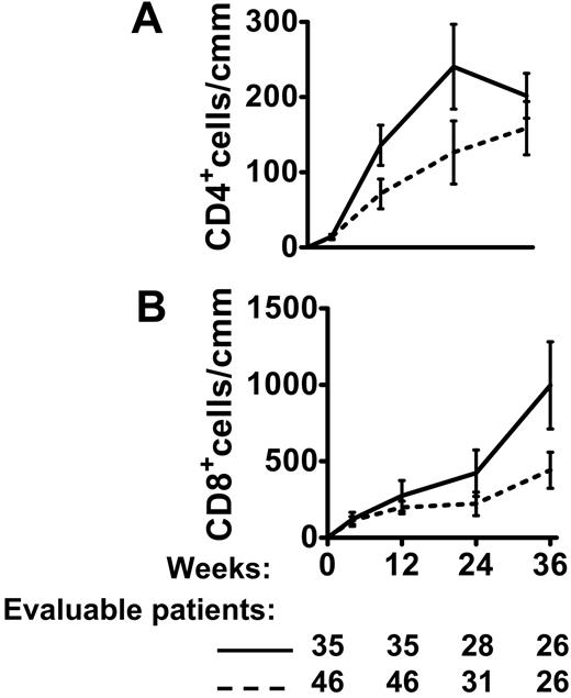
Figure 2 shows recovery of absolute CD4+ and CD8+ T-cell counts as a function of time after transplantation. Compared with the 46 controls, CD4+ and CD8+ cell counts were higher in the 35 immunotherapy-treated patients, reaching significance at 12 weeks (P < .01 and P < .02, respectively) and 24 weeks (P < .05 for both). A significant difference persisted in CD8+ counts at 36 weeks (P < .02 vs controls).
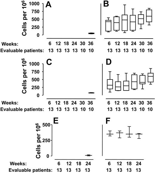
Aspergillus and CMV-specific T-cell frequencies in donors and in immunotherapy and control groups were assessed by limiting dilution analyses of proliferating cells in response to Aspergillus- or CMV-antigen stimulation. As all clones exhibited the CD3+/CD4+ phenotype, frequency results are expressed as the number of CD3+/CD4+ growing clonal cultures in 106 plated CD3+/CD4+ cells. In the 81 donors, the frequency of specific CD4+ T-cell clones was 763 ± 158 cells per 106 cells for Aspergillus and 2623 ± 654 cells per 106 cells for CMV. In the 46 haploidentical transplant recipients who did not receive adoptive therapy, spontaneous recovery of CD4+ T-cell clones specific for Aspergillus or CMV was observed in very low frequency (Aspergillus: 72 ± 5 cells per 106 cells; CMV: 44 ± 5 cells per 106 cells) from 36 weeks after transplantation onward. Within 3 weeks of the immunotherapy infusion, Aspergillus-specific CD4+ T-cell clones were 353 ± 119 per 106 cells; CMV-specific CD4+ T-cell clones were 404 ± 124 per 106 cells (P = .004 vs noninfused patients). Frequencies remained high for as long as they were monitored (36 weeks after transplantation) (Figure 3A-D). After infusion, all pathogen-specific clones detected in patients responded to donor pathogen-pulsed PBMCs and approximately 50% also responded to recipient pathogen-pulsed PBMCs (data from 3 separate analyses in 3 recipients, not shown), indicating their potential to respond in vivo to pathogens presented by donor- and recipient-type APCs.
As CD8+ cells are pivotal in defense against viral infections,26 we monitored CMV-specific CD8+ cells. In controls, IFN-γ-producing CMV-specific CD8+ cells were first detected 24 weeks after transplantation at a frequency of 10 ± 9 per 106 cells (Figure 3E). After adoptive therapy with CMV-specific CD4+ clones, IFN-γ-producing CMV-specific CD8+ cells were detected within 3 weeks of infusion in normal frequencies (Figure 3F) (400 ± 50 cells per 106, vs donor values: 500 ± 60 in 106 cells) (P = .003 vs noninfused controls).
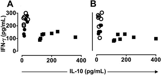
In donors, Aspergillus- and CMV-specific T-cell clones produced 217 ± 56 pg/mL (mean ± SD) IFN-γ and 15 ± 6 pg/mL IL-10. In noninfused patients, spontaneous Aspergillus- and CMV-specific clones were detected 9 to 12 months after transplantation. They produced 100 ± 19 pg/mL IFN-γ (P = not significant [ns] vs donors) and 216 ± 95 pg/mL IL-10 (P < .001 vs donors). After immunotherapy, specific T cells produced 171 ± 32 pg/mL IFN-γ (P = ns vs noninfused patients) and 19 ± 7 pg/mL IL-10 (P < .001 vs noninfused patients) (Figure 4).
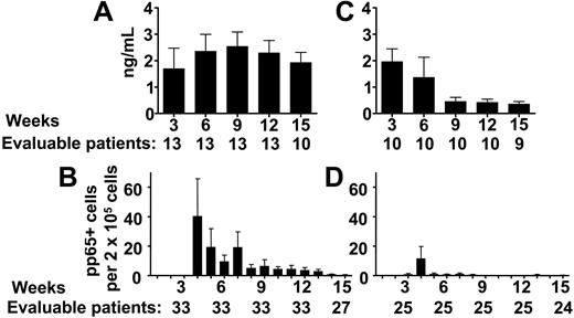
The 13 controls for the anti-Aspergillus trial all displayed positive galactomannan antigenemia and/or other signs of invasive aspergillosis (Table 2).22,23 Their positive galactomannan antigenemia persisted for as long as monitored (Table 2; Figure 5A). Six of the 13 patients succumbed to invasive aspergillosis in 4.8 ± 1.2 weeks of diagnosis and 7 cleared the disease in 5 ± 0.7 weeks of diagnosis (Table 2).
The 10 recipients of anti-Aspergillus adoptive therapy all had Aspergillus pneumonia and positive galactomannan antigenemia at the time of infusion (Table 3). Galactomannan antigenemia fell within 6 weeks of infusion to the normal range (< 1 ng/mL) (P < .002 vs controls) where it persisted for as long as monitored (Table 3; Figure 5B). Nine of the 10 patients who received anti-Aspergillus adoptive therapy cleared invasive aspergillosis in a mean of 7.8 ± 3.4 weeks (with a trend toward a significant difference between survivors after immunotherapy vs controls, P = .062). The other patient died of Aspergillus pneumonia within 2 weeks of diagnosis (Table 3).
Thirty of the 33 control patients underwent repeated CMV reactivation for as long as monitored (Table 4; Figure 5C). Twelve developed CMV disease and 9 died (ie, 6 of the 7 pneumonia cases, 1 of the 2 hepatitis cases, and both patients with pneumonia plus encephalitis) in a mean of 3.2 ± 0.9 weeks of diagnosis (Table 4). Of the 25 patients who received CMV-specific adoptive therapy, only 7 underwent CMV reactivation (Table 5). Episodes of CMV reactivation were fewer in number with significantly lower antigenemia levels (P < .004 vs controls) (Figure 5D). Five patients developed CMV disease: 1 pneumonia, 1 pneumonia plus encephalitis, 2 encephalitis, and 1 enteritis. CMV disease was fatal in 2:1 case of encephalitis and 1 case of enteritis (after 4 and 2 weeks of diagnosis, respectively) (Table 5). A trend toward a significant difference was observed between survivors after immunotherapy (P = .062 vs controls).
Posttransplantation CD4+ and CD8+ T-cell counts in 35 transplant patients who received T-cell therapy and in 46 control transplant recipients. X axes: weeks after transplantation and number of evaluable patients at each time point. (A) CD4+ cells in immunotherapy (straight line) and control cohorts (broken line). (B) CD8+ cells in immunotherapy (straight line) and control cohorts (broken line). Compared with controls, CD4+ and CD8+ cell counts were higher in immunotherapy-treated patients, reaching significance at 12 weeks (P < .01 and P < .02, respectively) and 24 weeks (P < .05 for both). A significant difference persisted in CD8+ counts at 36 weeks (P < .02 vs controls).
Posttransplantation CD4+ and CD8+ T-cell counts in 35 transplant patients who received T-cell therapy and in 46 control transplant recipients. X axes: weeks after transplantation and number of evaluable patients at each time point. (A) CD4+ cells in immunotherapy (straight line) and control cohorts (broken line). (B) CD8+ cells in immunotherapy (straight line) and control cohorts (broken line). Compared with controls, CD4+ and CD8+ cell counts were higher in immunotherapy-treated patients, reaching significance at 12 weeks (P < .01 and P < .02, respectively) and 24 weeks (P < .05 for both). A significant difference persisted in CD8+ counts at 36 weeks (P < .02 vs controls).
Total weeks of liposomal amphotericin therapy for invasive aspergillosis were not significantly different in the control and the immunotherapy cohorts (5.7 ± 3.5 vs 7.5 ± 3.5 weeks, P = ns). Due to uncontrolled invasive aspergillosis, mean duration of therapy in the 6 of 13 controls who died was 4.8 ± 1.2 weeks. Nine of 10 survivors in the immunotherapy group were treated for 7.8 ± 3.4 weeks, until invasive aspergillosis resolved.
Total weeks of gancyclovir treatment for positive CMV antigenemia were 6.9 ± 3.4 in the controls and 2.0 ± 1.5 in the immunotherapy group (P < .005), reflecting the fewer episodes of CMV reactivation and the significantly lower antigenemia levels in the latter group.
Infections, other than invasive aspergillosis and CMV disease, were fatal in 20 patients. Nine of 25 anti-CMV and 2 of 10 Aspergillus immunotherapy-treated patients died of other infections, such as Epstein-Barr virus (EBV) and human herpesvirus 6 (HHV-6). After a median follow-up of 24 months (range, 3-77 months), cumulative incidence of overall infectious mortality (invasive aspergillosis, CMV disease, and any other infection-related death) was 0.53 in control cohorts versus 0.41 in patients receiving immunotherapy. Overall, 10 patients died of relapse. Event-free survivors are 15 of 46 patients in control cohorts and 18 of 35 immunotherapy-treated patients.
Providing CMV immunity after transplantation from naive donors into CMV-positive recipients
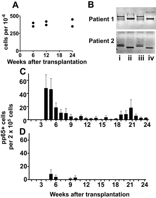
In 2 haploidentical transplants from a CMV-negative donor into a CMV-positive recipient (1 AML in CR and 1 MM), we used recipient cells taken before transplantation to generate CMV-specific T-cell clones for posttransplantation adoptive therapy. All clones displayed a CD4+ phenotype and a predominant type-1 cytokine pattern (not shown). Clones were screened for reactivity against donor cells. Those displaying alloreactivity were discarded as they could have caused rejection upon infusion. Nonreactive clones were pooled and infused at a dose of 5 × 10⁵ cells/kg. Infusion did not cause graft rejection, cytopenia, or other adverse effects. CMV-specific T-cell clones were detected in PBMCs 3 weeks after infusion (Figure 6A), and their frequencies have remained stable for as long as they were monitored (6 months). DNA polymorphism pattern analysis indicated that these anti-CMV responses were of recipient origin (Figure 6B). In 8 control CMV-positive patients who received transplants from CMV-negative donors, T-cell responses to CMV were not detectable for 1 year after transplantation (not shown), and patients underwent repeated CMV reactivation for up to 24 weeks after transplantation (Figure 6C). Remarkably, the infusion of recipient-type CMV-specific clones in 2 CMV-positive recipients who received transplants from CMV-negative donors resulted in satisfactory control of CMV reactivation (Figure 6D).
Transfer of donor pathogen-specific clones across the HLA barrier. X axes: weeks after transplantation and number of evaluable patients at each time point. (A-D) Frequencies of Aspergillus- and CMV-specific T cells were monitored by limiting dilution analyses of proliferating pathogen-specific T cells for 36 weeks after transplantation (“Patients, materials, and methods”). As all clones exhibited the CD3+/CD4+ phenotype, frequency results obtained by limiting dilution analyses are expressed as the number of CD3+/CD4+ growing clonal cultures in 106 plated CD3+/CD4+ cells. (A,C) Frequencies of CD4+ T-cell clones specific for Aspergillus (A) and for CMV (C) in haploidentical transplant recipients who did not receive adoptive therapy (13 and 33 patients, respectively). (B,D) Frequencies of CD4+ T-cell clones specific for Aspergillus (B) and for CMV (D) in haploidentical transplant recipients who received adoptive therapy (10 and 25 patients, respectively). Box and whisker plots indicate mean, SD, and range of frequencies. Frequencies of Aspergillus- and CMV-specific CD4+ T-cell clones in infused patients versus controls: P < .005 at all time points. (E-F) Frequencies of CMV-specific CD8+ T cells as detected by ELISPOT in noninfused patients (E) and in infused patients (F) for 24 weeks after transplantation. Box and whisker plots indicate mean, SD, and range of frequencies. Frequencies of CMV-specific CD8+ T cells in infused patients versus controls: P < .005 at all time points.
Transfer of donor pathogen-specific clones across the HLA barrier. X axes: weeks after transplantation and number of evaluable patients at each time point. (A-D) Frequencies of Aspergillus- and CMV-specific T cells were monitored by limiting dilution analyses of proliferating pathogen-specific T cells for 36 weeks after transplantation (“Patients, materials, and methods”). As all clones exhibited the CD3+/CD4+ phenotype, frequency results obtained by limiting dilution analyses are expressed as the number of CD3+/CD4+ growing clonal cultures in 106 plated CD3+/CD4+ cells. (A,C) Frequencies of CD4+ T-cell clones specific for Aspergillus (A) and for CMV (C) in haploidentical transplant recipients who did not receive adoptive therapy (13 and 33 patients, respectively). (B,D) Frequencies of CD4+ T-cell clones specific for Aspergillus (B) and for CMV (D) in haploidentical transplant recipients who received adoptive therapy (10 and 25 patients, respectively). Box and whisker plots indicate mean, SD, and range of frequencies. Frequencies of Aspergillus- and CMV-specific CD4+ T-cell clones in infused patients versus controls: P < .005 at all time points. (E-F) Frequencies of CMV-specific CD8+ T cells as detected by ELISPOT in noninfused patients (E) and in infused patients (F) for 24 weeks after transplantation. Box and whisker plots indicate mean, SD, and range of frequencies. Frequencies of CMV-specific CD8+ T cells in infused patients versus controls: P < .005 at all time points.
IFN-γ and IL-10 produced by sets of T-cell clones that are representative of results in 81 haploidentical transplant donors, 46 control transplant recipients, and 35 transplant recipients who received adoptive therapy. (A) Aspergillus specific. (B) CMV specific. Donor clones (○) displayed protective high IFN-γ and very low IL-10 patterns. Spontaneously recovering clones in control haploidentical recipients (▪) displayed nonprotective lower IFN-γ and higher IL-10. After adoptive therapy, clones (•) displayed protective high IFN-γ and very low IL-10, overlapping with the pattern observed in their healthy donors (P < .001 vs noninfused patients; P = ns vs donors).
IFN-γ and IL-10 produced by sets of T-cell clones that are representative of results in 81 haploidentical transplant donors, 46 control transplant recipients, and 35 transplant recipients who received adoptive therapy. (A) Aspergillus specific. (B) CMV specific. Donor clones (○) displayed protective high IFN-γ and very low IL-10 patterns. Spontaneously recovering clones in control haploidentical recipients (▪) displayed nonprotective lower IFN-γ and higher IL-10. After adoptive therapy, clones (•) displayed protective high IFN-γ and very low IL-10, overlapping with the pattern observed in their healthy donors (P < .001 vs noninfused patients; P = ns vs donors).
Aspergillus and CMV antigenemias in 35 transplant patients who received T-cell therapy and in 46 control transplant recipients. X axes: weeks after transplantation and number of evaluable patients at each time point. (A-B) Aspergillus galactomannan antigenemia in haploidentical recipients with evidence of aspergillosis (values > 1 ng/mL indicate invasive aspergillosis). In 13 control patients who did not receive adoptive therapy (A) galactomannan antigenemia remained high. In 10 patients who had received adoptive therapy (B) antigenemia normalized within a few weeks (P < .002 vs controls). Results are expressed as Aspergillus galactomannan content (ng/mL). (C-D) CMV antigenemia in haploidentical recipients (positive cell number per 200 000 total cells). In 33 control patients who did not receive adoptive therapy (C) CMV continued reactivating for several weeks after transplantation. In 25 patients who had received adoptive therapy (D) CMV antigenemia normalized soon after infusion (P = .002). All data are presented as mean ± SD.
Aspergillus and CMV antigenemias in 35 transplant patients who received T-cell therapy and in 46 control transplant recipients. X axes: weeks after transplantation and number of evaluable patients at each time point. (A-B) Aspergillus galactomannan antigenemia in haploidentical recipients with evidence of aspergillosis (values > 1 ng/mL indicate invasive aspergillosis). In 13 control patients who did not receive adoptive therapy (A) galactomannan antigenemia remained high. In 10 patients who had received adoptive therapy (B) antigenemia normalized within a few weeks (P < .002 vs controls). Results are expressed as Aspergillus galactomannan content (ng/mL). (C-D) CMV antigenemia in haploidentical recipients (positive cell number per 200 000 total cells). In 33 control patients who did not receive adoptive therapy (C) CMV continued reactivating for several weeks after transplantation. In 25 patients who had received adoptive therapy (D) CMV antigenemia normalized soon after infusion (P = .002). All data are presented as mean ± SD.
Discussion
This paper describes the first use of, and clinical response to, adoptive immunotherapy for CMV in haploidentical stem-cell transplants and the first use of adoptive therapy for Aspergillus infection in any form of human hematopoietic-cell transplantation.
In the haploidentical transplant setting, extensive T-cell depletion of the hematopoietic stem-cell graft is essential for the prevention of GvHD, but it delays immune recovery and is the major cause of transplant-related mortality.1-3 Indeed, with the more than 90% primary engraftment rate, the 10% or lower incidence of GvHD, and the satisfactory graft-versus-leukemia effect,1-3,14 the high incidence of infection-related deaths remains the most pressing clinical issue.
In the present study, we generated large numbers of donor pathogen-specific T-cell clones, screened them individually for alloreactivity against recipient cells, identified and deleted those cross-reacting against recipient alloantigens, and infused them soon after haploidentical transplantation. We report successful transfer of immunity to Aspergillus and CMV without triggering GvHD in high-risk adults with diverse advanced-stage, heavily pretreated hematologic malignancies who had undergone haploidentical hematopoietic transplantation.
One could argue that cloning pathogen-specific T cells and deleting recipient-reactive clones were redundant steps because ex-vivo culture and pathogen specificity would have reduced the frequency of recipient-reactive T cells. In fact, in matched (T-cell-replete) transplants, adoptive therapy with EBV- or CMV-specific bulk-cultured T cells is not reported to cause GvHD.27-31 However, these transplant recipients are protected by posttransplantation immune-suppressive treatment for GvHD prophylaxis, which none of our patients ever received. Consequently, in our view, administration of non-allodepleted bulk-cultured T cells to haploidentical transplant recipients would have caused GvHD. Indeed, haploidentical grafts containing more than 104 T cells/kg body weight cause GvHD. Our present analyses demonstrate that pathogen-specific T-cell repertoires display a relatively high degree of cross-reactivity against disparate major histocompatibility complex (MHC) alloantigens. Despite our “allodeletion” method, one patient who received 3 × 10⁶ “allodeleted” clonal cells developed GvHD, clearly indicating that even undetectable frequencies of alloreactive T cells in the donor repertoire are capable of causing GvHD when given across the HLA barrier.
In haploidentical transplant recipients who did not receive adoptive therapy, spontaneous pathogen-specific T cells occurred in low frequency as late as 9 to 12 months after transplantation and displayed a nonprotective, type-2 cytokine profile. One single infusion (dose range of 105 to 106 cells/kg) of donor pathogen-specific clones, all of which were CD4+ and displayed a protective type-1 (high IFN-γ/low IL-10) cytokine pattern, was associated with early, long-lasting recovery of high-frequency, pathogen-specific T-cell responses. These clones displayed the same protective type-1 cytokine pattern as those infused.
Postimmunotherapy T-cell clone specificity, surface phenotype, cytokine pattern, and the brief interval between infusion and the in vivo detection of T-cell responses all indicate that the ex-vivo-generated Aspergillus and CMV immune cells were successfully transferred and expanded in vivo. This is particularly impressive, considering that long-term in vitro expansion in high-dose IL-2 might have interfered with in vivo survival/growth potential of infused cells.
Apparently other factors must have contributed to expansion and stability of the transferred repertoire. First, donor and recipient share one HLA haplotype. Consequently, 100% of pathogen-specific donor clones proliferated in vitro in response to restimulation with pathogen-loaded donor APCs, and approximately 50% responded when stimulated with pathogen-loaded recipient APCs, apparently recognizing epitopes presented by the shared HLA haplotype. Thus, while 100% of the transferred donor pathogen-specific repertoire was potentially responsive to antigen presentation by the reconstituting donor-type APCs, a large fraction was also capable of responding in vivo to recipient-type APCs, which remain in tissues after conditioning and that represent the bulk of tissue APCs for some time after transplantation.32,33
Furthermore, clones were selected on the basis of proliferation in response to antigen. The proliferative advantage may have facilitated expansion, particularly in a T-cell-depleted environment, where infused T cells undergo spontaneous homeostatic proliferation.34,35 Additionally, in our patients clonal growth/survival in vivo was not hindered by immune-suppressive drugs for GvHD prophylaxis. Thus, our safe system for deleting alloreactive cells from the pathogen-specific ones transformed lack of pharmacologic protection against GvHD from a “weakness” into a “strength,” as it most probably played a major role in the brisk in vivo expansion and long-term stability of the infused pathogen-specific T-cell clones.
Infusion of Aspergillus-specific type-1 CD4+ clones controlled Aspergillus antigenemia and helped to clear invasive aspergillosis in 9 of 10 patients. The impressive efficacy of our approach is consistent with findings that type-1 helper T cells are involved in control of aspergillosis in humans and mice8,17 and, indeed, have been shown to protect mice that underwent bone marrow transplantation from the disease.18
Infusion of CMV-specific CD4+ clones largely prevented CMV reactivation and reduced CMV mortality. Since clearance of virally infected cells is mediated by specific CD8+ cytotoxic cells,36-38 the infused CD4+ cells might have conditioned APCs39 to stimulate the CMV-specific CD8+ T cells transferred with the graft, thus promoting their clonal expansion. In fact, unlike noninfused control patients, CMV-specific CD8+ cells were detected shortly after infusing CMV-specific CD4+ clones.
Among patients receiving T-cell therapy, total CD4+ and CD8+ T-cell counts were significantly higher. Expansion of pathogen-specific CD4+ cells that had been infused may well explain the high CD4+ count. Pathogen-specific CD4+ cells might have primed APCs39 to present other antigens occurring in vivo and thus to expand T cells of other specificities. Whether infusing Aspergillus- or CMV-specific cells impacted on general immune reconstitution in our patients remains to be established.
Adoptive therapy with recipient-type CMV-specific T-cell clones in CMV-negative donor/CMV-positive recipient transplants. CMV-specific CD4+ T-cell clones were obtained from 2 CMV-positive patients before transplantation from CMV-negative donors. Clones were tested for alloreactivity against donor alloantigens. Those cross-reacting against donor were discarded, and the nonreactive were infused after transplantation. (A) Frequencies of CMV-specific T cells detected in these 2 patients at various time points after adoptive therapy infusion. (B) Donor-versus-recipient chimerism by DNA polymorphism in the 2 transplants. Lane i: donors' PBMCs; lane ii: recipients' pretransplantation PBMCs; lane iii: recipients' posttransplantation PBMCs; and lane iv: recipients' anti-CMV T-cell clones after adoptive therapy. Note, anti-CMV responses (lane iv) were of recipient origin (compare pattern with lane ii), unlike the bulk of the PBMCs (lane iii), which were of donor origin (compare pattern with lane i). (C) CMV antigenemias in 8 control CMV-positive patients who received transplants from CMV-negative donors showing prolonged CMV reactivation. (D) CMV antigenemias in the 2 CMV-positive patients who received transplants from CMV-negative donors who had received adoptive therapy. Note control of CMV reactivation. All data presented as mean ± SD.
Adoptive therapy with recipient-type CMV-specific T-cell clones in CMV-negative donor/CMV-positive recipient transplants. CMV-specific CD4+ T-cell clones were obtained from 2 CMV-positive patients before transplantation from CMV-negative donors. Clones were tested for alloreactivity against donor alloantigens. Those cross-reacting against donor were discarded, and the nonreactive were infused after transplantation. (A) Frequencies of CMV-specific T cells detected in these 2 patients at various time points after adoptive therapy infusion. (B) Donor-versus-recipient chimerism by DNA polymorphism in the 2 transplants. Lane i: donors' PBMCs; lane ii: recipients' pretransplantation PBMCs; lane iii: recipients' posttransplantation PBMCs; and lane iv: recipients' anti-CMV T-cell clones after adoptive therapy. Note, anti-CMV responses (lane iv) were of recipient origin (compare pattern with lane ii), unlike the bulk of the PBMCs (lane iii), which were of donor origin (compare pattern with lane i). (C) CMV antigenemias in 8 control CMV-positive patients who received transplants from CMV-negative donors showing prolonged CMV reactivation. (D) CMV antigenemias in the 2 CMV-positive patients who received transplants from CMV-negative donors who had received adoptive therapy. Note control of CMV reactivation. All data presented as mean ± SD.
We also overcame the high risk of developing lethal CMV disease after transplantation from a CMV-negative (naive) donor into a CMV-positive patient by using PBMCs collected before transplantation from 2 patients (AML and MM) as a source of CMV-specific T-cell clones. Infusion of recipient cells after transplantation may present ethical issues relating to relapse potential. Both patients were free of neoplastic cells in the blood when PBMCs were collected for generation of pathogen-specific clones, and the relatively long culture period made survival/expansion of malignant cells most improbable. Finally, clones were accurately screened for contaminating cells and all displayed a 100% CD3+/CD4+ phenotype. After depletion of donor-reactive clones that could have caused rejection, clones were safely reinfused after transplantation. CMV reactivation was successfully controlled (unlike controls who underwent repeated CMV reactivation for up to 24 weeks after transplantation) and neither patient developed CMV disease.
Our success in raising CMV-specific T-cell clones from all donors may have depended on our use of a CMV preparation containing antigens from all parts of the virus cycle of replication including early and late antigens, nuclear antigen, cytoplasmic antigen, and structural and nonstructural proteins (“Patients, materials, and methods”). One could object that our results might not be reproducible in future studies of cell therapy that will use CMV peptides. However, since we started this trial, a recent report has shown that certain CMV peptide pools can be used to raise CMV-specific T cells from different individuals, irrespective of the HLA class haplotype.40
In conclusion, this paper describes the first use of adoptive immunotherapy for CMV in haploidentical stem-cell transplants and first ever use of adoptive immunotherapy for Aspergillus infection. We achieved rapid pathogen-specific immune recovery associated with significantly reduced rates of CMV reactivation and galactomannan in serum. This pilot study opens new perspectives for reducing transplant-related mortality after allogeneic hematopoietic transplantation.
Prepublished online as Blood First Edition Paper, August 25, 2005; DOI 10.1182/blood-2005-05-1775.
Supported by a Translational Research Grant from the Leukemia and Lymphoma Society, and by grants from the Italian Association for Cancer Research, the Italian Ministry of Further Education, and the Italian Ministry of Health, by the European Community “Allostem” Project (contract number: LSHB-CT-2004-503319), and by the National Institutes of Health (project number 1 PO1 CA 100265-01A1). E.B. is the recipient of a fellowship from the Italian Foundation for Cancer Research. F.T. is the recipient of a fellowship from “Città della Speranza,” University of Padova, Padova, Italy. A.M. is a student of the International PhD Program of Molecular Medicine of Vita Salute San Raffaele University, Milan, Italy.
K.P. developed the pathogen-specific T-cell cloning procedure and postinfusion immune assessment. A.T., E.B., and F.T. performed the pathogen-specific T-cell cloning procedure, alloreactive clone deletion, and the in vitro postinfusion immune assessment. L. Ruggeri and A.C. monitored patients before and after infusion, collected, analyzed, and interpreted clinical data. M.C., E.U., and A.M. performed the cytokine ELISA and ELISPOT assays and antigenemia assays. F.A., as the Head of the BMT Unit, was responsible for clinical diagnoses and care of infectious complications, GvHD, transplantation procedures, and posttransplantation care. M.F.M., as Head of Haematology, made a major intellectual and critical contribution to the study design and drafting of this paper. L. Romani, as an expert in T-cell immunity against fungi in preclinical models, made a major contribution to the study design and supervised the Aspergillus fumigatus strain preparation. A.V. devised the study and wrote the protocol, critically evaluated the data at regular intervals, and drafted the paper. A.V. takes responsibility for integrity of the work as a whole.
An Inside Blood analysis of this article appears at the front of this issue.
The publication costs of this article were defrayed in part by page charge payment. Therefore, and solely to indicate this fact, this article is hereby marked “advertisement” in accordance with 18 U.S.C. section 1734.
We would like to thank Dr Mauro Di Ianni for DNA polymorphism analyses, Dr Antonella Santucci for statistical analyses, Dr Geraldine Anne Boyd for editorial assistance, and Kevin Koole (Microbix Biosystems) for the CMV-antigen preparation protocol.

This feature is available to Subscribers Only
Sign In or Create an Account Close Modal